
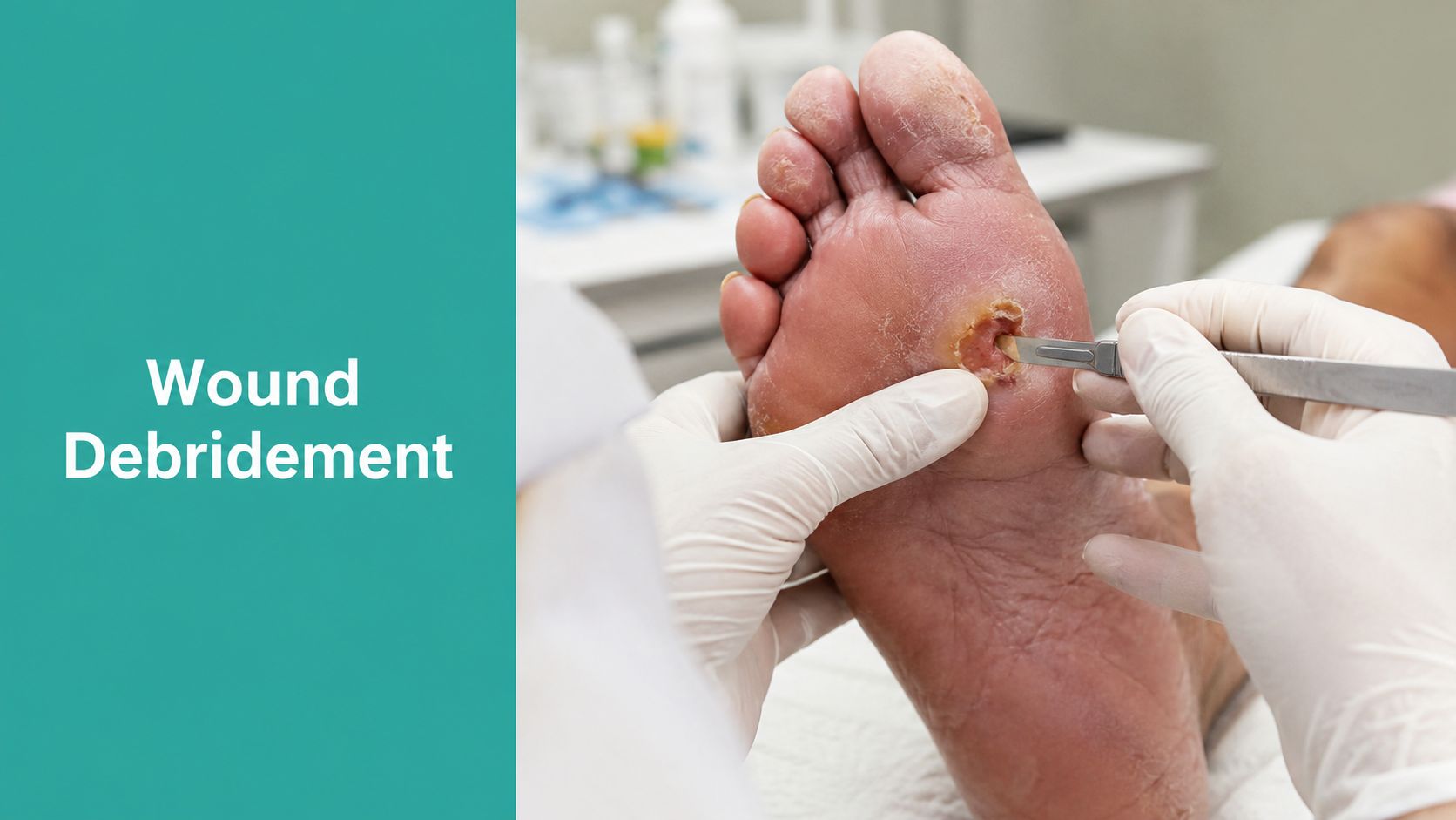
The plantar ulcer is open again, the callus rim is thick, and the dressing pulled away with a string of fibrinous slough. That's the moment when the room gets honest. The wound isn't choosing to heal on schedule, so the team has to choose the right sharp work, the right cadence, and the right note.
A good debridement visit has two jobs at once. It resets the wound bed and it leaves a record that can survive a coder, a payer, and a surveyor. If either part is weak, the next visit gets harder.
What Debridement Looks Like at the Bedside
The patient on the table is the one you've seen before, a 62-year-old with a Wagner 2 plantar diabetic foot ulcer. The edges are rolled with a dense callus rim, the base is mottled with mixed slough, and the drainage is serous enough to keep the dressing wet by lunch. The granulation you wanted last week is still stalled.
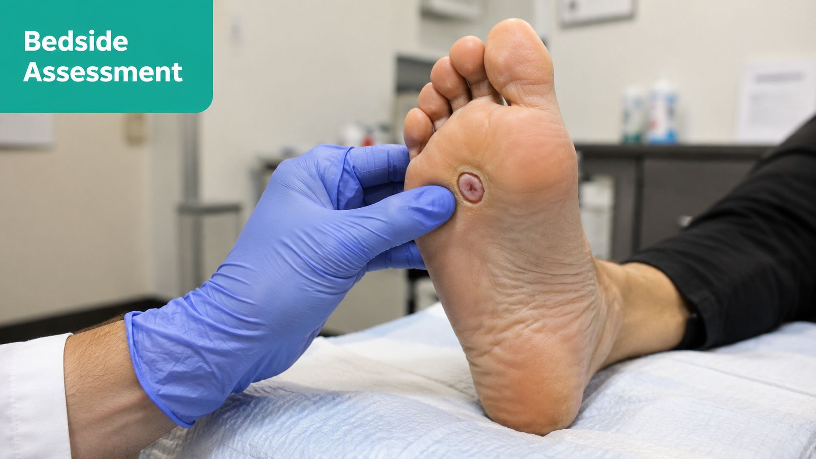
That's the bedside tension. The wound didn't agree to heal on your clinic cycle, so serial sharp debridement has to be treated like an active therapy, not an occasional cleanup. The note has to show what was removed, what was left behind, and why the patient needed you to do it now.
Practical rule: if the wound still has a callus shelf, adherent slough, or epibole after the visit, the problem wasn't “messy tissue,” it was ongoing wound biology that needed source control and edge restoration.
The chart should read like a procedure note, not a dressing note. That distinction matters for CPT 11042–11047, for severity linkage, and for the next clinician who opens the chart and needs to know whether the wound bed is improving or just being dressed over. In a busy clinic, that discipline is the difference between repeatable care and vague documentation.
Defining Wound Debridement in Clinical Practice
Wound debridement means removing devitalized tissue, slough, eschar, biofilm, foreign material, and barriers that keep the wound from building healthy granulation and epithelial migration. The target is viable tissue, often with a bleeding capillary base when sharp techniques are used. That is the clinical act, not a cosmetic cleanup.
The useful distinction is selective versus nonselective. Selective methods aim to remove nonviable tissue while sparing healthy tissue, which is why sharp conservative work is so valuable when the wound has adherent slough or callus and the margin needs precision. Nonselective methods are less tissue-specific, so they may suit different wound biology, different pain tolerance, or different care settings, but they don't give you the same bedside control.
What the wound bed needs from you
The goal is wound bed preparation. That usually means getting the surface ready for dressings, compression, cellular products, or a staged closure plan. Necrotic tissue and biofilm don't sit there neutrally. They sustain inflammation and bacterial load, and necrotic tissue has been described as delaying pressure-ulcer healing by interfering with healthy granulation and creating a better environment for bacteria and sepsis (NCBI Bookshelf).
A sharp pass, though, isn't magic. A single debridement may not measurably shift the wound microbiome, so if slough keeps re-forming, look harder at ongoing necrosis, moisture imbalance, ischemia, or poor offloading. Recurrent debris usually means the wound environment is still hostile, not that the first visit failed.
Debridement is a process, not a cosmetic event. If the wound keeps rebuilding slough, your plan needs more than a fresh gauze wrap.
Choosing the Right Method for the Wound in Front of You
The method should match the biology, anatomy, pain profile, and setting. That sounds obvious until a clinic default turns into the wrong tool for the wound. A thick plantar callus with adherent slough behaves nothing like a shallow, well-perfused ulcer with light fibrin.
Method by method
Sharp debridement is the workhorse when the wound has adherent slough or eschar and you need a clean tissue endpoint. It gives the best control over depth and edge work, especially when callus is part of the problem.
Enzymatic debridement fits thinner slough, lower-exudate wounds, and patients who can't tolerate the scalpel. It's slower, but it can be the right maintenance move when sharp work would be too painful or too aggressive for the current setting. If you need a practical reference point for this method in wound workflows, see selective debridement and collagenase use.
Autolytic debridement works best in shallow, moist, well-perfused wounds with a lighter burden of devitalized tissue. Think hydrogels, hydrocolloids, and appropriate foams when the wound can safely hold moisture and the goal is gentle separation rather than immediate source control.
Mechanical debridement includes wet-to-dry and irrigation, with irrigation often described around 8 to 15 psi in technical references (PMC review). It can dislodge loose debris, but it's low-selectivity and usually not the first choice in chronic care when you need precision.
Biologic debridement has a place too. In the right wound, and with patient access, it can be a credible selective option for cavities and highly exudative wounds. It's not fringe care when the anatomy and goals line up.
| Method | Best-Fit Wound | Selectivity | Pain Burden | Setting Notes |
|---|---|---|---|---|
| Sharp | Adherent slough, callus, eschar | High | Variable | Clinic or OR, depending on depth |
| Enzymatic | Thin slough, low-exudate wounds | Moderate | Usually lower | Good for maintenance or when sharp isn't tolerated |
| Autolytic | Shallow, moist, well-perfused wounds | Moderate | Low | Best when the wound can stay adequately moist |
| Mechanical | Loose debris, surface contamination | Low | Often higher | Useful selectively, not usually first-line |
| Biologic | Cavities, heavy exudate, selective tissue needs | High | Variable | Access and patient acceptance matter |
Technique choice should stay flexible. The newer U.S. consensus frame calls for an escalation and de-escalation approach, because a wound that starts with sharp work may need enzymatic or autolytic maintenance later, and a stalled wound may need more intensive source control again (WoundSource summary of the consensus).
Sharp Debridement Technique Pearls
Start before the blade touches the wound. Check perfusion, infection status, anticoagulation risk, and whether the anatomy in front of you can tolerate sharp work. A vascular assessment belongs in the decision path, because ischemic tissue does not forgive aggressive debridement (NCBI Bookshelf).
Lighting, instrumentation, and patient positioning matter more than people admit. If you cannot see the edge, you will miss undermining. If you do not have the right curette, scissors, or scalpel, you will either under-treat or over-cut.
The sequence that keeps notes clean
Work from the least ambiguous tissue to the most stubborn. Eschar comes off in controlled passes when it is appropriate, then slough, then the biofilm-impregnated surface tissue that keeps the bed stalled. After that, probe the margins for undermining, tunnels, and hidden depth, because those structures often change the apparent size of the wound once they are opened.
Edge work matters. A rolled or epibolic margin needs restoration, not just removal of debris. On the plantar foot, pare the hyperkeratotic rim enough to reduce pressure concentration and let the wound edge re-epithelialize. That is pressure redistribution and biology restoration, not callus shaving for appearance.
The biofilm problem belongs in the same conversation. Once a wound has a persistent slime layer and stalled granulation, sharp work often needs to be paired with a plan for repeat cleansing and local wound-bed control, including the practical steps described in this biofilm on skin guide.
Documentation habit: record the deepest tissue removed, the bleeding endpoint, hemostasis method, and the post-debridement dimensions. If the wound got bigger after the callus and undermining came off, say so plainly.
That last point is where many notes fall apart. Hidden cavities, ridge abscess pockets, and tunneling can make the wound look modest before debridement and larger afterward. If you do not chart the post-procedure wound dimensions, the record looks inconsistent when it is really accurate. A note that tracks depth, exposed structure, and hemostasis is the one that survives review.
For a cleaner coding workflow around selective sharp work, keep the documentation aligned with selective debridement CPT coding.
How Often to Debride and Why Frequency Drives Outcomes
Cadence changes outcomes, and the bedside proof is hard to ignore. In a 2013 retrospective cohort study, 70.8% of chronic wounds healed overall, with a median of 2 debridements per wound and a range from 1 to 138. More frequent debridement was strongly associated with faster healing, with the association reaching P < .001 (PubMed).
The practical message is straightforward. Chronic wounds rebuild slough, biofilm, callus, and drainage between visits, so intermittent cleanup leaves the same devitalized tissue in place and the wound keeps paying for it. Serial debridement is a clinical lever, and the note should justify that lever just as clearly as the blade choice.
What the cadence looks like in real clinics
Weekly-or-more debridement has been associated with much faster closure in commonly cited wound datasets. For diabetic foot ulcers, median time to heal was 21 days with weekly-or-more debridement, compared with 64 days when debridement happened every 1 to 2 weeks, and 76 days when it was no more frequent than every 2 weeks. For venous leg ulcers, the medians were 14, 42, and 49 days, respectively, with P < .001 (MedPage Today summary).
Complete healing rates also favored the more frequent schedule, with 30% vs 13% for DFUs and 50% vs 28% for VLUs in that same summary. That is enough spread to affect clinic flow, SNF follow-up, and mobile wound routing. It also helps explain why recurring sharp work is often tied to persistent surface burden, including the biofilm layer described in this skin biofilm guide(https://ekagrahealth.ai/biofilm-on-skin/).
The billing side follows the clinical side. Repeat visits need a documented medical reason, not just a standing habit. If the wound keeps rebuilding devitalized tissue, the note should say so. If the team is increasing frequency because the wound stalled, document the stall and the active findings, not just the calendar interval.
Anesthesia and Pain Control During Sharp Work
Pain tells you a lot. If the patient is guarding hard, the wound may be more inflamed than it looks, the debridement may be too aggressive for the setting, or deeper pathology may be hiding under the slough. Pain is also a charting issue, because uncontrolled discomfort makes the procedure look poorly selected even when the indication was sound.
For shallow bedside work, topical lidocaine or LET can help. Local infiltration is the usual move for selective sharp debridement when you need more control. Toe and finger work often needs a digital block, and broader field blocks make sense when the wound and callus extend beyond a small focal area.
Matching analgesia to depth
Oral pre-medication with NSAIDs or acetaminophen can take the edge off inflammatory pain before the visit. Short-acting opioids belong only when the work crosses into deeper tissue or bone and the setting can monitor the patient appropriately. Patients on anticoagulation, with significant neuropathy, or with severe PAD often need a change in method rather than a harder push with the same blade.
If the patient can't tolerate the plan, the plan needs revision. That may mean enzymatic or autolytic care today, with sharp work later when the wound and the patient are ready.
Document the pain plan the same way you document the procedure. A note that records the anesthetic used, the patient's tolerance, and the rationale for choosing a less invasive method reads like clinical judgment, not improvisation.
Coding and Documentation That Survives an Audit
CPT selection starts with the deepest tissue removed, not the superficial look of the ulcer. The 11042–11047 family tracks debridement of skin, subcutaneous tissue, muscle, fascia, and bone, and the payment level depends on the deepest layer debrided. That means a wound that looks small but exposes deeper tissue after debridement can land in a different code path than the chart initially suggests (WoundSource coding discussion).
The record has to support the code choice. Surveyors and payers look for the tissue level removed, the method used, wound size, anesthesia, hemostasis, and medical necessity. They also look for ICD-10-CM linkage that fits the wound type, whether that's diabetic ulcer specificity, pressure injury staging, or a chronic non-healing wound pathway.
Common denial triggers
- Missing depth: If the note doesn't say what tissue was removed, the code can fall apart.
- Missing size: Surface area after debridement needs to be clear enough to support the claim.
- Callus-only care: Paring hyperkeratosis without tissue removal is not automatically a debridement service.
- Missing method: Sharp, mechanical, enzymatic, or other methods need to be named.
- Missing necessity: The chart should show why the wound needed debridement today, not just that it happened.
There's also a clean documentation trap around cleansing. Removing non-tissue-integrated fibrin exudate, crust, or surface material without removing tissue doesn't meet the definition of a debridement code. That's why notes that blur cleansing, dressing removal, and callus care create audit risk.
For transcription-heavy programs, it helps to protect patient data transcription while still capturing wound depth, measurements, and procedure details in real time. Clear words in the note make cleaner claims later.
Case Examples From Real Practice
A patient with a Wagner 2 plantar DFU comes in with thick hyperkeratotic rim, mixed slough, no exposed bone, and perfusion that supports salvage. This is the outpatient path. The wound gets serial conservative sharp debridement, callus paring, and offloading, with the note documenting the tissue removed, the post-debridement surface area, and the fact that the wound bed was better exposed after edge work. The usual code path lands in the 11042 family when the deepest tissue removed is skin or subcutaneous tissue, and the ICD-10-CM linkage needs to reflect the diabetic ulcer site and severity clearly.
A different patient presents with a Wagner 3 plantar wound, probe-to-bone, MRI-confirmed osteomyelitis, and enough perfusion to attempt salvage. That patient usually needs operative surgical debridement in the OR, not just clinic-level sharp work. The code pathway shifts to 11044 or higher when bone is debrided, and the plan usually includes IV antibiotics and staged closure, because the wound biology has crossed into a different problem set.
Safety boundaries that don't get enough respect
Absolute and relative contraindications matter. Untreated PAD can make sharp debridement dangerous. Stable heel eschar in a non-infected patient is another situation where aggressive sharp removal can be the wrong move. Complications include bleeding, bacteremia, and injury to deep structures, especially when tunnel tracts or bone exposure are present.
A clean note on either case should include:
- Pre-procedure assessment: perfusion, infection, and pain status.
- Deepest tissue removed: skin, subcutaneous tissue, muscle, fascia, or bone.
- Surface area after debridement: not the pre-cleanup estimate.
- Anesthesia and hemostasis: what was used and how bleeding was controlled.
- Changed dimensions: if debridement exposed hidden depth, say it.
- Plan forward: offloading, dressing choice, antibiotics, or OR escalation.
- ICD-10-CM linkage: enough specificity to justify the medical necessity.
Keep that checklist in mind on the next clinic day. If the wound is a DFU, a pressure injury, or a chronic leg ulcer, the same discipline applies. The note should tell the story of the tissue, the method, the depth, and the reason the patient needed your hands on that visit.
EkagraHealth AI helps wound teams capture debridement details at the point of care, including tissue removed, depth, instruments, and pre- and post-procedure dimensions. If you're trying to make sharp work easier to document and easier to code, visit EkagraHealth AI and see how it fits into a wound clinic, mobile program, or SNF workflow.
