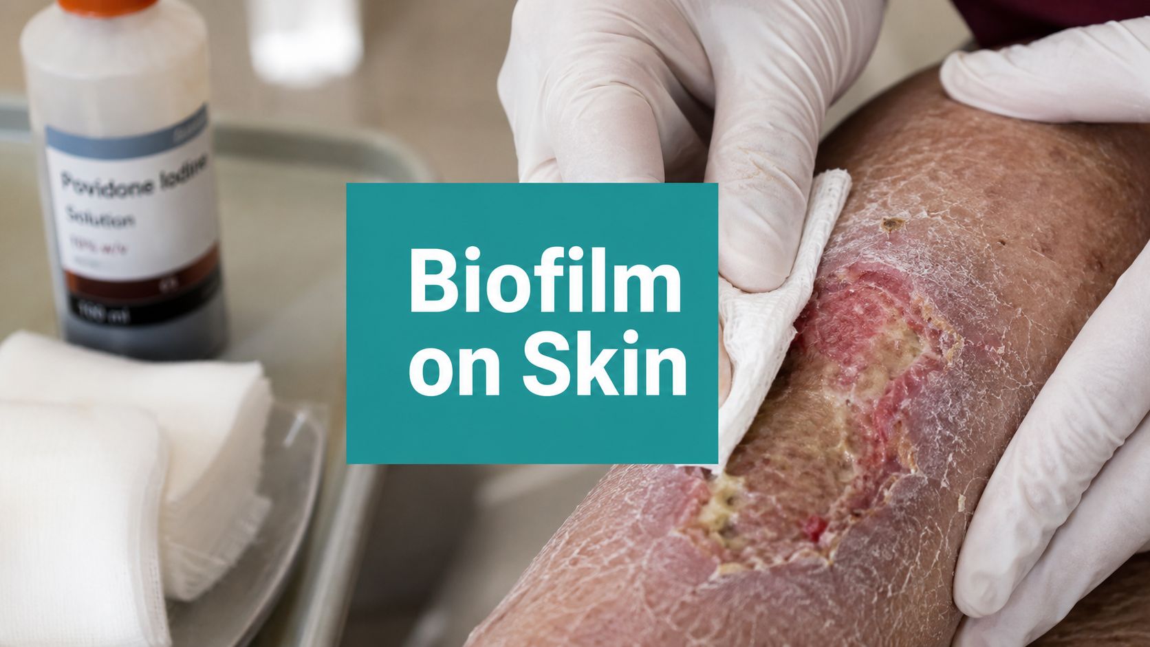
You debrided a Wagner 2 diabetic foot ulcer last week. The bed looked cleaner at the end of the visit, the drainage was manageable, and the family left thinking things had finally turned a corner. Then the patient comes back, and the wound looks familiar in the worst way. Thin yellow slough again. A slick surface. Pale granulation. More drainage than you expected. Not frankly cellulitic. Not truly improving.
That pattern is where a lot of wound programs lose time, and where a lot of claims get denied.
The mistake is treating the wound as vaguely “slow to heal” instead of naming the problem. In many of these cases, you're dealing with biofilm on skin and in the wound bed, not simple surface contamination. Once you recognize that, the care plan changes. Your documentation should change too. Payers don't reimburse “it still looked bad.” They reimburse medically necessary, clearly justified interventions tied to a wound that has stalled despite appropriate care.
Table of Contents
- That Stalled Wound Is Probably Biofilm
- The Microbiology Behind the Slime
- How to Recognize Biofilm Without a Microscope
- The Stepwise Approach to Biofilm Management
- Debridement Deep Dive Codes and Techniques
- Documentation and Billing for Biofilm-Driven Care
- Clinical Vignettes and Final Takeaways
That Stalled Wound Is Probably Biofilm
Monday's wound looks better after debridement. By the next visit, the same ulcer has drifted back to a familiar pattern. Thin slough. More drainage than expected. Fragile granulation. Periwound inflammation that never fully settles. In practice, that repeated rebound should push biofilm high on the differential.
The key point is behavior over time. A wound can be cleaned regularly, offloaded appropriately, compressed correctly, and still fail to build durable forward progress. That pattern matters because it changes both treatment and documentation. If the wound keeps reverting after apparently appropriate care, the note cannot just say “continue local wound care.” It needs to describe the recurring tissue changes, the reason serial debridement remains medically necessary, and why the plan is being adjusted instead of repeated out of habit.
I see this most often in diabetic foot ulcers, venous leg ulcers, and pressure injuries with several competing barriers to healing. Edema, neuropathy, ischemia, pressure, friction, and poor glycemic control may all be present at once. Biofilm does not replace those problems. It sits on top of them and helps explain why the wound looks briefly improved after treatment, then stalls again before the next encounter.
That distinction has administrative consequences.
If you suspect biofilm, the chart should support active management rather than passive observation. Document the pre-debridement wound bed, the immediate post-debridement change, and the short-interval recurrence that justifies repeat intervention. That language helps later when you bill serial debridement, defend visit frequency, or answer a payer request for records. For teams building chronic wound care workflows, frequent reassessment is not just good medicine. It also produces cleaner support for medical necessity than vague weekly notes that all read the same.
A simple SOAP example is often enough to tighten the record:
S: Patient reports persistent drainage and no meaningful interval improvement despite adherence to offloading/compression.
O: Wound bed with recurrent adherent slough, friable granulation, moderate exudate, and delayed size reduction since prior visits.
A: Stalled chronic wound with recurrent surface contamination pattern concerning for biofilm, contributing to delayed healing.
P: Sharp debridement performed to remove nonviable tissue and disrupt suspected biofilm. Continue moisture balance, pressure or edema control, and close follow-up due to repeated wound bed deterioration between visits.
That kind of note gives you something usable later if serial debridement is billed under CPT 97597, 97598, 11042, or related codes, depending on depth and tissue removed. It also shows why another antibiotic is not automatically the right next step. For a plain-language explainer you can share with staff, this overview of how biofilms develop is useful.
The Microbiology Behind the Slime
Biofilm isn't just bacteria sitting on top of skin. It's a structured community attached to a surface and protected by an extracellular polymeric substance, or EPS. Think of free-floating bacteria as scattered individual cells. Think of biofilm as a fortified settlement built onto the wound bed.

Why the wound keeps relapsing
The matrix is the whole problem. It physically shelters organisms, slows penetration of antimicrobials, and blunts immune access. That's why the wound can seem calm on the surface while still acting chronically inflamed underneath.
In vivo, biofilm can colonize and cover a surface within 4 to 8 days, and if a wound plateaus for about 3 to 4 weeks, biofilm should move high on your differential even when classic infection signs are absent, as summarized by DermNet's review of bacterial biofilm. Clinically, that timing matters. It explains why a wound that looked improved right after debridement can look stalled again at the next visit.
If you want a simple, non-clinical explainer to share with staff or newer clinicians, this overview of how biofilms develop is useful because it walks through attachment, microcolony formation, and maturation in plain language.
Why swabbing and washing fall short
Routine cleansing helps. It does not solve the structural issue.
A surface rinse can remove loose debris and reduce obvious bioburden, but it often leaves the organized matrix behind. Standard swabs have the same limitation. They sample what's accessible, not necessarily what's driving the chronicity. That's why a wound can have a modest culture result and still behave aggressively, or show organisms that don't fully explain the clinical picture.
Biofilm is less a “which bug is it” problem and more a “what architecture is protecting it” problem.
That distinction should shape treatment. If the problem is structural, the first effective move is structural disruption. This is why mechanical debridement remains the center of care and why prolonged empiric antibiotics without source control so often disappoint.
How to Recognize Biofilm Without a Microscope
You see the patient on Monday. The wound is cleaned up, the surface looks better, drainage is controlled, and the plan seems reasonable. By the next visit, the bed has that same slick coating, the tissue looks dull again, and your note starts sounding like last week's note. That repeat pattern is often the bedside clue.
What the wound bed is telling you
Biofilm is usually a clinical recognition problem, not a microscopy problem. The diagnosis often comes from wound behavior over time, especially when standard care elements are already in place and the wound still cycles back to the same appearance.
Common bedside findings include:
- Recurrent surface film: After cleansing or debridement, the wound looks cleaner for a short interval, then a shiny, viscous, or slimy layer returns.
- Poor-quality granulation: Tissue appears pale, dusky, friable, or easy to disrupt instead of healthy and progressive.
- Exudate that does not fit the rest of the picture: Drainage may be heavier than expected, or thicker and more adhesive.
- Persistent odor after cleaning: The wound never quite smells clean even when obvious debris has been removed.
- Minimal measurable progress: Offloading, compression, edema control, and moisture management may all be appropriate, yet the wound dimensions barely change.
As noted earlier in the article, chronic wounds carry biofilm far more often than acute wounds. A commonly cited estimate is about 60% of chronic wounds versus 6% of acute wounds. Organisms in biofilm can also be far less responsive to antibiotics than free-floating forms. Clinically, that helps explain why a wound may look only mildly inflamed while still resisting closure.
Slough can confuse the picture. Some wounds have both slough and a recurrent biofilm pattern, and the two are not interchangeable. If you need a quick refresher on what slough suggests at the bedside, this guide on slough in a wound bed is a useful reference.
What not to over-trust
Swab results have limits. A low-yield culture does not rule out a structurally protected microbial burden, and a positive culture does not prove it is the reason healing has stalled.
A brief improvement on oral antibiotics can also mislead the team. If the wound quiets down during the course, then returns to the same baseline once antibiotics stop, that supports the need for source control and repeat mechanical disruption. It does not automatically support another empiric prescription.
Documentation matters as much as pattern recognition. If you suspect biofilm, chart the recurrence clearly: film re-forms within days, granulation remains friable, odor persists after cleansing, exudate stays disproportionately heavy, and wound area reduction remains negligible despite adherence to the broader plan. That language helps justify why serial debridement is still medically necessary, especially when you later bill repeated wound debridement services and need to show that each session addressed a persistent barrier to healing rather than routine cleaning.
A SOAP note can make that easier. In the objective section, record tissue type, percent nonviable surface material, exudate character, odor after cleansing, and whether the wound bed deteriorated between visits. In the assessment, state that wound behavior is concerning for biofilm-driven chronicity. In the plan, tie debridement frequency to the observed rebound pattern. Payers respond better when the note shows failed interval progress and a specific reason the wound needs repeated disruption.
Dressings support this work, but they do not replace recognition of the pattern. If the wound bed is dry after debridement and needs moisture balance to support autolysis or patient comfort, DME Superstore's hydrogel guide gives a practical summary of where hydrogels fit. The trade-off is simple. Too much moisture can worsen maceration, and too little can leave the surface desiccated and harder to manage.
The Stepwise Approach to Biofilm Management
A familiar clinic problem goes like this. The wound looks cleaner right after treatment, drainage eases for a day or two, then the surface turns dull again, slough returns, and progress stalls. At that point, the plan has to do two jobs at once. It has to improve the wound bed, and it has to create a record that explains why repeated skilled intervention remains medically necessary.
For biofilm-suspected wounds, management works best as a sequence. Debride. Apply a topical strategy that can contact the wound surface after disruption. Choose dressings that support moisture balance and protect the periwound. Then reassess on a short enough interval to see whether the wound rebounds.
Debridement
Serial debridement is usually the anchor of the plan because biofilm is a surface-level barrier that keeps re-forming. In practice, the question is rarely whether to debride. The question is how aggressively, how often, and whether the patient and setting can support that schedule.
Sharp debridement is often the most efficient option when the wound has recurrent slough, a glossy or slimy surface after cleansing, or a pattern of short-lived improvement. It removes nonviable tissue and disrupts the matrix that shields organisms from topical contact. If the wound is ischemic, very painful, anticoagulated, or anatomically risky, the trade-off changes. You may need a more conservative approach, but the note should still explain why the method was limited and what barrier remains.
That matters clinically and administratively. If you expect serial debridement, document the wound behavior that justifies it at each visit. A practical plan might read: reassess in 3 to 7 days due to rapid re-accumulation of surface debris, persistent exudate, and limited interval healing. That language supports the care plan now and helps later if repeated procedure codes are questioned.
Topical agents
Topical therapy has a narrower role than many notes suggest. It supports debridement. It does not replace it.
After the wound bed is mechanically disrupted, topical antiseptics or antimicrobial dressings have a better chance of reaching exposed organisms and reducing the surface burden. Selection depends on drainage, tissue tolerance, peri-wound skin condition, depth, and whether the wound bed is dry, balanced, or overloaded with exudate. A wet venous ulcer and a dry neuropathic wound should not get the same dressing logic.
The trade-offs are practical:
- Over-drying the wound can slow granulation and increase pain at dressing change.
- Under-managing exudate encourages maceration and lets surface debris build back up.
- Reaching for systemic antibiotics without signs of deeper infection can add adverse effects while leaving the local barrier in place.
If the wound repeatedly develops adherent yellow material, call it what it is and describe the extent. Clear tissue identification improves both care and documentation. This guide on slough in a wound bed is a useful refresher for teams that tend to chart every yellow surface finding the same way.
Dressing strategy
Dressings should match the wound after debridement, not the wound from last month.
A heavily draining ulcer usually needs absorbency, edge protection, and a change schedule that prevents periwound breakdown. A cleaner wound with a dry surface may need moisture donation so the bed does not desiccate between visits. If you need a quick refresher on where that fits, DME Superstore's hydrogel guide gives a practical summary.
Keep the dressing plan simple enough that staff and caregivers can carry it out consistently. Complex regimens fail quickly in home settings, and payer denials often follow when supply use looks out of proportion to the documented wound characteristics. If serial debridement is part of the strategy, the between-visit dressing should support that goal by controlling exudate, limiting trauma, and preserving the progress you created at the bedside.
A workable post-debridement checklist:
- Match absorbency to actual drainage level.
- Protect fragile wound edges and surrounding skin.
- Avoid dressings that tear up fresh granulation at each change.
- Write the change frequency so it fits the site of care and available hands.
- Document why the selected dressing supports the current debridement plan.
Debridement Deep Dive Codes and Techniques
A lot of chronic wound care gets under-documented because clinicians describe the wound but don't describe the work. Biofilm management is one of the clearest examples. The procedure is often medically necessary, but the note reads like routine dressing care.
A major review states that approximately 80% of all human infections are associated with biofilms, and that when a lower-extremity ulcer fails to shrink after 2 to 4 weeks of standard care, biofilm becomes a primary suspect. That shift in understanding is one reason repeated debridement and topical antisepsis often outperform prolonged antibiotics alone as summarized here.
Choosing the right debridement method
For suspected biofilm, sharp debridement is usually the workhorse because it is immediate and targeted. It lets you remove slough, nonviable tissue, and the adherent surface layer that keeps reforming. Curette, scalpel, or scissors can all be appropriate depending on wound location, depth, pain, and clinician skill.
Selective debridement has a narrower role. It can make sense when tissue preservation is critical and the surface burden is mostly loose fibrin or slough. Enzymatic and autolytic approaches can support ongoing management between visits, but they are usually adjuncts in a wound that is clearly stalled. Mechanical methods can help in some settings, but if the wound keeps cycling back, many cases still need sharper source control.
Debridement methods for biofilm and associated CPT codes
| Debridement Method | Clinical Application for Biofilm | Primary CPT Codes |
|---|---|---|
| Sharp excisional debridement to subcutaneous tissue | Stalled wound with recurrent slough or nonviable tissue requiring active removal of biofilm-laden tissue and wound bed reset | 11042, 11045 |
| Sharp excisional debridement to muscle and/or fascia | Deeper wound with devitalized tissue extending beyond subcutaneous level | 11043, 11046 |
| Sharp excisional debridement to bone | Osteal exposure or necrotic bone requiring debridement | 11044, 11047 |
| Selective debridement | Removal of devitalized tissue without excision of viable deeper tissue, often for surface fibrin or slough | 97597, 97598 |
The coding point is simple. Bill to the depth of tissue debrided, not the deepest tissue visible in the wound. Your procedure note should include instrument used, tissue removed, pre- and post-debridement measurements when required by your workflow, hemostasis, patient tolerance, and the medical reason repeat debridement was necessary.
Documentation and Billing for Biofilm-Driven Care
Monday morning clinic. The wound measures about the same as last week, the surface film is back after prior debridement, drainage has picked up again, and the patient's insurer is now asking why another procedure was needed. That is the biofilm problem in practice. You are treating a biologically stubborn wound and building a record that has to justify each step.
What payers need to see
A defensible note shows a pattern over time, not a one-line procedure entry. If serial debridement is part of care, the chart should explain why the wound keeps stalling, what recurring findings support suspected biofilm, what conservative treatment was already in place, and what changed after intervention.
Document the wound as a chronic wound that is failing to progress despite appropriate standard management for its cause. That may include offloading for a diabetic foot ulcer, compression for a venous ulcer, edema control, moisture balance, pressure redistribution, or topical antimicrobial management. Then document the features that keep returning. Adherent slough, slick surface film after cleansing, friable or poor-quality granulation, recurrent malodor at dressing change, increased exudate, and a short-lived response after prior debridement all help explain why repeat source control is medically reasonable.
Payers also look for operational details that are often missed. State the interval change since the last visit. Record tissue type removed, depth of tissue debrided, instrument used, bleeding and hemostasis, pain control, patient tolerance, and the post-procedure plan. If the procedure was selective, your note should read differently than an excisional debridement note.
The coding side has to match the note. Use 97597 and 97598 for selective debridement when you are removing devitalized tissue without excising viable deeper tissue. Use 11042 and 11045 for subcutaneous excisional debridement, 11043 and 11046 for muscle or fascia, and 11044 and 11047 for bone. The common error is obvious. Clinicians document exposed structures but fail to document that those structures were debrided. Billing then outpaces the procedure note.
SOAP language that actually helps
Generic wording creates denials because it hides medical necessity. A better SOAP note links the wound's behavior to the decision to debride again.
Subjective: Patient reports persistent drainage and odor at dressing changes. No fever, chills, or new spreading erythema. Prior oral antibiotics gave no sustained change in wound appearance.
Objective: Chronic plantar diabetic foot ulcer with minimal interval size improvement. Wound bed shows recurrent adherent yellow slough, surface film returning after prior cleansing, pale friable granulation, and moderate serosanguinous drainage. No systemic signs of infection documented today.
Assessment: Stalled chronic diabetic foot ulcer with suspected biofilm burden based on repeated relapse pattern, poor granulation quality, and failure to progress despite offloading, local wound care, and prior debridement.
Plan: Sharp excisional debridement of nonviable subcutaneous tissue performed today using curette to disrupt recurrent surface burden and remove devitalized tissue. Hemostasis achieved with pressure. Continue offloading and moisture-balanced antimicrobial dressing. Short interval follow-up due to recurrent wound bed deterioration between visits.
That format does two jobs. It supports care, and it shows why repeat debridement was not routine scheduling.
Be explicit about why you did not lead with antibiotics alone. If there is no cellulitis, no systemic illness, and the wound repeatedly relapses after temporary improvement, say so. The chart should show that the working problem is a chronic wound with recurrent surface burden requiring source control, not an untreated acute soft tissue infection.
For payer review, I also recommend one sentence that ties serial procedures to observable recurrence: “Repeat debridement is indicated due to rapid reaccumulation of devitalized tissue and recurrent wound bed stagnation since the last visit, limiting progress toward closure.” That sentence helps when you are justifying ongoing treatment over several weeks.
Teams tightening documentation can use SOAP note templates and wound charting examples to standardize what gets captured at each visit. If your clinicians struggle with omissions, Simbie AI SOAP notes also gives a general view of structured SOAP workflows that can reduce missed elements in procedure documentation.
Clinical Vignettes and Final Takeaways
The pattern becomes obvious when you see it enough times.
Vignette one
A plantar diabetic foot ulcer, Wagner 1 moving toward 2 in practical management terms, had decent offloading and no dramatic surrounding cellulitis. The wound still plateaued. Each visit showed recurrent yellow slough, a slick surface after cleansing, and granulation that never looked healthy. The mistake would've been calling it “stable.”
The better read was a stalled chronic wound with suspected biofilm. The note focused on recurrent slough, nonprogressing measurements, tissue quality, and rationale for repeat sharp debridement. That kind of chart supports both care and billing.
Vignette two
A venous leg ulcer came in with heavy exudate, periwound maceration, low-grade odor after dressing removal, and a wound bed that looked superficially clean but biologically stagnant. Compression was part of the answer, but not the whole answer.
The plan worked only once the team stopped treating drainage as the sole problem. Serial debridement, topical control after disruption, and more disciplined documentation of exudate type, periwound condition, and recurrent surface film made the case make sense clinically and administratively.

A few takeaways are worth keeping blunt:
- Assume biofilm early in a stalled chronic wound. Don't wait for the chart to collect weeks of vague disappointment.
- Debridement is usually the pivot point. Topicals and dressings matter more after the matrix is disrupted.
- Don't let the note lag behind the care. If you're doing serial debridement, your documentation should show the recurring pattern that justifies it.
- Describe the wound like a clinician, not a template. Tissue type, exudate character, periwound status, and short-interval recurrence are what make the record credible.
The wound bed and the claim form are connected more tightly than is often acknowledged. When you identify biofilm on skin and in chronic wounds accurately, treat it aggressively, and document the pattern clearly, outcomes improve and denials get harder to justify.
EkagraHealth AI helps wound care practices turn visits into cleaner SOAP notes, tighter CPT and ICD-10 coding, and fewer documentation gaps that trigger denials. If your team is spending too much time charting serial debridements, wound measurements, and medical necessity language by hand, it's worth seeing how a documentation and billing backbone built for wound care can lighten the load.
