
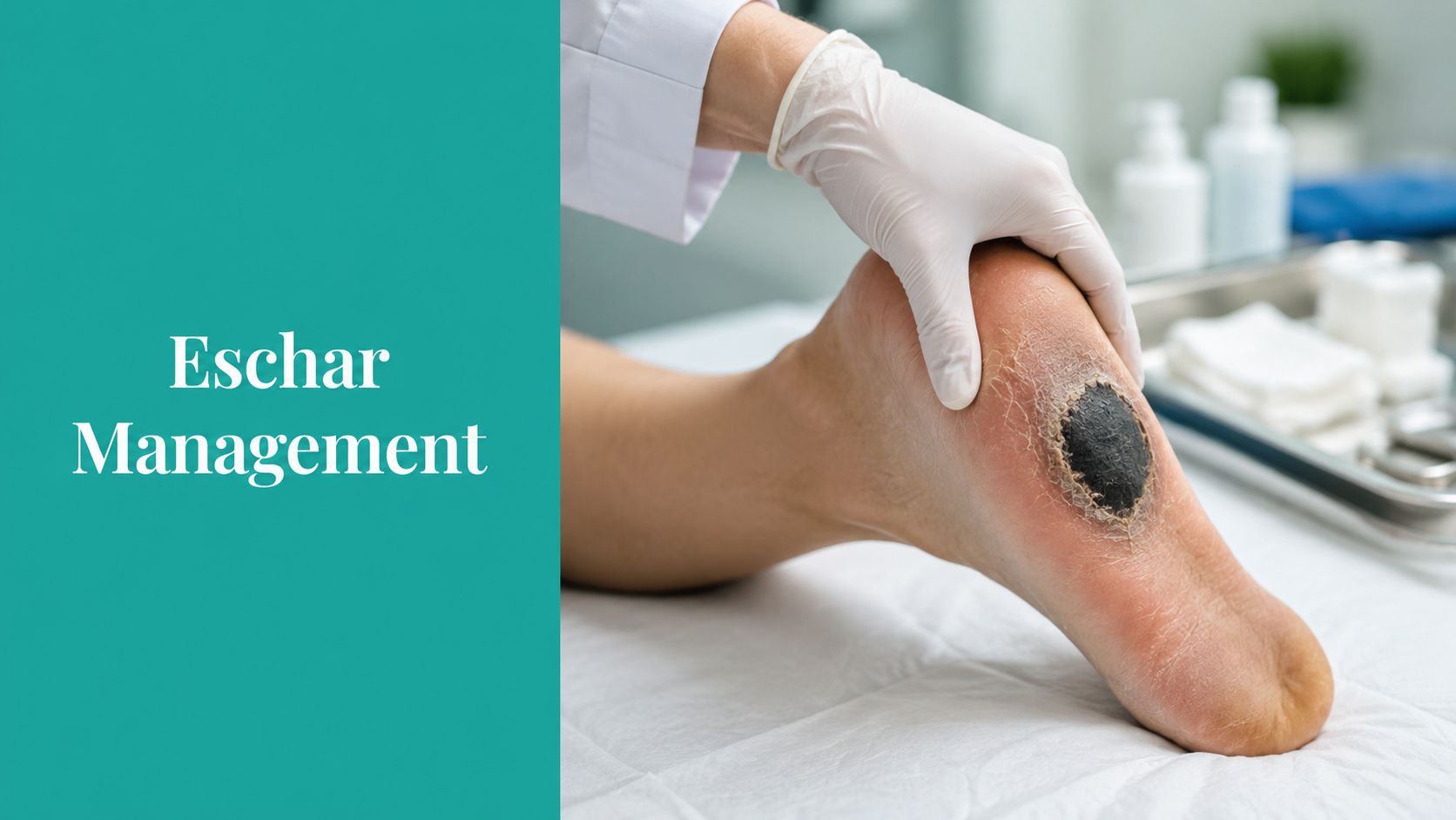
A black heel on a diabetic patient usually lands on your list at the worst time, a home visit, a late charting catch-up, or a rushed rounding block. The lesion may be dry and hard as a shell, or it may hide drainage and erythema at the edges. That first look matters because eschar can be a protective cover in one patient and a sign of deeper necrosis or infection in another.
The practical problem is that clinicians often see one dark surface and stop there. That's where both patient safety and billing get messy. If you treat a stable heel cap like routine slough, or document it as a generic scab, you can miss perfusion issues, miss infection, and create a chart that won't support the decision you made.
Introduction to Eschar Assessment
A home health nurse walks into a house and finds a black crust on the heel of a patient with diabetes. The wound is quiet, the foot is cool, and the family wants to know whether it needs to be cleaned off right away. That's the right moment to slow down, not speed up.
Start with three observations. Is the tissue dry and firmly adherent? Is there pain, warmth, odor, drainage, or erythema? Does the location suggest pressure or ischemia rather than surface contamination? Those details drive whether you protect the area, image it, culture it, or escalate. They also decide whether your note supports conservative care or debridement.
Practical rule: A black heel surface is not automatically “dirty tissue.” In an ischemic limb, removing it too early can enlarge the defect and make healing harder.
The clinical stakes are bigger than the wound bed. In wound medicine, eschar is not a diagnosis by itself. It is a sign of tissue death that may sit on top of pressure injuries, ischemic ulcers, burns, infections, or rickettsial disease, so the history has to come first, not the instrumentation.
A clean opening note usually includes the appearance, location, surrounding tissue, and the perfusion context. If the patient has a pressure point wound, document the offloading plan. If the lesion doesn't fit the usual pressure pattern, your differential has to widen quickly.
Understanding Eschar and Related Wound Coverings
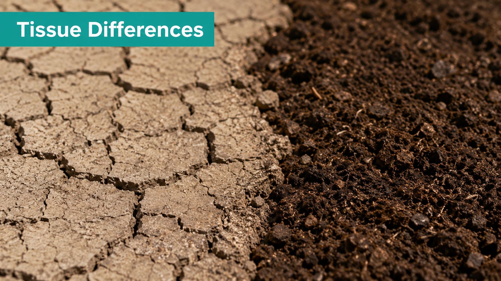
At the bedside, eschar, scab, and slough are not interchangeable. A scab is dried exudate on the surface. Eschar is devitalized tissue, often dark, adherent, and firm. Slough tends to be moist, stringy, or yellow-gray, and it usually reads as nonviable tissue in a wetter wound environment.
The easiest documentation mistake is calling any dark crust “scab” because it sounds less serious. That can understate the problem. If the tissue is leathery, fixed, and sitting on a full-thickness wound bed, call it eschar and describe what you can see around it, not what you wish the wound looked like.
The distinction also matters for claims. Shared Health Services notes that crust is dried exudate and not devitalized tissue, while eschar is devitalized tissue. If only crust is removed and no wound remains underneath, that does not support debridement billing. That's a common denial path in routine chart review and post-payment audit.
A tight SOAP note should use consistent language across the exam, photo caption, and plan. Write the color, moisture, adherence, odor, periwound condition, and whether the lesion obscures the base. If the wound bed can't be staged because the surface is covering deeper tissue loss, say that plainly.
For more on the gray zone between nonviable material and wound-bed debris, see the related discussion of slough in a wound bed.
Charting tip: If the edge is dry but the border is red or boggy, don't bury that in a general “wound stable” phrase. Periwound change changes the whole interpretation.
Clinical Importance of Eschar in Wounds and Infections
The same dark lesion can send you in two very different directions. A dry heel eschar over a pressure point often points toward ischemia and protection first. A necrotic scab at an arthropod bite site points toward infectious workup and exposure history. If you mix those up, you either over-treat a stable wound or under-treat a systemic illness.
The CDC describes an eschar-associated rickettsiosis lesion as the inoculation site for pathogens transmitted by ticks or mites, so necrotic scabs after arthropod exposure should trigger travel and exposure histories (CDC eschar-associated rickettsioses fact sheet). That same morphology can also show up in other serious conditions. The point is not to force a diagnosis from appearance alone. The point is to let the lesion push you toward the right history.
The pattern matters in burns too. If you're thinking about infection risk in a burn with necrotic tissue, a useful companion resource is managing infection risks from burns, especially when you're deciding whether to widen the differential beyond pressure injury. Use that mindset when the lesion doesn't match a simple heel ulcer.
A practical triage sequence helps:
- Exposure history first: Tick, mite, trauma, burn, surgery, or pressure exposure all point you in different directions.
- Systemic clues next: Fever, regional adenopathy, pain out of proportion, or malaise should move infection up the list.
- Local wound behavior last: Stable, dry, adherent tissue is different from a lesion that is draining, malodorous, or inflamed.
In a suspected infection, the lesion is not just a wound-bed issue. It becomes a diagnostic clue, a source-control question, and sometimes a public health issue.
Assessing Depth Perfusion and Coding for Eschar
Depth and perfusion come before code selection. If the tissue is black but the limb is poorly perfused, you need to know whether debridement is even appropriate. If the lesion is on a heel or over an arterial territory, assess arterial flow, not just surface appearance. That's the difference between a wound that can close and one that will declare itself worse after an aggressive procedure.
For bedside work, document the measurable findings. Record wound dimensions, tissue depth if visible, drainage type, odor, and periwound temperature or color. If you have vascular data, include that. If you don't, say why the wound was not fully opened or staged. That level of honesty is what protects the claim and the patient.
Practical rule: If perfusion hasn't been checked, aggressive sharp debridement on a stable heel eschar is a decision, not a default.
The billing distinction is just as important. Shared Health Services notes that removing only crust without exposing viable tissue cannot be billed as debridement. That's why the note has to prove you removed devitalized tissue, not just surface debris.
CPT and ICD-10 Codes for Eschar Assessment and Debridement
| Code | Description | When to Use |
|---|---|---|
| 11042 | Debridement, subcutaneous tissue, first 20 sq cm or less | Use when debridement reaches subcutaneous tissue and the note supports depth and area |
| 11043 | Debridement, muscle and/or fascia, first 20 sq cm or less | Use when the wound is debrided to muscle or fascia and the record shows that depth |
| 11044 | Debridement, bone, first 20 sq cm or less | Use when bone is involved and actually debrided |
| 11045 | Each additional 20 sq cm, subcutaneous tissue, add-on | Use only with the base debridement code when extra area is treated |
| 11046 | Each additional 20 sq cm, muscle and/or fascia, add-on | Use only with the corresponding base code |
| 11047 | Each additional 20 sq cm, bone, add-on | Use only with the corresponding base code |
| ICD-10-CM for pressure injury staging | Stage and location must match the lesion status | Use the exact pressure injury code that fits the documented stage or unstageable status |
| ICD-10-CM for diabetic foot involvement | Match the diabetic foot ulcer code set to depth and laterality | Use when diabetes and foot ulceration are both documented |
| ICD-10-CM for ischemic or arterial disease context | Use vascular disease codes when perfusion is the driver | Use when poor blood flow is central to the wound's cause |
If the lesion is unstageable because eschar obscures the base, that language must be in the chart. The related guidance on unstageable pressure ulcer documentation helps align the wound description with the coding path.
Management Options for Eschar Removal and Preservation
Management starts with a blunt question. Is this tissue acting like a cap or acting like a problem? In pressure injuries and arterial insufficiency, dry, stable eschar can function as a biologic cover. Once it becomes boggy, draining, malodorous, or erythematous, debridement is generally indicated.
The leave-it-alone approach has a real place. Keep the area dry, offload pressure, protect the periwound, and document the stability of the cap. This is common on ischemic heels, where premature sharp debridement can enlarge the defect and worsen nonhealing. If the cap is stable, the goal is surveillance, not heroics.
When removal is appropriate, the method should match the wound. Conservative autolysis works when the tissue is stable enough and the wound environment can safely support moisture balance. Enzymatic or mechanical methods can be used when sharp removal isn't appropriate. Sharp or surgical debridement belongs when the lesion is unstable, infected, or clearly preventing source control. A partial eschar that has lifted and revealed a deeper cavity may also need subsequent wound-bed management, including negative-pressure therapy when the bed and exudate profile fit.
Common treatment logic
- Autolytic care: Favor when the goal is gradual softening and the tissue is not acutely infected.
- Sharp or surgical debridement: Favor when devitalized tissue is loose, infected, or blocking assessment.
- Mechanical cleansing: Use carefully when surface debris needs removal but the wound can't tolerate aggressive sharp work.
- Protective offloading and dry care: Use when perfusion is marginal and the cap is stable.
- Post-debridement wound support: Use dressings and adjuncts that match the depth, exudate, and bioburden.
The code range matters, but only after the clinical choice is made. Document depth before the procedure, the tissue removed, the post-debridement base, and why that method was chosen over preservation. If you're writing for surveyors and payers, the note has to show medical necessity, not just activity.
For a focused breakdown of code-level technique, the internal coding reference on selective debridement CPT code is the right companion to this kind of charting.
Real Cases and Documentation Templates
A heel eschar in a diabetic patient with peripheral arterial disease and an inoculation eschar from a tick bite may look similar at a glance. They are not managed the same way, and they shouldn't be documented the same way either. Eschar can be a key diagnostic clue for infectious, toxic, embolic, or vasculitic disease, and too many wound notes skip the timing and exposure details that decide the next move (MedlinePlus on eschar).
Case 1, ischemic heel eschar
The heel is dry, adherent, and non-fluctuant. The patient has known diabetes, diminished pulses, and pain only with manipulation. The right note emphasizes perfusion status, offloading, and the decision to preserve the cap.
SOAP snippet
- S: Heel sore without drainage. Denies fever.
- O: Dry black adherent heel eschar, no odor, no fluctuance, periwound cool, limited capillary refill.
- A: Stable eschar over ischemic pressure point, perfusion concern.
- P: Offload heel, protect dry eschar, vascular assessment, avoid sharp debridement until perfusion clarified.
Case 2, tick-related inoculation eschar
The lesion is a necrotic scab with surrounding inflammation after outdoor exposure. The chart needs travel, insect exposure, fever history, adenopathy, and timing. If those details aren't there, the note reads like generic wound care when it should read like infectious triage.
SOAP snippet
- S: Recent tick exposure, fever, malaise.
- O: Necrotic lesion with surrounding erythema at bite site, regional tenderness.
- A: Eschar-associated infectious lesion, rickettsial disease in differential.
- P: Exposure history documented, prompt systemic evaluation, consider infectious workup.
If the visit is moving fast, voice capture can help preserve the exact sequence of findings and decisions. A practical reference is WhisperAI's guide to medical documentation, especially when you need the chart to reflect the exposure history and the wound decision clearly.
Key Takeaways for Practice and Billing
Eschar is a finding, not a diagnosis. The first job is to decide whether it's stable cover, unstable necrosis, or a clue to infection or ischemia. That decision affects staging, debridement, imaging, and whether the chart supports a procedure claim.
Billing gets cleaner when the note proves depth, tissue type, and medical necessity. Crust is not eschar, and removing only crust doesn't support debridement. If the wound is unstageable, say so. If perfusion is the limiting factor, say that too. Clean documentation protects the patient and the claim.
Use EkagraHealth AI when you want wound notes, code mapping, and procedure documentation captured in one workflow, with the eschar description, staging language, and CPT logic recorded in a way that supports both clinical care and revenue cycle review.
