
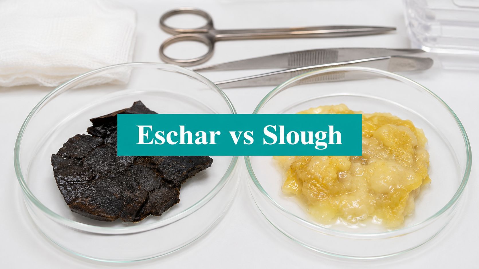
At the bedside, the wound doesn't always tell you what it is right away. A dark heel, a yellow fibrous base, a drainage-heavy sacral ulcer, all of them can look like “dead tissue” until you slow down and separate eschar from slough with a real management plan in mind. That distinction changes staging, debridement, billing, and whether the wound is even safe to touch.
| Tissue type | Typical look | Texture and adherence | What it usually means | Usual management direction |
|---|---|---|---|---|
| Eschar | Black or dark brown | Dry, hard, leathery, firmly adherent | Full-thickness necrosis, often tied to ischemia or pressure | Leave stable heel eschar intact, or debride if unstable |
| Slough | Yellow, white, or gray | Moist, soft, stringy, loosely adherent | Nonviable debris in an inflammatory wound bed | Remove when appropriate so granulation can progress |
For clinicians working across bedside care, documentation, and revenue integrity, practical wound judgment is essential. A wound note that captures moist vs dry, stable vs unstable, and adherent vs loose tissue does more than describe the bed. It supports the treatment plan, the debridement choice, and the story you'll need if the chart gets reviewed. Teams using digital healthcare solutions to organize wound documentation often find that the primary challenge isn't seeing necrotic tissue. It's classifying it consistently enough that the whole team acts on it the same way.
Introduction to Eschar vs Slough
A patient rolls in with a heel pressure injury, or a diabetic foot ulcer that's been “cleaned” but still looks wrong. The wound bed is dark in one spot, yellow and stringy in another, and the notes from the prior visit don't line up with what's in front of you. That's the moment where eschar vs slough stops being a textbook comparison and becomes a clinical decision with consequences.
The wrong label can push a wound into the wrong pathway. If the base can't be seen because necrotic tissue is covering it, the injury may be unstageable until enough devitalized tissue is removed to expose the true depth, which can later change the stage and the care plan once the base is visible. That matters for documentation, prognosis, and whether debridement is even appropriate.
A clean, realistic note also helps downstream teams. Surveyors and coders both care whether the tissue was described as stable, loose, dry, moist, or draining, because those words carry management implications. If the bed is clearly ischemic and covered in stable heel eschar, the plan is not the same as a draining ulcer coated with slough and fibrin.
Practical rule: if you can't describe the tissue honestly and specifically, you probably can't defend the treatment choice either.
The point isn't just identification. It's deciding whether the wound needs protection, debridement, perfusion workup, or a more conservative path while the vascular picture gets sorted out.
Definitions of Eschar and Slough
The simplest working definition is still the one that helps at the bedside. Eschar is dried-out dead tissue. In AHRQ pressure ulcer guidance and related wound references, it's the more fixed phenotype, usually dry, black or dark brown, and firmly adherent. Slough is liquefied or wet dead tissue, often yellow to white, moist, soft, and loosely adherent, and it tends to sit in a wound bed that is still biologically active but stuck in inflammation. The distinction matters because a wound can look superficially similar while hiding very different depths and risks.
A stable dry heel eschar is managed differently from a sloughy sacral wound with drainage. The Agency for Clinical Innovation notes that stable, dry, adherent heel eschar should not be removed because it acts as a natural biologic cover. That is the part many new clinicians miss. Not every dark wound bed is a debridement target.
If you need a quick internal refresher on terminology, the internal reference slough meaning in medical documentation is a useful companion. It lines up with the bedside reality that slough is not just “bad tissue,” it's a moisture-rich, inflammatory burden that often needs removal, while eschar can function as a protective cap when it's stable and dry.
The practical documentation habit is to write what you see, not what you assume. Use words like firmly adherent, loosely adherent, dry, moist, stringy, leathery, black, yellow, and draining. Those details make the note useful to wound nurses, physicians, coders, and surveyors alike.
Morphology and Healing Implications
What the tissue is telling you
Morphology tells you more than color. Eschar is classically dry, black or dark brown, and firmly adherent, reflecting full-thickness necrosis. Slough is usually yellow to white, moist, soft, and often loosely adherent. That difference isn't cosmetic. It points to different wound physiology and, often, different perfusion status.
Eschar often shows up where blood flow has been poor, pressure has been prolonged, or infection has already done damage. In plain terms, it's a deeper necrotic phenotype, and the bedside question is whether the tissue is stable enough to leave alone or unstable enough to remove. Slough usually means the wound is producing exudate, fibrin, and inflammatory debris but hasn't fully desiccated. That's why it often looks stringy, wet, and yellow instead of black and crusted.
Why perfusion changes the answer
A black heel in a patient with severe PAD, diabetes, or a high-risk Wagner picture is not the same problem as slough in a venous ulcer that still bleeds and granulates after cleaning. Ischemic tissue doesn't tolerate aggressive debridement well, because removing a stable cover can expose deeper nonviable tissue and worsen the defect. Moist slough, by contrast, usually signals a wound bed that's stalled and needs a plan to clear debris so granulation can start.
A wound bed can change phenotype over time. What begins as slough can dry out and behave more like unstable eschar if the surface desiccates.
That transition matters in day-to-day practice. If the base gets firmer, drier, or more leathery, the documentation should reflect that the tissue is no longer the same as it was on the last visit. Persistent moisture, odor after cleansing, and loose fibrin should push you to think about bioburden, drainage control, and whether the wound is underperforming because it's inflamed, not just because it's dirty.
The clinical habit that pays off is simple. Tie appearance to perfusion, not just to color. If the tissue looks fixed, dry, and dark, think ischemia or stable barrier. If it looks moist, stringy, and yellow, think inflammatory burden and stalled healing.
Infection Considerations and Staging
A wound covered with necrotic tissue can hide a lot. According to AHRQ pressure ulcer guidance, slough or eschar can completely obscure the true depth of full-thickness tissue loss, which is why these wounds are labeled unstageable until the devitalized tissue is removed and the base is visible. That's not just a staging technicality. It changes how the wound is documented, how prognosis is framed, and what the next clinical step should be.
The infection issue is just as important. A dark, dry eschar may be stable and safe to leave in place, especially on the heel, but once it becomes wet, loose, boggy, draining, or red around the edges, the story changes. That's when the tissue is no longer just a biologic cover. It may be hiding a deeper infectious process or tissue breakdown that needs debridement and closer inspection.
Slough also deserves caution. It's often mistaken for harmless debris when it may reflect persistent inflammation, bacterial burden, or moisture imbalance. If a wound smells worse after cleansing, has increasing erythema, or looks more drained and stringy than it did before, don't assume the answer is “more scrubbing.” Reassess the wound bed, the periwound, and the patient's systemic picture.
Bedside rule: if necrotic tissue is masking the base, you don't truly know the stage, the depth, or the urgency until the wound is opened up enough to inspect it properly.
That's why the distinction between stable and unstable tissue matters. Stable heel eschar often stays intact. Unstable tissue, especially when it loosens or starts to wet, moves you toward removal and a clearer look at what's underneath.
Comparing Debridement Modalities
Choosing a debridement method is less about habit and more about tissue type, perfusion, pain, and risk. The first decision is whether you're dealing with slough, stable dry eschar, or a mixed wound bed that needs more than one approach. The second decision is whether the patient can tolerate the procedure and whether the wound can heal once the debris is removed.
| Modality | Indications for Slough | Indications for Eschar | Key CPT Codes |
|---|---|---|---|
| Sharp debridement | Useful when slough is loose, adherent, or clearly blocking granulation | Appropriate for unstable eschar, not stable dry heel eschar | 11042–11047 |
| Enzymatic debridement | Can help with residual slough when sharp removal isn't ideal | Limited role, usually only with selected thin or scored necrotic tissue | 11042–11047 only if sharp/excisional work is actually performed |
| Autolytic debridement | Often reasonable for noninfected slough when a slower, gentler approach fits the patient | Usually not the first choice for thick dry eschar | No debridement CPT if only dressing-based management |
| Surgical debridement | Reserved for complex, contaminated, or extensive necrotic burden | Used when eschar is thick, unstable, or tied to deeper tissue compromise | 11043–11047 depending on depth |
The useful clinical split is this, slough is generally treated as nonviable wound debris that needs removal to allow granulation, while stable dry eschar on ischemic heels is commonly left intact because debridement can convert it into an open wound with higher infection risk. That's the pivot point. The wrong move on a stable heel can turn a closed barrier into a bigger problem.
Sharp debridement is fast and direct, but it needs a wound that can tolerate it. If the bed is well perfused and slough is preventing progress, sharp removal often makes sense. If the heel is dry, dark, and adherent, that same impulse can be harmful. Enzymatic debridement has a narrower role. It can help with slough and some thinner necrotic tissue when sharp work isn't ideal, but it is not a substitute for good judgment about ischemia or infection.
I keep the practical question simple in mixed settings. Is this tissue blocking granulation, or is it serving as a protective cover over a poorly perfused area? The answer drives the modality.
For teams that want a structured way to document and code debridement decisions, the internal reference wound debridement guidance pairs well with bedside workflow. It's also where the note has to stay precise enough to support the actual service performed, not just the intent.
Coding and Documentation Best Practices
Coding starts with the tissue description, not the CPT book. If the note says only “necrotic tissue removed,” that's thin. If it says adherent slough removed from the wound base, stable dry heel eschar left intact, or unstable eschar became loose and was debrided, the chart has substance. That language helps justify 11042–11047 when sharp or excisional debridement was performed, and it also protects you when no debridement code is appropriate because the wound was managed conservatively.
A good note distinguishes the tissue type and the reason for the plan. For slough, say whether it was loose, stringy, moist, yellow, or associated with drainage and odor after cleansing. For eschar, say whether it was dry, black, firmly adherent, and stable or whether it was wet, loose, boggy, or draining. The words matter because they tell the payer and the surveyor that the clinical decision was reasoned, not reflexive.
Documentation habit that holds up well: record what the wound looked like before cleansing, after cleansing, and after any tissue removal.
That sequence matters more than many clinicians realize. Some material that looks like slough is purulence, fibrinous exudate, or mixed necrotic debris, so the note should show that you assessed the bed carefully rather than assuming every yellow film is the same thing. If you defer debridement, write why. If the heel was stable, intact, and dry, say that. If perfusion is poor, say that too.
For note structure, a SOAP format works best when it stays specific. Subjective should capture pain, odor, drainage, and tolerance. Objective should include tissue type, adherence, moisture, exudate description, and periwound condition. Assessment should connect tissue phenotype to healing status, staging, and perfusion concerns. Plan should say whether the wound will be observed, debrided, offloaded, referred, or reassessed after vascular workup.
A clear documentation system also helps the back office. If a clinician writes that stable heel eschar was intentionally not removed, the coding team has a defensible record for why no debridement code was billed. That same precision reduces denials and keeps surveyors from guessing at intent. If your team wants a stronger way to convert bedside language into defensible notes, a workflow that combines documentation support with Typist's guide to NLP can be useful in practice, especially when staff members chart the same wound differently from visit to visit.
For a focused internal reference on tissue phenotype, the page on eschar is worth keeping close by. It reinforces the distinction that matters most for coding, stable tissue should be documented as stable, and unstable tissue should be documented as unstable.
Clinical Scenarios and Recommendations
A Wagner 2 diabetic foot ulcer with stringy slough and moderate exudate usually points toward a wound bed that still has life in it, but is stalled by inflammatory debris. After cleansing, I'd describe the tissue, remove loose slough if perfusion and pain allow, and document the exudate type and periwound condition clearly. If debridement is done, the note should support the appropriate 11042–11047 code choice based on depth and tissue removed.
A stable heel eschar in a patient with PAD is a different case. If it's dry, intact, and adherent, leaving it alone is often the safer move, because aggressive debridement can open a barrier the patient can't heal. If the eschar turns wet or loose, the plan changes fast, and vascular evaluation should be part of the discussion before anyone reaches for a sharp instrument.
A sacral pressure injury with mixed necrotic burden is usually a staging problem first and a debridement problem second. Once the devitalized tissue is cleared enough to see the base, the wound may reclassify, which affects the care plan and the record. In the UK, 3.8 million people had a wound managed in 2017/18, and healing was only 49% for chronic wounds, rising to 59% without infection versus 45% when infection was suspected, which is a good reminder that slough and infection often travel together in the wounds that linger (wounds international consensus document).
If the tissue is stable, protect it. If it's loose and blocking healing, remove it. If perfusion is questionable, get the vascular picture before making the wound larger.
If your team wants cleaner wound notes, better tissue classification, and documentation that lines up with debridement decisions, visit EkagraHealth AI and see how it supports wound-bed documentation, coding alignment, and workflow at the point of care. It's built for the kind of charting pressure that follows every eschar versus slough decision.
