
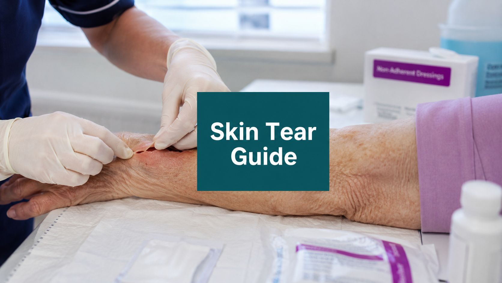
You're doing a routine skin check before rounds, and the CNA says, “It's just a little skin tear on her forearm.” The flap is folded over, the bed linen is dotted with blood, and the last note says “abrasion.” That's how small wounds turn into bad charts, delayed healing, and payer questions. Skin tears are common, but they're not casual wounds. If you work with older adults, SNF residents, home health patients, or anyone with fragile skin, you need to know both the bedside management and the documentation work that follows it.
Most articles stop at the definition. That's not enough. In practice, what matters is whether you can identify the wound correctly, preserve the flap if it's viable, select a dressing that won't create more trauma, and write a note that supports the care you gave.
Table of Contents
- Core Definitions and Classification Systems
- Identifying At-Risk Patients and Common Locations
- Immediate Assessment and Bedside Management
- Definitive Treatment Cleansing Flap Management and Dressing Choices
- Documentation Coding and Billing for Skin Tears
- Proactive Prevention Strategies in Practice
- Key Takeaways for the Wound Care Professional
Core Definitions and Classification Systems
A true skin tear usually announces itself the moment you expose it. The skin looks fragile, the wound is traumatic rather than pressure-related, and the flap tells you whether you still have tissue worth saving.
Start with what it is not
A skin tear is not a pressure injury. It's an acute traumatic wound caused by friction, shear, or blunt force. In older adults, the burden is large enough to affect daily operations. A 2024 systematic review found pooled prevalence of 6.0% and pooled incidence of 11.0% across older populations, with long-term care facilities reaching 11.0% prevalence according to this systematic review on skin tear epidemiology in older adults.
It's also not just a “superficial wound” in the lazy charting sense. The mechanism matters. A skin tear is a traumatic separation of skin layers caused by mechanical force. That puts it in a different bucket from pressure injuries, and often a different bucket from simple abrasions or clean lacerations. If you need a quick refresher on how clinicians distinguish shallow traumatic wounds more broadly, this overview of superficial wounds in clinical practice is useful background.
Use classification to guide care
At the bedside, classification should help you decide what to do next. If it doesn't change management, it's just paperwork.
The ISTAP framework is what most clinicians should know and use:
- Type 1 means no skin loss. The flap can be repositioned to cover the wound bed.
- Type 2 means partial flap loss. Part of the flap can cover the wound bed, but not all of it.
- Type 3 means total flap loss. The flap is absent, and the wound bed is fully exposed.
That simple sequence tells you the core clinical question. Is the flap viable, and can it function as protection?
Practical rule: The first thing I want documented is not “small skin tear.” I want flap status. Viable, nonviable, partial loss, complete loss. That's what drives treatment.
You'll still see the older STAR terminology in legacy charts. Read it, but don't build your current practice around it. For day-to-day use, ISTAP is cleaner and easier to teach across nursing, wound, and provider teams.
Abrasions usually scrape the epidermal surface. Lacerations usually imply a deeper or more sharply incised traumatic cut. Skin tears sit in their own lane because fragile skin separates, often with a flap that can still be preserved. If you miss that distinction, the note gets vague fast, and vague notes create bad follow-up.
Identifying At-Risk Patients and Common Locations
Some patients are skin-tear patients before the wound ever happens. You can often identify them during the first handshake, transfer, or dressing change.
The patient profile is usually obvious once you know what to look for
The classic presentation is older, frail skin with poor structural support. You'll see elastosis, ecchymosis, thin epidermis, and skin that doesn't tolerate friction well. Add edema, limited mobility, cognitive impairment, poor transfer mechanics, chronic disease, or adhesive exposure, and the risk goes up fast in practical terms.
Mechanical causes are usually mundane. A forearm drags against a bedrail. A shin hits a wheelchair footplate. Someone removes an adhesive too quickly. Staff pull a patient up in bed instead of using proper handling technique. None of this looks dramatic in the moment, but fragile skin doesn't need dramatic force.
A related workflow issue is chart review. Risk is often sitting in the record before you see the patient. Prior traumatic wounds, repeated adhesive injuries, edema history, mobility limitations, anticoagulant use, and recurrent falls matter. If your team needs a faster way to organize that history, tools that let you chat with patient records using AI can be useful for surfacing patterns before bedside assessment.
Where skin tears show up most often
Skin tears cluster in frail skin, especially on the hands, arms, and lower extremities. They aren't fringe wounds in facility practice either. In North American skilled nursing facilities, real-world documentation showed skin tears made up 10.3% to 12.8% of all wounds, with median healing times of 15 to 27 days depending on classification, as summarized in this Wounds International review of skin tears.
That distribution makes sense clinically. Extremities are exposed to transfer trauma, falls, furniture strikes, wheelchair contact, and adhesive removal. The lower legs are especially unforgiving in patients with edema or vascular disease. The forearms and hands get injured during routine care more often than many new clinicians expect.
A useful mental map during rounds:
- Forearms and hands often reflect transfer friction, bumps, and tape trauma.
- Shins and lower legs often signal frailty plus edema, venous disease, or recurrent environmental trauma.
- Any repeated site should make you think system failure, not bad luck.
If a resident has two “accidental” skin tears in the same month from routine care, I stop calling it accidental. Something in handling, environment, or skin protection isn't working.
That mindset changes prevention. You stop reacting to isolated wounds and start identifying a pattern.
Immediate Assessment and Bedside Management
The first bedside encounter matters more than people think. It allows you to save the flap, reduce additional trauma, and create a note that matches the wound.
Use a simple bedside sequence
I teach new staff a quick wound sequence: Hemostasis, Exudate, Assessment, Dimensions.
-
Hemostasis
Start with gentle pressure using sterile gauze. Don't keep lifting the gauze to check every few seconds. Let pressure work. If the extremity can be raised safely, do it. -
Exudate
Describe what's there before you flood the area. Is it scant serous drainage, serosanguinous drainage, or active bleeding? “Drainage present” is a weak note. Character and amount matter. -
Assessment of flap and bed
Check flap color, attachment, viability, and whether it can be re-approximated. Pink and viable is not the same as dusky, desiccated, or necrotic. Assess the wound bed depth and exposed tissue. Then look at the periwound. Fragile, ecchymotic, macerated, edematous, and dry all mean different things. -
Dimensions
Measure length, width, and if appropriate, depth. For irregular tears, don't guess. Document the actual shape if needed, and note percentage of flap loss when that's clinically clear.
What not to do in the first encounter
A lot of avoidable damage happens because people rush.
- Don't scrub aggressively. Fragile tissue won't tolerate it.
- Don't yank the flap open to “get a better look.” You may convert a salvageable flap into tissue loss.
- Don't default to closure strips or traumatic adhesive fixation on paper-thin skin.
- Don't write “skin tear dressed” and move on. That note won't help the next clinician.
The bedside exam should answer four things. What caused it, what the flap looks like, what the wound is draining, and what you did right away.
A short example of useful wording: traumatic skin tear to right dorsal forearm after transfer, partial flap loss, flap viable but fragile, serosanguinous drainage scant, periwound ecchymotic without warmth, wound cleansed, flap gently re-approximated, non-adherent dressing applied.
That's a real clinical note. It tells the next person what happened and why your management made sense.
Definitive Treatment Cleansing Flap Management and Dressing Choices
The default approach for most skin tears should be conservative and flap-preserving. Too many wounds get treated like the flap is debris. If the flap is viable, it's not debris. It's useful tissue.
Preserve the flap when you can
ISTAP defines a skin tear as a traumatic wound where the residual skin flap can function as a biologic dressing if viable. The first-line move is gentle re-approximation, avoiding staples or suture-like closure methods that increase trauma to fragile skin, as outlined in this clinical education summary on ISTAP-based skin tear care.
That principle should drive your technique. Cleanse gently with saline or another non-cytotoxic cleanser. If the flap is folded back, ease it into place with a moistened applicator or gloved hand. Don't stretch it. Don't force exact edge perfection. The goal is tissue protection, not cosmetic closure.
If the flap is clearly nonviable, dark, dry, or detached to the point that it cannot serve as coverage, then you document that accurately and manage the wound bed accordingly. Debridement decisions need to match tissue status, not habit. New clinicians often get in trouble here by charting debridement language that overstates what was done, or by coding procedural work when they really performed cleansing and flap repositioning only.
A practical point on care planning. Healing depends on wound status, moisture balance, and minimizing repeat trauma. This overview of the wound healing process in clinical practice is a useful framework when you're deciding whether the wound is moving as expected or stalling.
Choose dressings based on the wound you have
Dressing selection for skin tears should be boring in the best way. Protect the flap. Maintain a moist wound environment. Avoid creating a second injury during removal.
A workable bedside approach looks like this:
-
Viable flap with low drainage
Use a non-adherent contact layer and a secondary protective dressing that won't strip skin on removal. -
Moderate exudate
Add absorbency, often with a foam-type secondary dressing, while still protecting the flap and periwound. -
Dry, fragile wound surface
Consider hydration support rather than drying it out further. -
Periwound at high risk from adhesives
Use skin protection strategies and avoid aggressive adhesive borders whenever possible.
If the dressing change causes a new tear, the dressing plan failed. I don't care how tidy it looked on day one.
What doesn't work well is selecting dressings by routine. “We always use this on skin tears” is not a clinical rationale. Exudate level, flap viability, wound location, edema, and the likelihood of traumatic removal should drive the choice.
Documentation Coding and Billing for Skin Tears
Good clinicians lose revenue and create compliance risk when they treat the wound correctly, then chart it like an afterthought.
What has to be in the note
A common problem is documentation and coding drift. Skin tears are often confused with lacerations or abrasions in charting, and that matters because the wound should heal on an acute timeline of about 14 to 21 days if managed well. The documentation has to support the medical necessity of the care provided, as noted in this clinical summary on skin tear documentation and healing expectations.
A defensible note should include:
-
Cause of injury
Transfer trauma, wheelchair strike, adhesive removal, fall, bedrail contact, or unknown traumatic event. -
Exact location
“Right forearm” is acceptable. “Upper extremity” is weak unless that's all you know during triage. -
Flap description
Viable or nonviable, approximate degree of loss, color, attachment, and whether it was re-approximated. -
Wound bed and depth
Partial-thickness appearance versus deeper tissue involvement if present. -
Exudate and bleeding
Serous, serosanguinous, sanguineous. Scant, small, moderate, heavy. -
Periwound
Fragile, ecchymotic, edematous, macerated, erythematous, dry, intact, or denuded. -
Treatment performed
Cleansed with saline, flap re-approximated, selective removal of nonviable tissue if actually performed, dressing type applied, patient tolerance.
A lot of teams are tightening dictation workflows because incomplete transcribed notes create their own compliance issues. If you're reviewing how voice documentation fits HIPAA requirements, HyperWhisper's HIPAA compliant guide is a practical place to start.
How coding and billing drift starts
Billing trouble usually begins with vague language. “Skin tear treated and dressed” won't support much. Neither will calling every traumatic wound a laceration. The code set has to match the injury, and the procedure code has to match the service performed.
If you document debridement, describe the devitalized tissue, the instrument or method, the tissue level addressed, and the post-procedure wound characteristics. If all you did was cleansing and flap repositioning, chart that. Don't inflate the note to fit a code. Payers and auditors look for internal consistency.
For procedure coding in wound practice, clinicians often think in families such as 97597 for selective debridement and 11042 to 11047 for debridement by depth and surface area when those services are performed and documented. The unwritten rule is simple. If your note doesn't clearly support tissue type, depth, and medical necessity, don't expect the code to survive review.
For teams standardizing this workflow, a structured wound care documentation template helps reduce the usual omissions. EkagraHealth AI is one example of a documentation workflow that drafts wound notes, maps coding, and supports image-based tracking in a wound care setting.
Surveyors and payers both notice the same thing. A chart that names the wound but never describes the wound.
That's the practical standard. Describe what you saw, what you did, and why it was necessary.
Proactive Prevention Strategies in Practice
The patient who tears her shin on the wheelchair pedal usually gives you plenty of warning beforehand. Dry skin. Edema. Frail gait. Repeated bumps during transfers. Tape marks on the forearms. Prevention starts long before the next wound opens.
Build a skin safety routine
A prevention plan works best when it's concrete enough for staff to follow on a busy day.
For a high-risk patient, I want the routine to include the basics:
- Daily skin inspection on exposed extremities, especially forearms, hands, and shins.
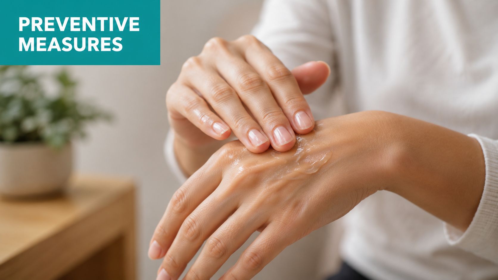
- Regular emollient use to reduce dryness and improve skin suppleness.
- Safer adhesive practice with gentle products, careful removal, and avoidance of unnecessary tape.
- Transfer discipline so staff lift, guide, and reposition without dragging skin across linen or equipment.
- Environmental cleanup including padded sharp edges and fewer strike hazards around beds, wheelchairs, and walkers.
- Protective clothing or sleeves when repeated trauma is a pattern.
This isn't glamorous work. It's routine work done consistently.
A quick example. The resident who keeps tearing the same forearm may not need a new dressing strategy first. She may need staff to stop anchoring wraps with aggressive tape, plus soft sleeves during the day, plus moisturizer after bathing, plus better transfer mechanics. The wound is the result. The system problem came first.
Lower leg tears need a broader plan
Lower-leg tears deserve more respect than they often get. In patients with edema, venous disease, and frailty, they may require elevation and compression therapy when appropriate. A shin tear in an older patient with chronic edema is not trivial. It can become a chronic wound if the biomechanical and vascular drivers are ignored, as noted in this NHS resource on lower-leg skin tear management.
That means bedside prevention has to expand beyond “cover the wound.”
Consider the broader checklist:
-
Edema control
If the leg stays swollen, the wound environment stays compromised. -
Protection from repeat trauma
Shins meet furniture, footplates, and transfer surfaces all day. -
Compression decisions
Appropriate only when clinically suitable, but often central to lower-leg management. -
Nutrition and hydration review
Fragile skin rarely improves on local care alone.
A lower-leg skin tear that lingers isn't always a dressing problem. Sometimes it's an edema problem wearing a wound-care label.
That's the part newer clinicians sometimes miss. Prevention is not one intervention. It's skin care, handling, environment, and management of the patient's underlying drivers.
Key Takeaways for the Wound Care Professional
A skin tear is easy to underestimate and expensive to mismanage. The wound may look small, but the clinical work around it is not small. You need accurate identification, flap-focused assessment, careful treatment, and documentation that can survive handoff, audit, and billing review.
The bedside priorities are straightforward. Recognize the mechanism. Assess flap viability early. Protect fragile tissue. Choose dressings that won't create another injury at the next change. If the lower leg is involved, think beyond the tear itself and address edema, vascular issues, and repeated trauma risk.
The charting priorities are just as important. Don't write “skin tear” as if that explains the wound. Document the cause, location, flap status, exudate, periwound condition, treatment performed, and the rationale for ongoing care. If debridement is performed, the note has to support the tissue level and medical necessity. If it wasn't performed, don't imply that it was.
Good skin tear care is really system care. Prevention, assessment, treatment, and documentation all have to line up. When they do, patients heal on an acute trajectory more often, handoffs get cleaner, and your billing team isn't left reconstructing the visit from fragments.
EkagraHealth AI helps wound care teams handle the part of skin tear management that often gets missed after the bedside exam: clean documentation, coding support, wound image tracking, and claims-ready workflow. If your practice is spending too much time fixing vague notes after the visit, EkagraHealth AI is worth a look.
