
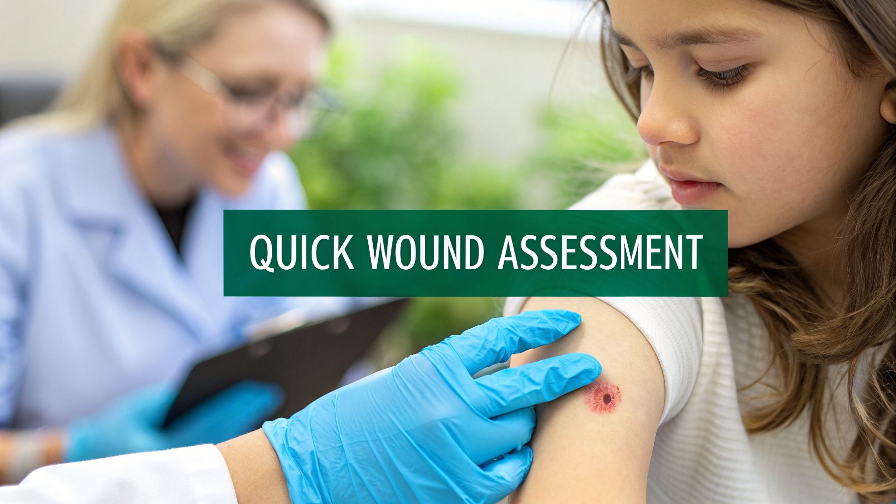
Superficial wounds are injuries that only scratch the surface, affecting the skin's outermost layer, the epidermis. Think of your everyday scrapes, minor cuts, and first-degree burns—the kind that don't break through the skin's main protective barrier. These seemingly minor injuries are a critical part of a clinician's daily practice, as their proper assessment can prevent significant complications down the line. This guide will delve into what superficial wounds are, how to assess them accurately, their common causes, and why proactive management is essential for both patient outcomes and the financial health of a healthcare practice.
Decoding Superficial Wounds and Skin Integrity

When you see a minor scrape or a bit of redness on a patient, it’s tempting to dismiss it as a simple fix. But to truly understand what are superficial wounds, we need to appreciate their role in overall skin health. Imagine the skin as a multi-layered shield. A superficial wound is like a scuff on that shield’s surface—it hasn’t punched all the way through, but it has definitely weakened that first line of defense. This initial damage is confined to the epidermis, which is the thinnest and most external layer of the skin. It serves as the primary barrier against environmental threats, including bacteria, chemicals, and UV radiation.
In some cases, the injury might just graze the papillary dermis, the layer sitting right underneath the epidermis, which is rich in nerve endings and blood vessels. Because these wounds are shallow, they usually heal up quickly and without leaving a scar, a process known as re-epithelialization. Still, their simple appearance can be deceptive. A failure to recognize the underlying cause or potential for complications can turn a minor issue into a chronic problem, particularly in vulnerable patient populations.
Differentiating Wound Depth
To really get a handle on a superficial wound's importance, it helps to see how it stacks up against deeper injuries. Skin isn’t just a single sheet; it’s a complex, organized structure. Knowing exactly where the damage stops is the absolute key to a good assessment and the right treatment plan. A correct diagnosis of wound depth dictates everything from dressing selection to the need for specialist referral.
It’s crucial to draw a clear line between the three main wound depths:
- Superficial Wounds: These injuries only involve the epidermis. They often look like a sunburn or a light abrasion (think "road rash") and typically don't bleed. The area may be red, painful, and tender to the touch, but the skin's deeper structures remain intact. Healing occurs through the regeneration of epithelial cells.
- Partial-Thickness Wounds: These go deeper, extending through the epidermis and into the dermis. You'll often see them as blisters or shallow ulcers with a moist, pink or red base. Because the dermis contains nerve endings, these wounds can be quite painful. Any bleeding is usually minimal, appearing as capillary oozing.
- Full-Thickness Wounds: This is the most serious category. The injury has penetrated through the epidermis and dermis into the subcutaneous tissue (fat). In the most severe cases, you might even see muscle, tendon, or bone. These wounds often have a more complex appearance, with various colors (yellow, black, white) in the wound bed, and may be less painful if nerve endings have been destroyed.
To help you quickly tell them apart in a clinical setting, here's a quick reference table.
Wound Depth at a Glance: Epidermis to Fascia
This table is a great cheat sheet for quickly distinguishing between superficial, partial-thickness, and full-thickness wounds based on the skin layers involved and what you see.
| Wound Depth | Key Skin Layers Involved | Common Clinical Appearance |
|---|---|---|
| Superficial | Epidermis only | Redness, light abrasions (like a scratch), no bleeding. Looks like a sunburn. Healing is rapid. |
| Partial-Thickness | Epidermis and part of the Dermis | Pink or red moist base, may have blisters. Can be painful and have minimal bleeding. Scarring is possible. |
| Full-Thickness | Epidermis, Dermis, and into the Subcutaneous Tissue (fat) | Wound base may appear white, yellow, or black. Can expose muscle, tendon, or bone. Always scars. |
Remember, this distinction is more than just academic. Getting the depth wrong can lead to the wrong treatment, which can delay healing and cause serious complications. For example, what looks like simple redness might actually be a Stage 1 pressure injury—a critical warning sign, especially for patients with limited mobility. This underscores the need for a precise and careful initial evaluation.
A superficial wound is the first sign that the skin’s integrity is at risk. It serves as a potential gateway for infection and can be an early indicator of more severe underlying issues, such as pressure-related damage or circulatory problems.
Why the Initial Assessment Is So Crucial
Identifying a wound as "superficial" is just the starting point, not the final word. These seemingly minor injuries demand a careful initial look because they can be the first domino to fall toward a major complication. A small, ignored wound, especially in an immunocompromised or diabetic patient, can quickly get infected, leading to cellulitis or a stubborn, non-healing ulcer.
For clinicians, this early stage is a golden opportunity to step in. A thorough evaluation can stop a small problem from snowballing into a painful and expensive chronic condition that requires advanced therapies and specialist intervention. This proactive mindset not only protects your patient’s health but also helps manage clinic resources by preventing the need for the extensive treatments that advanced wounds require. For those using digital tools, this detailed initial assessment is foundational for accurate tracking in platforms like WoundReference, creating a reliable baseline for monitoring progress.
A Clinician's Guide to Assessing Superficial Wounds

Accurately assessing a superficial wound is about more than just a quick glance. To really understand what you're dealing with, you need a systematic approach that confirms the injury's nature, rules out deeper damage, and sets a clear baseline for treatment. It’s a hands-on, methodical process that starts with the basics of visual and tactile examination.
Your assessment should always begin with a detailed visual inspection. Get up close and look at the wound’s color, size, and exact location on the body. Is the redness even, or is it patchy? Are the wound edges well-defined or diffuse? Look for subtle signs of trauma, like minor scrapes or scratches, that can help confirm your initial diagnosis of a superficial wound. Documenting these initial observations with precise measurements is critical for tracking changes over time.
After looking, it's time to feel. Gently perform a careful palpation of the area around the wound to check its temperature and texture. Does the skin feel warmer than the surrounding tissue, suggesting inflammation? Is it firm, or does it feel boggy and soft, which could indicate fluid accumulation or deeper tissue damage? These tactile clues give you critical information about inflammation and what might be happening beneath the surface.
The Blanching Test: Differentiating Redness
One of the most valuable techniques in your assessment toolbox is checking for non-blanchable erythema. This is a simple but powerful test that helps distinguish between benign inflammation and the early stages of a pressure injury. Just press down gently on the red area with your finger for a few seconds, then lift it away and watch what happens.
- Blanchable Redness: If the skin whitens (blanches) when you press it and then quickly flushes red again, the tiny capillaries underneath are still intact and blood flow is fine. This is what you'd expect to see with simple skin irritation or a very minor abrasion.
- Non-Blanchable Redness: If the area stays red even after you release the pressure, it’s a warning sign. This indicates capillary damage and is the classic sign of a Stage 1 pressure injury—a more serious issue that needs immediate action to stop it from getting worse. This finding signals that tissue is being deprived of oxygen and is at high risk for breakdown.
Distinguishing between a simple friction abrasion and a Stage 1 pressure injury is one of the most critical assessment skills in wound care. While both appear as superficial redness, their causes and clinical implications are vastly different, directly impacting treatment protocols and reimbursement.
Understanding this difference is everything. A superficial scrape might just need a light protective dressing. A Stage 1 pressure injury, on the other hand, requires a full-scale offloading plan, a schedule for repositioning the patient, and frequent checks to prevent it from deteriorating into a deep, painful ulcer.
Taking a Comprehensive Patient History
The physical exam only gives you part of the picture. To get the rest, you have to dig into the patient's history to uncover the mechanism of injury. Ask specific, open-ended questions: How did this happen? Was it from a fall? Was there prolonged pressure on the area from sitting or lying in one position, or was it caused by friction or moisture?
Knowing the root cause is fundamental to building a care plan that actually works. For example, you’d manage a friction wound completely differently from redness caused by moisture-associated skin damage (MASD). A friction injury requires protection and reducing shear forces, while MASD demands moisture control and barrier creams. This history gives you the context you need to make a confident and defensible clinical decision.
Leveraging Technology for Objective Assessment
Modern technology is changing the game for how we assess and document wounds. AI-powered tools can add a much-needed layer of objectivity and consistency to your workflow, moving beyond subjective visual assessments. Some platforms, for instance, can analyze a wound photo to give you precise, automated measurements and objectively assess color variations within the wound bed, such as the percentage of erythema.
This creates a reliable, data-driven baseline that’s far more accurate than just eyeballing it. With objective data, it’s much easier to track healing over time and build a strong case to justify your clinical interventions and support billing. As technology evolves, knowing the latest wound assessment tools for nurses is more important than ever for providing top-tier care. A repeatable, defensible assessment is the bedrock of good wound management.
Where Do Superficial Wounds Come From? Common Causes and Scenarios

Knowing what a superficial wound is is one thing. But to be truly effective in practice, you need to understand the how and why behind them. Each wound has an origin story, and that story is the single most important clue for creating a successful care plan and preventing recurrence.
These injuries aren't random. They're the direct result of a specific force meeting the skin. By digging in to find the root cause—be it pressure, friction, moisture, or thermal energy—you shift from simply patching a problem to actually solving it. That's the difference between basic wound care and true clinical expertise.
Pressure and Shear: The Silent Culprits
Pressure is easily one of the most dangerous and common causes, particularly for patients with limited mobility, such as those who are bedridden or use a wheelchair. When a bony area like a heel, elbow, or the sacrum bears weight for too long, blood flow gets cut off. That lack of oxygen starts to damage the tissue, leading to a Stage 1 pressure injury—a perfect example of a superficial wound that serves as a major red flag for deeper trouble.
Think about an elderly patient who spends hours in a wheelchair. The unrelenting pressure on their sacrum can create a patch of non-blanchable erythema. This isn't just a red spot. It’s a loud-and-clear warning sign that the tissue is in distress and on the verge of breaking down completely into a partial- or full-thickness wound.
Understanding a wound's origin story is vital. A superficial wound caused by pressure requires a completely different intervention (offloading, repositioning) than one caused by moisture or friction. The cause dictates the care plan.
Shear is pressure's destructive partner. This happens when the skin stays put but the skeleton slides, like when a patient slumps down in bed. This movement stretches and tears the tiny blood vessels feeding the skin, resulting in oddly shaped red or purple areas that can worsen very quickly. Recognizing the signs of shear is crucial for implementing proper patient positioning techniques.
Friction and Minor Bumps
Friction wounds, which we often call abrasions, happen all the time. They’re created when skin gets scraped against a surface, literally rubbing away the epidermis. A "sheet burn" from being pulled across linens is a classic example, as is the "road rash" an athlete gets from a fall.
Though they might seem minor, these scrapes can be surprisingly painful because they expose so many nerve endings. And for vulnerable patients, a small friction wound can become a huge problem.
Just look at these real-world examples:
- The Patient with Diabetes: Someone with diabetes wears new shoes that rub their heel raw. For most people, it's an annoyance. For them, it's a serious risk. Neuropathy might prevent them from feeling it, and poor circulation can stop it from healing, opening the door to a chronic foot ulcer.
- The Post-Surgical Patient: A weak patient recovering from surgery might bump their shin on a bed rail. In their fragile, deconditioned state, that simple scrape is now a perfect entry point for an infection that could derail their entire recovery.
Burns and Moisture Damage
First-degree burns are another primary source of superficial wounds. These injuries only damage the epidermis, causing pain, redness, and some swelling without any blisters. A mild sunburn or a quick splash from a hot liquid are textbook cases. While they usually heal on their own, they still represent a compromise to the skin's first line of defense and require proper care to prevent infection and promote comfort.
Finally, never underestimate the damage caused by moisture. Constant exposure to wetness can cause Moisture-Associated Skin Damage (MASD). We see this all the time in patients with incontinence, where the skin's protective barrier is broken down by urine or feces. It becomes red, raw, and incredibly vulnerable, appearing as diffuse erythema often accompanied by satellite lesions if a fungal infection is present. This proves once again why finding the cause is the most critical first step.
The Clinical and Financial Imperative of Proactive Care
It’s all too easy to glance at a superficial wound and dismiss it as a minor issue—something to quickly clean and cover. But thinking this way misses a massive clinical and financial risk. Ignoring these seemingly simple injuries is like seeing a small crack in a dam and walking away; what starts small can quickly lead to a much bigger, more destructive problem.
A superficial wound can escalate into a chronic, painful, and costly condition faster than many clinicians realize. That journey from a bit of red skin to a full-thickness ulcer isn't just a possibility; it's a common reality in high-risk populations. This is why acting early and decisively isn’t just good practice—it's one of the highest-impact things you can do to improve patient outcomes and control healthcare costs.
The True Cost of a "Wait and See" Approach
When a superficial wound is ignored or isn't assessed properly, the fallout can be severe. That initial break in the skin is an open invitation for bacteria, paving the way for infections like cellulitis. For a patient who is already frail or has other health problems like diabetes or vascular disease, this can set off a disastrous chain reaction leading to hospitalization, systemic infection (sepsis), and a sharp decline in their quality of life.
The financial hit is just as serious. Treating a chronic wound costs a fortune compared to what it takes to prevent one in the first place. Think about it: advanced dressings, specialist visits, debridement procedures, antibiotics, and potential hospital stays all add up, creating a huge burden for both the patient and the healthcare system. The investment in prevention is minimal compared to the staggering cost of treating complications.
Why Prevention Is Smart Business
For any healthcare organization, from a small private clinic to a large hospital network, proactive wound care makes perfect business sense. By stepping in early to manage superficial wounds effectively, you're hitting several crucial goals at once:
- Better Patient Outcomes: First and foremost, you stop a wound from getting worse. This means less pain for the patient, a lower risk of serious complications, and a better chance of maintaining their mobility and independence.
- Lower Overall Costs: The cost of a simple protective dressing and a good offloading schedule is pocket change compared to the tens of thousands of dollars needed to manage a single advanced pressure ulcer.
- Fewer Hospitalizations: Good preventative care keeps patients out of the hospital. This frees up beds for sicker patients and sidesteps the high costs of inpatient stays for wound complications.
- A Stronger Reputation: Facilities that are known for excellent, proactive care build incredible trust with patients and referring providers. This attracts more patients and cements your reputation as a leader in the community.
A proactive mindset shifts wound care from a reactive, expensive service into a value-generating strategy. Investing in the proper management of superficial wounds is a clear sign that you're committed to both your patients' health and your organization's financial stability.
Understanding the Scale of the Problem
The slide from a superficial lesion into a chronic wound is happening everywhere. Superficial wounds, especially those that begin as Stage 1 pressure injuries, are a huge—and often preventable—problem. A staggering number of people suffer from chronic wounds, and many of those wounds started as minor, manageable skin damage that was not addressed in time.
For example, look at decubitus ulcers (pressure injuries). The global incidence of these wounds, which often start superficially as non-blanchable erythema, more than doubled between 1990 and 2021, jumping from 300,442 cases to 645,588. You can dive deeper into the data on the global burden of pressure injuries00155-2/fulltext) to see just how widespread this is.
This isn't just a statistic; it's a call to action. We have to treat every superficial wound, particularly on a vulnerable patient, as the potential start of a chronic condition. By seeing the risk and acting right away, clinicians can stop this progression before it even gets a foothold. This isn't just an option—it’s a clinical and financial imperative.
Evidence-Based Management and Healing Timelines

Once you've identified a superficial wound and assessed the risks, it's time to act. Your goal isn't just to slap a bandage on it; it's about creating the perfect environment to help the body's own healing machinery do its job effectively and efficiently.
The first step is always gentle cleansing. Use a mild soap and water or sterile saline to carefully wash away any dirt, debris, or bacteria. The trick is to be thorough without causing more damage to the delicate tissue that’s already been injured. Avoid harsh antiseptics like hydrogen peroxide or alcohol on open wounds, as they can be cytotoxic and impede healing.
After cleaning, the next priority is protecting the wound. With the skin barrier broken, the area is vulnerable to infection and friction. A simple protective layer can prevent a minor scrape from turning into a much bigger problem. This might involve a dressing, a barrier cream, or simply offloading pressure from the area.
Choosing the Right Dressing
In modern wound care, we've moved away from the old "let it air out" approach. We now know that creating a moist healing environment is key. This simple strategy speeds up re-epithelialization, reduces pain, and stops hard scabs from forming, which can actually get in the way of new skin cells moving across the wound.
Of course, not all dressings are the same. The right one depends entirely on the wound's specific needs—namely, its location, the amount of drainage (if any), and the underlying cause.
- Transparent Films: These are fantastic for very shallow scrapes, skin tears, or Stage 1 pressure injuries. Think of them as a second skin. They are semi-permeable, letting oxygen in but keeping water and bacteria out, and because they're clear, you can check on the wound without having to take the dressing off.
- Hydrocolloids: These are a step up in absorbency. When they come into contact with wound fluid, they form a soft gel that creates a moist environment. This makes them perfect for superficial wounds that might be weeping just a little, as they keep the area moist and even help gently clear away any dead tissue (autolytic debridement).
Understanding why you choose one over the other is what really elevates your practice. A transparent film is perfect for a dry abrasion needing protection, whereas a hydrocolloid provides that extra bit of moisture management for a wound with minimal drainage.
Expected Healing Timelines
For a healthy person with a straightforward superficial wound, healing is usually a quick affair. The body's repair crew gets to work right away, and you can expect the skin to be fully restored within 7 to 14 days. This process involves inflammation, proliferation (cell growth), and maturation.
The whole point of evidence-based care is to guide the wound smoothly through its natural healing phases. The right dressing and proper care can make a huge difference in recovery time and how the skin looks afterward.
This timeline is your benchmark. If a wound isn't showing signs of improvement within this timeframe, it's an early signal that something else might be going on, such as an underlying comorbidity, infection, or persistent pressure. For a closer look at each recovery phase, you can explore the wound healing process timeline in our detailed guide.
Red Flags and When to Refer
While most superficial wounds heal just fine, you have to stay vigilant for signs that things are going sideways. Catching these "red flags" early allows you to step in or get a specialist involved before a small problem becomes a chronic one.
Be on high alert for any of these warning signs:
- Signs of Infection: Redness that spreads beyond the wound margins, increasing pain or warmth, swelling, purulent (pus-like) drainage, or a bad smell.
- Stalled Healing: The wound looks exactly the same—no smaller, no better—after two weeks of appropriate care.
- Deterioration: The wound is actually getting bigger, deeper, or breaking down in new spots.
- Disproportionate Pain: The patient is in a lot more pain than you would expect for such a minor wound.
If you spot any of these signs, it's time to rethink your care plan. It could point to a brewing infection, poor blood flow (arterial insufficiency), or an underlying health issue like uncontrolled diabetes. In these situations, referring the patient to a wound care specialist is the right move to prevent long-term complications and ensure they get the advanced help they need.
How Better Documentation Boosts Your Bottom Line
Think of your clinical documentation as more than just a patient record. It's the financial engine of your entire wound care practice. Getting reimbursement right means creating detailed, accurate notes that tell a clear and compelling story, especially for injuries that start out as superficial.
This process is what connects your clinical work directly to your revenue cycle. Every detail you note down—from the wound's size and characteristics to what you think caused it—has to back up the ICD-10 and CPT codes you bill. Without that solid connection, payers have an easy reason to deny claims, which means lost revenue and a mountain of frustrating administrative work for your team.
Building an Audit-Proof Patient Record
Creating documentation that can stand up to a payer audit all comes down to being specific. A quick note like "redness on back" is a guaranteed recipe for a denied claim because it lacks medical necessity and clinical detail. A truly defensible note is packed with structured details.
- Precise Location: Pinpoint the exact anatomical spot using proper medical terminology. Instead of just "hip," write "over the left ischial tuberosity."
- Accurate Measurements: Always record dimensions in centimeters (length x width x depth). For superficial redness, be sure to measure the entire affected area.
- Wound Characteristics: Describe what you see and feel—the wound bed, its edges, and the surrounding skin. Use objective terms like "non-blanchable erythema," "intact skin," or "warm to the touch."
- Suspected Cause: Connect the dots. Link the wound to a specific cause, like "secondary to prolonged pressure from sitting" or "friction from medical device."
These details work together to paint a clear picture for the payer, justifying medical necessity and locking in your coding choices. For a deeper dive into creating solid notes, check out our comprehensive wound care documentation template.
The High Cost of Inefficient Workflows
When a superficial wound gets worse, the financial fallout is massive. Take pressure ulcers, which often begin as simple redness. They end up costing the U.S. healthcare system a staggering $2.1 billion every year. For both small clinics and large hospital systems, clunky, disconnected workflows just pour fuel on the fire, leading to missed documentation, coding errors, and lost revenue. You can learn more about the public health impact of chronic wounds in this Wounds Asia journal article.
This is where today’s technology can make a real-world difference. An integrated AI platform can handle the entire "voice to claim" process in just a few minutes. It uses AI to analyze and track Stage 1 injuries, automates measurements, and helps ensure your coding is spot-on for better revenue and compliance.
Technology that closes the loop between clinical observation and billing isn't a luxury anymore—it's essential. By automating documentation and coding, clinics can cut administrative time by up to 70%. That frees up clinicians to focus on what matters most: high-value, preventative patient care.
This kind of automation drastically reduces coding errors, strengthens the argument for medical necessity, and ultimately means you get paid faster with fewer denials. It’s a solution that scales, working just as well in an outpatient clinic as it does in home health or a large hospital.
By rethinking how you capture and process patient encounters, you can transform documentation from a time-sucking chore into a powerful driver of financial health. With voice-to-chart technology, clinicians can narrate their findings during an exam and let the system automatically turn that conversation into structured data and the right codes. It's an approach that doesn't just save time—it builds an ironclad foundation for every single claim you submit.
Frequently Asked Questions About Superficial Wounds
Even when you have a solid grasp of superficial wounds, real-world questions always pop up in the middle of a busy clinic day. This quick FAQ section tackles some of the most common challenges, offering practical answers to reinforce what you know and give you some immediate, actionable advice.
How Can You Reliably Distinguish a Superficial Wound from a Partial-Thickness Wound?
The real difference is all about how deep the injury goes. I like to use an analogy of sanding a piece of wood. A superficial wound is like a very light sanding that just scuffs the top layer of paint—that's your epidermis. Clinically, this looks like reddened skin, maybe like a mild sunburn or a shallow scrape, with minimal to no bleeding. The key is that the skin is still intact, even if it's red and inflamed.
A partial-thickness wound, on the other hand, is like sanding past the paint and into the wood itself. This injury reaches down into the dermis. You’ll often see a blister (intact or ruptured) or a shallow, open wound with a moist, pink base. A blister or any visible break in that top layer of skin is your classic sign that the injury has gone deeper than just superficial.
When Should a Simple Superficial Wound Be Referred to a Wound Care Specialist?
Most superficial wounds heal just fine on their own with proper care, but it's crucial to know when to call for backup. You should get a specialist involved if you notice any red flags.
Think about referring a patient if:
- The wound shows no signs of healing within about two weeks, despite appropriate interventions.
- You see signs of infection, like redness that's spreading, pus, a bad odor, or the patient has systemic signs like a fever.
- The patient is dealing with major health issues that get in the way of healing, such as uncontrolled diabetes, peripheral artery disease, or is on immunosuppressive therapy.
- The wound gets worse quickly or the pain seems much more severe than the injury would suggest.
Any one of these signs tells you there's a more complex problem under the surface that needs an expert eye to diagnose and manage properly.
A superficial wound that fails to progress through the normal healing stages is no longer a simple injury. It is a warning sign of an underlying barrier to healing that needs to be identified and addressed by a specialist to prevent chronic complications.
What Is the Biggest Documentation Mistake Made with Superficial Wounds?
Hands down, the most common and costly mistake I see is a lack of specificity. Just writing "redness on back" is far too vague for good clinical tracking and will almost certainly be kicked back by payers. It simply doesn't paint a clear picture of what's going on or justify the need for skilled care.
Good documentation is all about the details. Make sure your notes always include:
- Precise location and dimensions (e.g., "a 3×4 cm area of erythema over the sacrum").
- Wound bed characteristics (e.g., "non-blanchable erythema" or "skin intact, warm to touch").
- Suspected cause (e.g., "appears related to pressure from prolonged sitting in a wheelchair").
This level of detail is absolutely vital. It justifies the care you’re providing, helps you track progress accurately, and makes your coding defensible and audit-proof. It turns a simple note into a powerful clinical and financial record that supports your practice's revenue cycle.
At Ekagra Health AI, we transform your clinical notes into clean claims in minutes. Our voice-first platform captures ambient conversations during patient encounters and automatically generates structured charts, assigns the correct codes, and submits for reimbursement. By reducing documentation time by up to 70%, we empower your team to focus on what matters most—delivering exceptional patient care. Discover how our end-to-end solution can streamline your workflow and accelerate your revenue cycle by visiting https://ekagrahealth.ai.
