
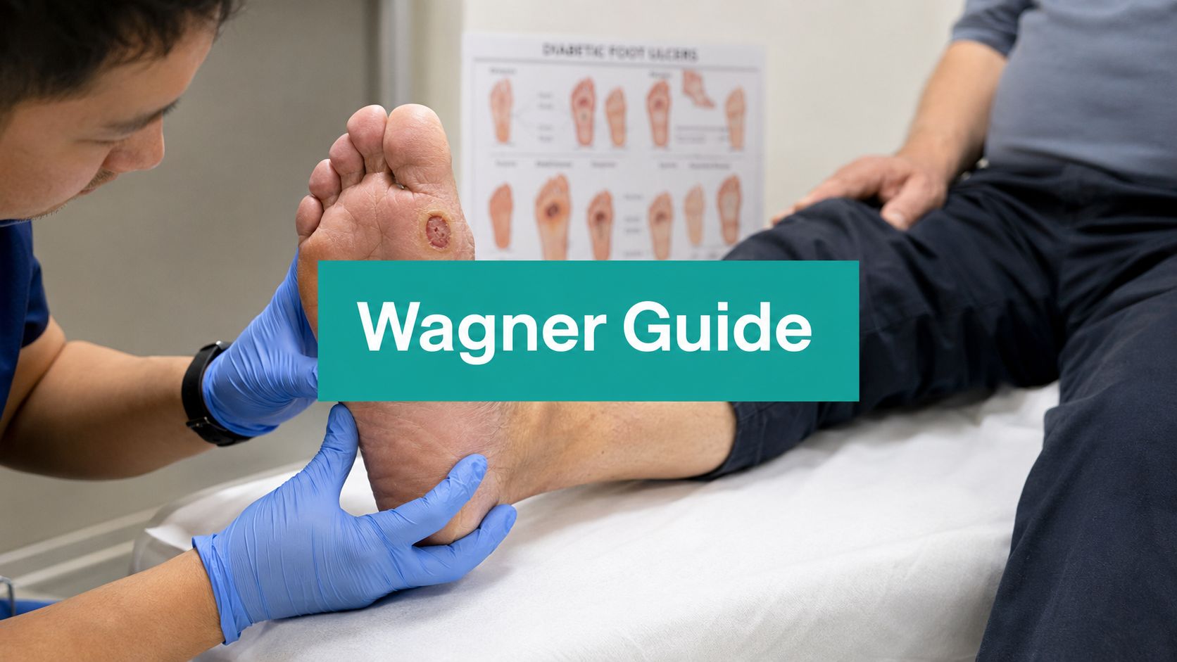
A Saturday-morning emergency department handoff comes with familiar gaps: a 62-year-old patient with diabetes, a swollen, erythematous forefoot, two previous toe amputations, and an outpatient note that says only “chronic diabetic wound.” Before debating dressings or discharge, the receiving clinician needs a severity shortcut. The first question is practical: what Wagner grade does the wound support?
The answer won't replace vascular assessment, infection staging, or clinical judgment. It does give the team a common starting point for triage, treatment intensity, documentation, and communication. Used correctly, Wagner classification turns a vague description into a defensible statement about depth and tissue loss.
Why Wagner Still Earns Its Place at the Bedside
Wagner survives because clinicians from different disciplines can understand it quickly. Podiatry, vascular surgery, infectious disease, nursing, home health, and hospital medicine may describe the same foot differently, but “Wagner 3 with suspected osteomyelitis” immediately signals a deep, complicated wound. The original purpose was to standardize diabetic foot ulcer severity by depth and gangrene, and later clinical work showed that grade tracks outcomes in a practical way. In a 200-patient cohort, the median healing time rose from 23 days for Wagner 1 to 50 days for Wagner 2, 54 days for Wagner 3, and 119 days for Wagner 4 in the reported cohort.
That spread matters during handoff. A superficial ulcer may fit outpatient offloading and close follow-up. A wound with an abscess, exposed bone, or gangrene may require imaging, operative evaluation, vascular input, or a goals-of-care discussion. Wagner doesn't make that decision alone, but it helps the team recognize when a routine clinic pathway no longer fits.
A shared shorthand with real triage value
The scale also works because it maps roughly to disposition pressure. Grades 0 and 1 usually point toward prevention, offloading, and local wound management. Grades 2 and 3 demand a closer look at deep structures and infection. Grades 4 and 5 move the conversation toward perfusion, surgical source control, limb salvage, or palliation.
Published cohorts reinforce that higher grades carry more limb-loss risk. In one study, 31 patients with Wagner 3, 4, or 5 ulcers, 68.9% of that high-grade group, ended with amputation or disarticulation as reported in the comparative analysis. That doesn't turn the grade into a prediction for an individual patient. It does justify treating a high grade as a prompt for escalation rather than a descriptive footnote.
Practical rule: Use Wagner to establish urgency quickly, then add the information Wagner leaves out before committing to a treatment or disposition plan.
Speed is the advantage, not completeness
Texas, PEDIS, and IDSA/IWGDF frameworks add axes for ischemia, infection, perfusion, sensation, or research stratification. That detail is valuable, especially for multidisciplinary planning and research. At the nurses' station, though, a six-grade depth-and-gangrene scale is often faster to communicate.
Its limits are equally clear. Wagner doesn't independently classify perfusion, neuropathy, ulcer area, or the full severity of infection. A Wagner grade is therefore a working shortcut, not a complete wound assessment.
The Six Grades Explained With Clinical Examples
The most useful way to apply the scale is to connect each grade to three things: what you see, what should change your next step, and what must appear in the note. The standard system runs from Grade 0 through Grade 5, progressing from an at-risk foot to extensive gangrene as summarized in this clinical classification review.
Grade 0
The skin is intact, but the foot is at risk. Look for callus over a bony prominence, deformity, Charcot changes, or a history of ulceration. The immediate work is prevention: remove pressure, address footwear, and establish a repeat foot-exam cadence.
A new warmth, callus breakdown, or discoloration should trigger reassessment rather than reassurance. Document the intact skin, deformity or callus location, prior ulcer history, pulses, sensation, and offloading plan.
Grade 1
This is a superficial ulcer limited to the skin, with no deep structure involvement. A clean base and absent infection signs support local wound care, sharp removal of devitalized surface tissue when clinically appropriate, offloading, and a vascular check.
Escalate when the base deepens, drainage changes, erythema expands, or probing suggests tendon, capsule, or bone involvement. Describe the wound bed, exudate type, periwound skin, measurements, and whether the ulcer remains confined to skin. A useful wound-bed description reference helps keep those observations specific rather than reducing the note to “wound stable.”
Grade 2
The ulcer extends to tendon, capsule, or bone, but the grade does not add abscess or osteomyelitis. Bedside versus operating-room management becomes a real decision at this stage. Probe depth, assess for cellulitis, obtain appropriate cultures when infection is suspected, and determine whether imaging or surgical evaluation is needed.
The note should identify the deepest exposed or probed structure and distinguish exposure from confirmed infection. “Deep ulcer” isn't enough. State whether tendon, capsule, or bone is involved and describe drainage, odor, undermining, and surrounding inflammation.
Grade 3
Grade 3 adds abscess, osteomyelitis, septic joint involvement, or another deep infection pattern. This isn't a simple local-care wound. The usual intent is urgent evaluation, often including admission, imaging for bone involvement, culture strategy, antibiotics, and surgical source control.
Document the evidence supporting the grade: fluctuance, purulence, exposed bone, imaging findings, joint involvement, systemic symptoms, or operative findings. The transition from Grade 2 to Grade 3 is the key inflection point because infection changes the wound from a mechanical problem into a limb-threatening one.
Grade 4
Grade 4 means localized gangrene, typically involving a toe or forefoot. Assess perfusion immediately, involve vascular and surgical services, and document the tissue boundaries that will guide amputation-level planning.
A dry, clearly demarcated area and a wet, draining, malodorous area aren't managed the same way. Record whether gangrene is dry or wet, the presence of drainage or systemic illness, pulse findings, and the proposed vascular workup.
Grade 5
Grade 5 describes extensive gangrene of the whole foot. The central decisions are whether revascularization is feasible, whether major amputation is appropriate, and how the patient's goals and overall condition shape the plan.
Document the extent of necrosis, infection burden, perfusion findings, pain, functional status, and discussions with the patient or surrogate. Don't let the grade become a substitute for a goals-of-care conversation.
| Grade | Bedside Picture | Trigger to Escalate | First Management Step |
|---|---|---|---|
| 0 | Intact skin with callus, deformity, or prior ulcer risk | New warmth, breakdown, or discoloration | Offload pressure and intensify foot surveillance |
| 1 | Superficial skin ulcer | Deepening, spreading erythema, or changed drainage | Local care, sharp surface debridement when appropriate, offloading, vascular check |
| 2 | Ulcer reaching tendon, capsule, or bone | Cellulitis, exposed bone, drainage, or failure to improve | Define depth, evaluate infection, consider imaging and surgical review |
| 3 | Deep infection, abscess, osteomyelitis, or joint involvement | Systemic signs, purulence, instability, or worsening tissue loss | Urgent imaging, antibiotics, admission or operative source control |
| 4 | Localized toe or forefoot gangrene | Wet gangrene, spreading necrosis, or poor perfusion | Vascular assessment and amputation-level planning |
| 5 | Extensive gangrene of the whole foot | Progressive necrosis or non-salvageable tissue | Revascularization and major amputation or palliation discussion |
Where Wagner Falls Short Compared to Other Systems
Wagner's strength is compression. Its weakness is compression. It reduces a complicated diabetic foot to depth, infection-related deep involvement, and gangrene, while other systems keep clinically distinct variables separate.
The University of Texas system adds separate dimensions for ulcer depth, infection, and ischemia. That distinction matters when two wounds reach the same structure but have different blood flow or infection burdens. PEDIS is more granular for structured assessment because it names perfusion, extent or size, depth and tissue loss, infection, and sensation independently. The IDSA/IWGDF approach focuses more directly on infection severity and helps distinguish a localized infection from a limb-threatening or life-threatening presentation. The classification review from the International Working Group on the Diabetic Foot describes why Wagner remains familiar while also highlighting its limited ability to capture infection and ischemia separately in its discussion of classification systems.

The perfusion problem
A Wagner 3 ulcer in a warm foot with palpable pulses doesn't carry the same revascularization question as a Wagner 3 ulcer in a cold, pulseless foot. Wagner can label both wounds the same because it doesn't independently grade ischemia. That is exactly where the amputation level, healing potential, and vascular plan diverge.
The same blind spot applies to neuropathy, wound surface area, and organism-specific concerns such as MRSA risk. Wagner can tell you that bone or deep infection is involved. It can't, by itself, tell you whether perfusion is adequate for closure or how severe the infection is across the full clinical picture.
Use Wagner for triage shorthand. Switch to a multidimensional system when the team is deciding on vascular intervention, amputation level, or trial enrollment.
Debridement Recommendations and CPT Coding by Grade
Wagner grade can help frame the clinical problem, but CPT selection follows the tissue removed, not the grade printed at the top of the note. CMS states that codes 11042 through 11047 are reported according to the deepest tissue removed and the wound surface area. For one wound, use the deepest level removed. For multiple wounds, add areas only when they share the same depth under CMS debridement coding guidance.
For a superficial Grade 1 wound, remove callus, slough, or devitalized tissue only to the level clinically necessary. If subcutaneous tissue is removed, the base code is 11042, with 11045 for each additional 20 square centimeters. Don't select a deeper code because the ulcer is labeled Grade 2 or Grade 3.
Grade 2 may require 11043 when the procedure removes muscle and/or fascia. Grade 3 can involve 11044 when bone is removed, but exposed bone alone doesn't prove that bone debridement occurred. The operative or procedure note must state what was removed.
Match the procedure to the documented tissue
Grade 4 wet or infected gangrene may require staged operative debridement, potentially involving muscle, fascia, or bone, alongside antimicrobial management and urgent vascular assessment. Grade 5 care is patient-specific. If the foot isn't revascularizable and the intent is odor or drainage control, the procedure may remain superficial, but the record must support the tissue level removed and the medical necessity.
CMS and Medicare guidance emphasize two denial-sensitive elements: post-debridement surface area and depth of tissue removed in the applicable documentation article. The code family divides by tissue depth and size: 11042 and 11045 for subcutaneous tissue, 11043 and 11046 for muscle and/or fascia, and 11044 and 11047 for bone, with the add-on codes applying to each additional 20 square centimeters as outlined in CPT coding guidance.
| Wagner Grade | Recommended Debridement | Primary CPT | Add-on CPT | Documentation Trigger |
|---|---|---|---|---|
| 0 | Callus or non-wound pressure management, if a procedure is clinically indicated | Depends on tissue removed | Depends on tissue removed | State whether tissue was actually removed |
| 1 | Sharp removal of devitalized superficial or subcutaneous tissue | 11042 when subcutaneous tissue is removed | 11045 | Post-procedure area and subcutaneous depth |
| 2 | Debridement to muscle or fascia when confirmed | 11043 | 11046 | Identify muscle or fascia removed |
| 3 | Deep source-control debridement, including bone only when removed | 11043 or 11044 | 11046 or 11047 | Link infection findings to the tissue removed |
| 4 | Often staged operative debridement for wet or infected gangrene | 11043 or 11044 when supported | 11046 or 11047 | Define gangrene, perfusion issue, and operative depth |
| 5 | Palliative superficial debridement when clinically appropriate | 11042 when subcutaneous tissue is removed | 11045 | State palliative intent, area, and tissue level |
A wound debridement documentation workflow can support consistency, but the clinician's assessment still has to establish why the procedure was necessary and what tissue was removed.
Documentation and Coding Pitfalls That Trigger Denials
Most denials don't arise because a clinician chose the wrong Wagner grade in isolation. They arise because the surrounding record doesn't prove the procedure, severity, or diagnosis. A reviewer can't reconcile a deep CPT code with a note that says only “ulcer debrided.”
What the procedure note must prove
The recurring failures are predictable:
- Missing surface area: Record the wound area after debridement in square centimeters. Without it, the reviewer can't validate the base code or any add-on code.
- Unclear tissue depth: State whether removal reached subcutaneous tissue, muscle and/or fascia, or bone. “Excisional debridement” without a tissue level is weak support.
- Stale wound data: Include a dated measurement and image close to the procedure. A copied-forward grade doesn't establish the wound's current condition.
- Unsupported deep coding: Don't report bone debridement when the record shows only soft-tissue removal. Exposed bone, suspected osteomyelitis, and bone removed are different statements.
- Disconnected medical necessity: Tie drainage, necrosis, infection, pressure, ischemia, or nonviable tissue to the need for debridement.
The medical necessity documentation resource is useful as a process check, but it can't replace a precise bedside examination.
ICD-10-CM pairing needs the same discipline
For diabetes with a foot ulcer, E11.621 generally needs a secondary ulcer code that identifies site and severity. A heel ulcer belongs in the L97.4- family, while an ulcer on another part of the foot belongs in L97.5-, with the full code selected according to the documented severity. A generic diagnosis paired with a detailed procedure leaves the claim vulnerable.
Osteomyelitis, peripheral arterial disease, and sepsis also need their own diagnostic treatment when present and supported. Don't assume the Wagner grade communicates those conditions to the payer. It doesn't.
Audit habit: Before signing, compare the assessment, wound measurements, procedure depth, CPT code, and ICD-10-CM pairing line by line. Any mismatch is a denial opportunity.
Point-of-Care Documentation Template and EkagraHealth AI Workflow
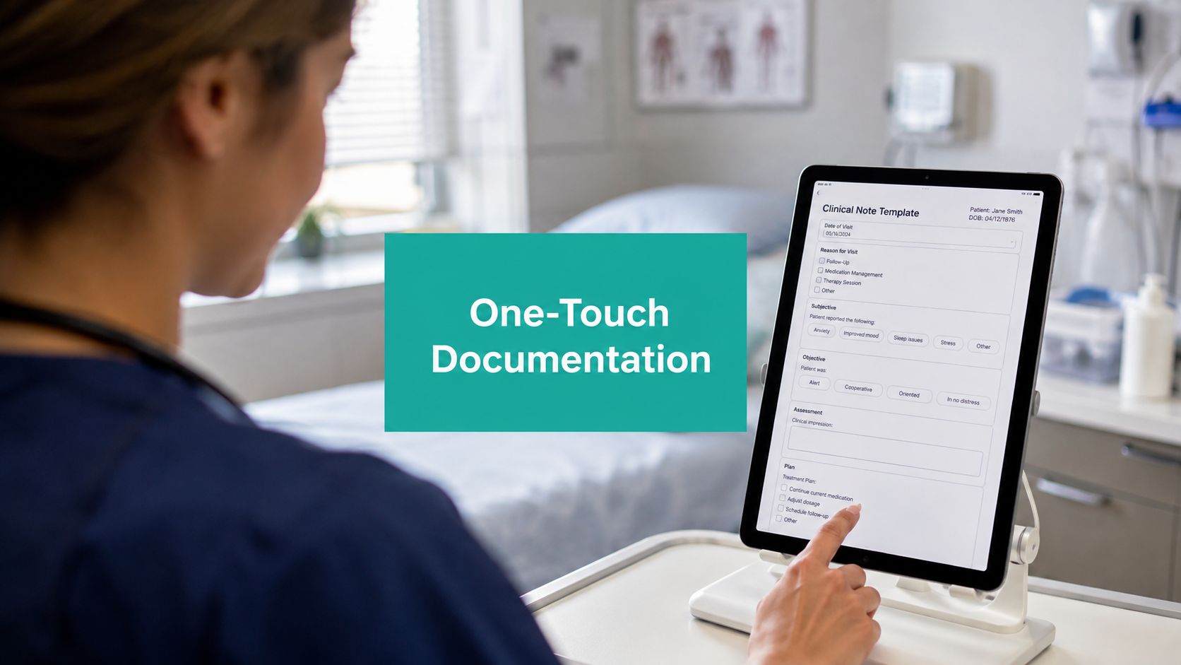
A reliable bedside note should capture the clinical picture once, in an order that supports both care and billing. Start with the Wagner grade, display the grade definition, and require confirmation rather than allowing an unexamined value to carry forward.
Next, record length, width, and depth in centimeters, plus undermining in centimeters. Add wound-bed tissue by zone, exudate type and amount, odor, periwound condition, pain, drainage, and infection signs. If the wound has slough, eschar, granulation, maceration, erythema, or callus, name it directly.
A usable point-of-care template
Assessment
- Classification: Wagner grade and the clinical finding that supports it.
- Location: Laterality, foot region, and anatomical site.
- Measurements: Length × width × depth in centimeters, post-debridement area when a procedure is performed.
- Structures: Skin, subcutaneous tissue, tendon, capsule, muscle, fascia, or bone.
- Wound bed: Granulation, slough, eschar, exposed structure, and tissue distribution.
- Exudate: Type, amount, and change from the prior assessment.
- Periwound: Erythema, warmth, edema, maceration, callus, induration, or breakdown.
- Infection and perfusion: Local or systemic signs, pulses, vascular findings, and tests ordered.
- Plan: Offloading, imaging, culture strategy, antibiotics, referral, and follow-up.
Close with the procedure details. State the deepest tissue removed, total area after debridement, instruments or method used, hemostasis, tolerance, and the medical necessity for the service. Then review the proposed CPT and ICD-10-CM pairing before signing.

Where structured assistance fits
EkagraHealth AI can fit this workflow by helping classify the Wagner grade from the wound image, estimate surface area from image pixels, suggest CPT and ICD-10-CM pairings, and push a structured draft into the EHR for clinician review. The clinician still confirms the grade, validates measurements, edits the assessment, and signs the final record.
That division of labor is the practical one. Automation can reduce retyping and surface missing fields. It can't independently resolve whether a cold foot needs revascularization, whether bone is infected, or whether palliation matches the patient's goals.
Prognosis Beyond the Grade and When to Escalate
A Wagner grade is a tissue snapshot, not a complete prognosis. Higher grades tend to carry longer treatment courses and greater amputation risk, but patient-level factors can change the trajectory substantially. A 2023 study found Wagner and Texas stage useful for prognosis, while dialysis status, CRP, albumin, and major adverse cardiac events materially changed predictive value; among patients on dialysis, inflammatory and nutritional markers outperformed wound stage itself in the reported prognostic analysis.
That means the same grade can represent very different patients. A Grade 2 ulcer in a patient receiving dialysis with peripheral arterial disease and poor nutrition deserves more urgency than the textbook label suggests. A stable, well-perfused outpatient with a Grade 3 wound may still be a salvage candidate, but only after deep infection and perfusion are addressed.
The escalation thresholds below are practical triggers for local protocols and specialist review. They aren't substitutes for examination or payer-specific requirements.
| Patient Factor | Threshold to Escalate | Override Starting At Wagner Grade | Referral Path |
|---|---|---|---|
| Renal disease | Dialysis status or markedly reduced kidney function | Any grade with deterioration or infection concern | Nephrology, vascular, wound team |
| Nutrition | Albumin under 2.5 g/dL | Grade 2 or higher, especially with tissue loss | Nutrition and wound team |
| Inflammation | CRP trending upward for 72 hours | Grade 2 or higher with infection findings | Infectious disease or hospital team |
| Perfusion | ABI under 0.5 or toe pressure under 30 mmHg | Any open ulcer | Urgent vascular assessment |
| Glycemic control | Glucose above 250 mg/dL and uncontrolled | Any active ulcer with delayed progress | Diabetes management and wound team |
| Pulses | ABI under 0.8 or non-palpable pulses | Any Wagner grade | Vascular surgery referral |
| Deep infection | Grade 3 or higher with systemic signs | Grade 3 | Infectious disease, admission, and surgical review |
| Offloading failure | No meaningful response after two weeks | Grade 1 or higher | Podiatry and offloading reassessment |
| Tissue defect | Grade 4 defect exceeding 2 square centimeters | Grade 4 | Vascular, surgical, and reconstructive review |
The bedside rule is simple: Wagner tells you about the wound. Labs and perfusion tell you about the patient. Keep both in the assessment, because a severity shortcut becomes unsafe when it hides the factors driving healing failure.
EkagraHealth AI helps wound teams structure Wagner grading, capture measurements and image-supported wound details, and connect documentation with CPT and ICD-10-CM review. Visit EkagraHealth AI to see how the workflow can support cleaner point-of-care documentation and more defensible debridement records.
