
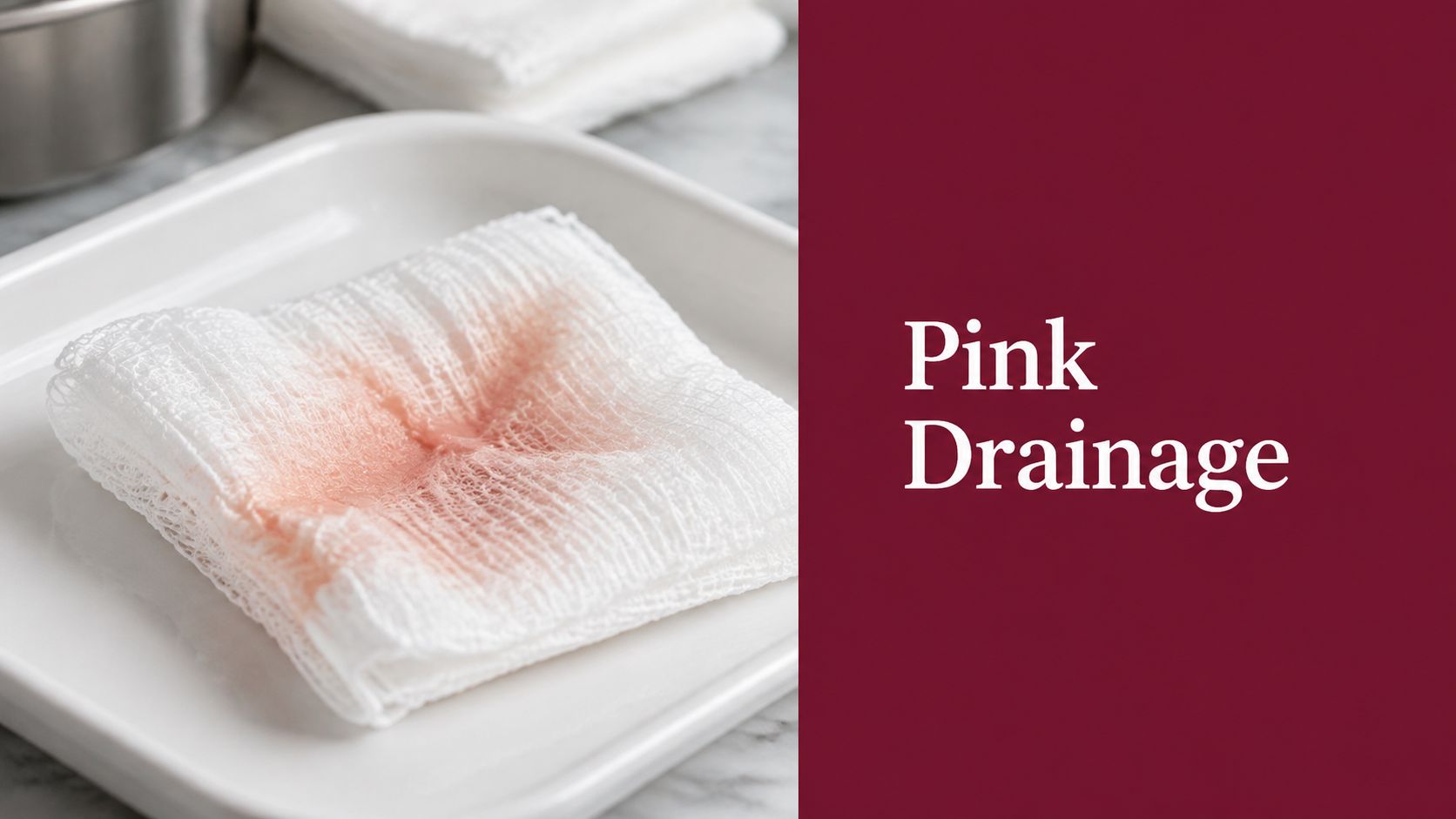
Monday morning, the foam dressing comes off a postoperative abdominal incision and leaves a faint salmon-pink stain across the contact layer. Under the overhead lamp, the clinician tilts the gauze and asks three questions: Is the color uniform? Does it have an odor? How long ago was the dressing changed?
That bedside pause is appropriate. Serosanguineous drainage color gives useful information, but color alone doesn't establish whether a wound is healing normally. Timing, amount, consistency, odor, periwound condition, pain, wound type, and the patient's vascular and bleeding risks turn a visual observation into a clinical judgment. The same pale pink fluid can be expected after a recent debridement, while a change toward bright red output may require immediate reassessment and more precise documentation.
The First Time You See Pink Drainage on a Gauze
A faint pink mark on gauze usually reflects serous fluid mixed with a small amount of blood. The fluid is generally thin and watery, not thick or sticky. Wound-exudate references describe this pattern as clear, pink to light red, and slightly thicker than water, with the color reflecting a small number of red blood cells in serous fluid (modern wound-exudate guidance).
The first mistake is calling it “drainage” and stopping there. A useful assessment starts with the dressing itself, then moves to the wound and the patient:
- Color: Is it pale pink, salmon, light red, or frankly bright red?
- Pattern: Is the staining diffuse, streaked, pooled, or continuing to accumulate?
- Consistency: Does it remain thin, or has it become cloudy, viscous, or clotted?
- Odor: Is there no odor, or is there a new sweet, pungent, or foul smell?
- Timing: How long did the dressing remain in place, and is the output increasing?
- Patient response: Has pain, warmth, swelling, or tenderness changed?
A postoperative incision may produce thin serosanguineous fluid during early healing. A recently traumatized wound or a wound after dressing removal can do the same. The practical question isn't whether pink is automatically normal. It's whether the drainage fits the wound's time course and clinical appearance.
Practical rule: Chart what you see on the dressing, then verify it against the wound bed, periwound skin, odor, symptoms, and trend.
That approach also protects the record. “Pink drainage noted” is a start. “Thin, pale pink, non-odorous exudate, evenly distributed on the contact layer, without active pooling or increased periwound warmth” gives the next clinician something they can evaluate.
Defining Serosanguineous Drainage Color
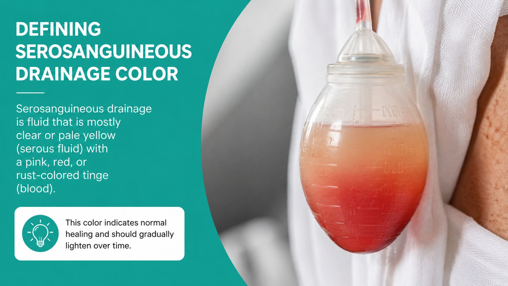
Serosanguineous drainage is a mixture of clear serous fluid and sanguineous fluid. Clinically, it appears pink to light red, often described as salmon, pale rose, or diluted red. Its consistency should remain thin and watery, with low viscosity and easy absorption into gauze. It shouldn't have the thick, sticky, cloudy quality associated with purulent material.
The color range matters because it separates serosanguineous output from two adjacent descriptions:
- Serous: clear, straw-colored, or light amber fluid with a watery texture.
- Serosanguineous: pink, salmon, or light red fluid with a watery texture.
- Sanguineous: bright red fresh blood, sometimes progressing toward darker red when bleeding is more substantial or has pooled.
- Seropurulent: cloudy, yellow, tan, gray, or green-tinged fluid that may be thinner than frank pus but is more concerning for infection-related change.
The requested color spectrum should be handled carefully in practice. Some educational materials describe the red-cell admixture as a small proportion of the total fluid, sometimes expressed as 5 to 25 percent, but that numerical estimate isn't something a bedside clinician can reliably measure by visual inspection. Document the visible descriptor instead of assigning a false precision. A surveyor can defend “pale pink, thin, watery” far more readily than an unsupported percentage.
Odor should be absent or neutral in uncomplicated serosanguineous drainage. Viscosity should stay low even when the amount is more than minimal. If the fluid becomes thick, cloudy, malodorous, or increasingly red, the descriptor needs to change and the assessment needs to follow it.
A strong chart entry includes the observable elements:
“Thin, watery, pale pink exudate. No odor. No visible clotting. Periwound skin intact without increased warmth.”
That language describes the exudate without claiming that color alone proves normal healing.
Why the Drainage Turns Pink in the First Place
The pink hue comes from the interaction between serous fluid and a small amount of blood during tissue repair. In the inflammatory phase, local mediators increase capillary dilation and permeability. Plasma moves into the wound bed, and a small number of red blood cells can escape through widened endothelial gaps. The result is diluted red fluid rather than a solid field of fresh blood.
Histamine and bradykinin contribute to vascular dilation and permeability during inflammation. As the wound enters the proliferative phase, angiogenesis adds another reason for light blood-tinged drainage. Fragile capillary loops form within new granulation tissue, and those vessels can leak with shear, cleansing, debridement, or removal of an adherent dressing.
Vascular endothelial growth factor, or VEGF, supports the formation of new vessels. Those early vessels aren't as structurally mature as established tissue vasculature. A small amount of oozing can therefore tint otherwise clear fluid, especially when a dressing has disturbed delicate granulation tissue.
The expected trajectory
A normal uncomplicated pattern often moves from more visibly bloody output toward pale pink serosanguineous fluid and then toward mostly serous fluid. This progression is especially useful in the first 48 to 72 hours after surgery, when thin, watery serosanguineous output can occur without concern (Cleveland Clinic's drainage overview).
The concern is a reversal of that trend. Pale pink becoming distinctly bright red can indicate renewed surface trauma, a disrupted capillary, an opened vessel, or another source of active bleeding. A change in color deserves more attention when it arrives with increased volume, thicker consistency, new clotting, worsening pain, or a dressing that is saturating more quickly.
Serous vs Sanguineous vs Seropurulent at a Glance
At the nurses' station, the quickest reliable differential uses four cues: color, consistency, odor, and clinical concern. Color gets the first look, but odor and thickness often prevent a misleading label. The wound exudate reference is useful for keeping those descriptors consistent across staff.
| Exudate Type Comparison | Color | Consistency | Odor | Clinical Concern |
|---|---|---|---|---|
| Serous | Clear, straw, or light amber | Thin and watery | Usually absent or neutral | Often consistent with early healing when the amount and trend fit the wound |
| Serosanguineous | Pink, salmon, or light red | Thin, watery, low viscosity | Usually absent or neutral | Common in early healing, after surgery, trauma, or dressing removal |
| Sanguineous | Bright red to dark red | Blood-like, may be thicker or contain clotting | Usually absent unless another process is present | Raises concern for active bleeding, vessel disruption, or renewed trauma |
| Seropurulent | Cloudy yellow, tan, gray, or green-tinged | Thin to moderately viscous, often cloudy | Sweet, pungent, or foul odor may occur | More concerning for infection-related exudate, especially with worsening local findings |
A clean, pale pink stain isn't the same as bright red blood. Likewise, cloudy yellow fluid isn't “serous with a little color.” The consistency has changed, and the assessment should reflect that.
Odor is frequently under-documented. A wound can have little visible change while developing a new unpleasant smell. Smell the removed dressing in a controlled clinical setting, not after it has been left near a bedside trash container or mixed with ambient room odors.
“Clear drainage” is another common charting shortcut that creates trouble. If the fluid is pink, write pale pink serosanguineous drainage. If it is translucent but has a red tint, “clear” is incomplete and may conflict with the wound photograph or the nursing note.
What Pink Drainage Looks Like by Wound Type and Wagner Grade
The same color has different implications depending on the wound's depth, tissue quality, and cause. A light pink output from clean granulation tissue after debridement isn't read the same way as pink fluid from a wound with undermining, necrotic tissue, or suspected infection.
Surgical incisions
Postoperative incisions commonly show light pink serosanguineous drainage as early capillary repair and angiogenesis progress. Many clinical sources consider thin, watery serosanguineous output from a surgical site acceptable for 48 to 72 hours, provided the trend moves toward lighter, more serous fluid rather than increasing blood-tinged output (postoperative drainage guidance).
Don't document the color without recording approximation of amount and dressing performance. A small stain that remains stable is different from repeated strike-through, pooling beneath the incision, or a sudden change from pink to bright red.
Venous and pressure wounds
Venous stasis ulcers can produce moderate exudate that mixes with hemosiderin staining. The result may look darker amber-pink or rusty, but the color must be interpreted alongside edema, maceration, wound-bed tissue, and the direction of the trend.
Stage 3 and Stage 4 pressure injuries may show pink drainage from granulation tissue. In these wounds, volume and tissue findings often matter more than hue. Increasing saturation, new odor, undermining, exposed structures, or a deterioration in periwound skin should move the assessment beyond “pink drainage present.”
Diabetic foot ulcers
A Wagner Grade 2 plantar ulcer with clean granulation tissue may lightly ooze serosanguineous fluid after sharp debridement. The note should identify the wound bed, depth, exudate, periwound status, and whether bleeding stopped with routine local measures.
A Wagner Grade 3 ulcer extends into deeper structures and may include abscess or deeper infection. If pink drainage becomes cloudy, yellow, tan, or green-tinged, the differential shifts toward seropurulent exudate. The Wagner classification reference helps align wound depth and clinical concern, but the drainage description still has to stand on its own.
At Wagner Grade 4 or Grade 5, drainage is secondary to vascular status and tissue viability. Assess perfusion, ischemic changes, necrosis, and systemic risk promptly. A pale pink stain shouldn't distract the team from a threatened limb.
Bedside Assessment Tips That Change the Reading
Start with the periwound skin, not the center of the gauze. Maceration means moisture has exceeded the skin's tolerance or the dressing interval isn't matching the output. The drainage may still be textbook pink, but the treatment plan is not working if the surrounding skin is whitening, softening, or breaking down.
Next, establish how quickly the dressing saturates. Foam, alginate, or another absorbent dressing can make a wound appear deceptively controlled until the fluid reaches the edges or strikes through. A dressing that saturates in under 24 hours represents heavier output than a small central pink stain suggests.

Read the wound in context
Use a consistent sequence:
- Inspect the periwound: Look for maceration, erythema, edema, blistering, and changes in skin temperature.
- Assess the wound bed: Identify granulation, slough, eschar, exposed structures, and active bleeding.
- Check the dressing: Record the distribution, saturation pattern, and whether fluid has reached the secondary layer.
- Evaluate symptoms: Compare current pain with the patient's recent baseline rather than relying on a single pain score.
- Compare sides when appropriate: Periwound temperature and limb findings are more meaningful when compared bilaterally.
- Assess odor correctly: Smell the removed dressing during removal and before it has been exposed to room or waste odors.
Escalating pain deserves attention even when drainage remains pink. Pain may reflect pressure, ischemia, infection, or tissue trauma, so reassurance based on color alone is unsafe.
A photograph can improve continuity when policy and consent permit it. Place the dressing or wound against a white gauze background, use consistent lighting, and avoid fluorescent color distortion. The goal isn't a dramatic image. It is a repeatable visual record that lets the team compare a true pale pink stain with a later, deeper red change.
Documentation and Coding for Serosanguineous Drainage
At the nurses' station, a note reading “pink drainage, moderate amount” may sound adequate, but it leaves several coding and clinical questions unanswered. A defensible entry connects the drainage to the wound assessment and states what the color does, and does not, establish. CMS expects a debridement procedure note to include wound characteristics such as diameter, depth, undermining or tunneling, color, exudate or necrotic tissue, and the level and type of tissue removed. These details support medical necessity for debridement services reported with CPT 11042 through 11047 (CMS wound-care billing guidance).
Use a wound-care documentation template or another structured SOAP workflow to keep the color description tied to the rest of the assessment. A practical entry might read:
- Subjective: “Patient reports the outer dressing became saturated sooner than usual and describes increased tenderness since the last dressing change.”
- Objective: “Thin, pale pink, non-odorous serosanguineous exudate. Dressing saturated in [document actual interval]. Wound dimensions, depth, undermining, tunneling, wound-bed tissue, and periwound findings documented separately.”
- Assessment: “Serosanguineous drainage remains consistent with the current wound type and recent treatment,” or, when findings are changing, “Increasing red output and pain require reassessment for bleeding or other complication.”
- Plan: “Continue ordered dressing regimen, protect periwound skin, reassess output trend, and escalate according to vascular, bleeding, or infection findings.”
Do not insert an invented dressing interval. Record the patient's report or the interval observed by staff.
| Documentation and Coding Map for Serosanguineous Drainage | Sample Language | ICD-10 / CPT Pair | Surveyor Flag |
|---|---|---|---|
| SOAP assessment | “Thin, pale pink, watery, non-odorous exudate” | Pair the wound diagnosis with the supported service code | “Moderate drainage” without color or consistency |
| Non-pressure ulcer | Identify site, laterality, severity, depth, and exudate | L97.- with the applicable wound diagnosis and service | Missing depth or anatomic specificity |
| Pressure injury | Record stage, location, dimensions, tissue, and drainage | L89.- with the applicable service | Stage not supported by the wound-bed description |
| Diabetic foot ulcer | Link diabetes, foot site, wound depth, and Wagner grade | E11.621 with the appropriate ulcer code and service | Diabetes documented without a clear ulcer linkage |
| Post-surgical wound | Describe incision status, drainage, and complication findings | Appropriate S/T code with the supported service | Postoperative status documented without current wound findings |
| Debridement | State the deepest tissue removed and treated surface area | CPT 11042–11047, selected by depth and surface area | Exudate alone used to justify surgical debridement |
The debridement code comes from the documented depth and tissue removed, the treated area, and the procedure details. Drainage color helps describe the wound, but it does not establish the code. “Debrided wound, moderate drainage” leaves the payer to infer key facts and can invite a denial.
Survey and billing problems often arise from copying an old exudate description forward, documenting “clear” when the fluid is pink, omitting odor, or failing to explain a dressing-frequency change. EkagraHealth AI can help clinicians capture wound findings in a structured SOAP workflow and connect documented characteristics with coding review. The treating professional remains responsible for the clinical assessment.
When Pink Becomes a Problem and What to Do Next
Pale pink, thin, non-odorous drainage often fits expected healing. The interpretation changes when the trend reverses or other findings appear. Treat a shift toward bright red or maroon fluid, a sudden increase in saturation frequency, new foul odor, increasing pain, or expanding periwound erythema as a reason to reassess and relabel the output.
A Wagner Grade 3 diabetic foot ulcer with pink drainage that persists beyond the expected healing trajectory without developing healthy granulation deserves a closer evaluation. The precise response depends on the patient's condition, wound findings, vascular status, medications, and local protocol.
Escalation sequence
- Repeat the wound and vascular assessment. Check pulses, perfusion, capillary refill where clinically appropriate, skin temperature, tissue viability, and changes from baseline.
- Consider ankle-brachial index or toe pressure for lower-extremity wounds when arterial status is uncertain or worsening.
- Order CBC and CRP when clinically indicated by concern for infection, inflammation, or systemic change.
- Obtain a wound culture when purulent conversion is suspected, using the method required by local practice and the treating clinician's judgment.
- Use plain film or MRI when osteomyelitis must be evaluated, particularly in a Wagner Grade 3 or higher wound with compatible findings.
Keep a short point-of-care checklist in the chart binder:
- Expected color: pale pink to light red, thin and watery.
- Red flags: bright red or maroon output, increasing volume, clotting, foul odor, worsening pain, cloudiness, or spreading erythema.
- Context: wound type, recent debridement, dressing-change trauma, anticoagulant exposure, and vascular status.
- Follow-up: document the trend and use the clinic's callback process, including the 24-hour callback rule when that is the established protocol.

Color is never interpreted alone. Trend, odor, amount, pain, periwound findings, and comorbidities determine whether pink is reassuring, a bleeding concern, an infection signal, or a documentation problem that needs correction.
If your team needs more consistent wound narratives, EkagraHealth AI can help structure SOAP documentation, capture exudate details, and support CPT and ICD-10 coding review. Visit EkagraHealth AI to see how a wound-care workflow can keep color, depth, treatment, and billing evidence together in the same clinical record.
