To really get a handle on wound care, you have to know what you’re looking at in the wound bed, and that starts with a clear understanding of granulation tissue. Simply put, this is the living proof that your patient's body is actively working to repair an open wound. Seeing it is a good sign—it means the healing process is on track. For clinicians, mastering the granulation tissue definition isn't just academic; it's a foundational skill for effective patient care, accurate documentation, and successful outcomes. Understanding what it is, what it looks like when healthy, and what can go wrong is crucial for guiding a wound from injury to closure.
What Is Granulation Tissue? A Clinician's View
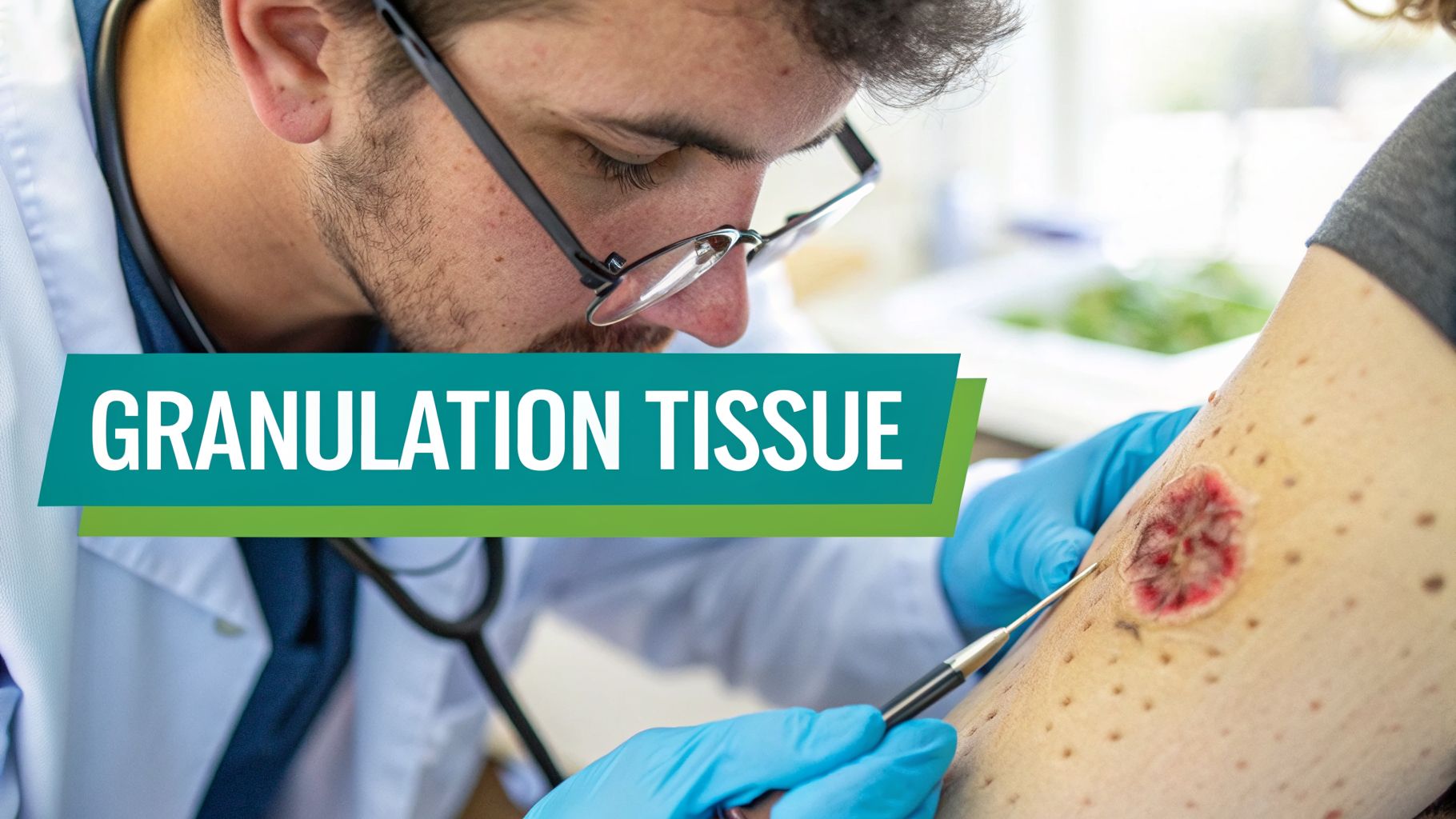
In clinical practice, the granulation tissue definition refers to the new, reddish, and slightly bumpy connective tissue that builds up from the base of a wound during the proliferative phase of healing. Its appearance is one of the first things we look for to confirm a wound has entered this critical stage and is healing properly. Think of it as the body's own biological scaffolding, being built from the ground up to fill the wound's empty space, creating the foundation upon which new skin will eventually grow. This process is a visible sign of progress and a key indicator that the body's repair mechanisms are functioning as they should.
This new tissue is a complex and fascinating mix of new blood vessels (capillaries), fibroblasts—which act like the construction crew of wound healing—and a loose extracellular matrix that holds everything together. Together, these components fill the wound defect, protect it from infection, and create the necessary foundation for new skin cells, or epithelium, to eventually migrate across and close the wound for good. Without the successful formation of a healthy granulation bed, the later stages of healing simply cannot occur.
Key Characteristics of Healthy Granulation Tissue
When we talk about healthy granulation tissue, we’re looking for specific visual and physical cues at the bedside. These signs tell us that the wound has what it needs to continue healing and is progressing correctly through the proliferative phase, which can last for several days or even weeks depending on the wound's size, depth, and the patient's overall health. A proper assessment hinges on recognizing these key features.
Healthy granulation tissue should have these features:
- Color: A vibrant, "beefy red" or bright pink hue is the classic sign of a rich, new blood supply (angiogenesis). This indicates robust oxygen and nutrient delivery to the healing site.
- Texture: The surface looks bumpy or granular, often described as having a "cobblestone" or "raspberry-like" appearance. Those little bumps are actually the loops of new capillaries growing up toward the surface.
- Moisture: It should appear moist but not overly wet. The new blood vessels are naturally permeable, allowing some plasma to exude and keep the surface hydrated, which is essential for cellular function.
- Viability: The tissue is firm and attached securely to the wound bed. It is resilient and won't wash away with gentle cleansing or irrigation. It represents new, living tissue.
Properly identifying granulation tissue is a fundamental skill in wound care. It helps you differentiate between healing and non-healing signs, such as slough or eschar. For a deeper dive into other tissues you might encounter, our guide on various https://ekagrahealth.ai/wound-bed-descriptions/ is a great resource.
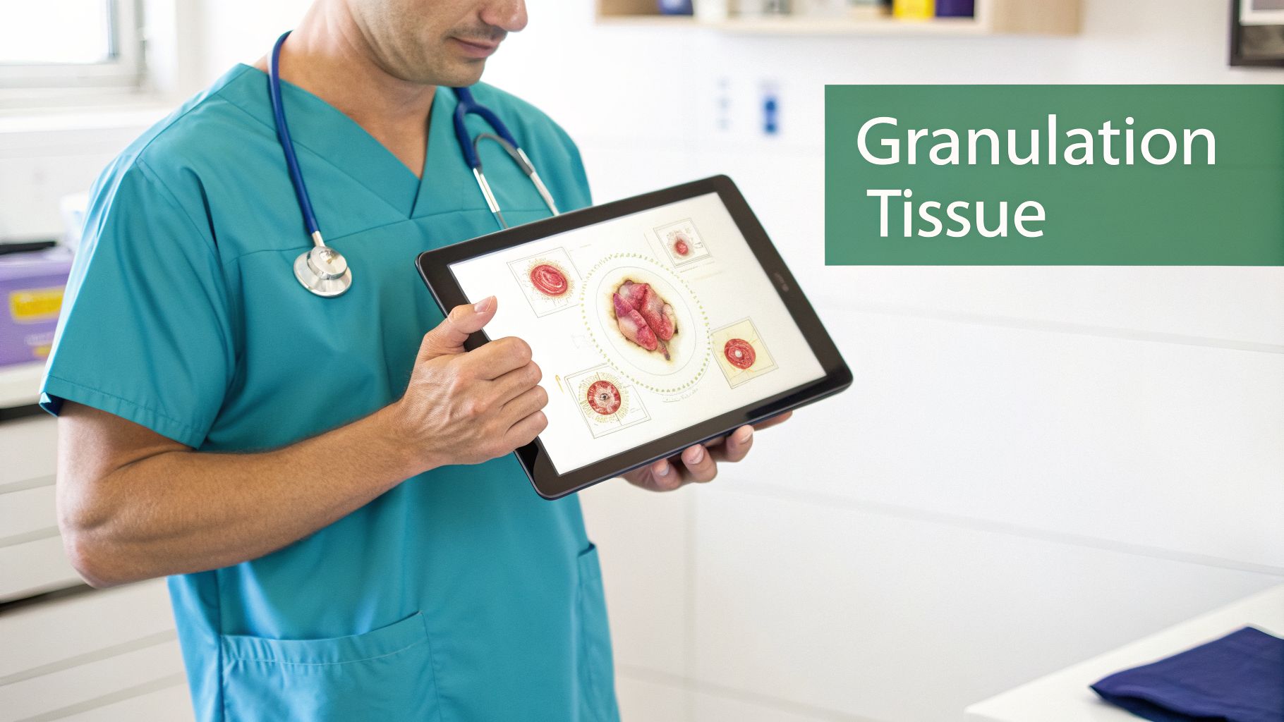
Healthy vs Unhealthy Granulation Tissue At a Glance
Of course, not all granulation tissue is a good sign. Sometimes, its appearance can signal a problem like infection, poor perfusion, or systemic issues that are impeding the healing process. This quick reference table can help you distinguish healthy from unhealthy tissue at the bedside, enabling faster intervention.
| Characteristic | Healthy Granulation Tissue | Unhealthy Granulation Tissue |
|---|---|---|
| Color | Bright, "beefy red" or pink | Pale pink, dusky red, or dark and discolored |
| Texture | Bumpy, granular, cobblestone-like | Smooth, gelatinous, or flat; may look fragile or "glassy" |
| Moisture/Exudate | Moist, with minimal serosanguinous drainage | Dry and dull, or excessively wet with purulent drainage |
| Bleeding | May bleed easily with minor trauma (e.g., debriding) | May bleed spontaneously or not bleed at all, even if poked |
Recognizing unhealthy granulation tissue early is critical. It’s a red flag that prompts you to reassess your treatment plan, look for underlying issues like infection or ischemia, and intervene before the wound stalls completely. This early detection is a hallmark of an expert clinician.
Modern tools and technologies are also changing how we collaborate on complex cases. For instance, using video conferencing for healthcare allows for remote consultations and specialist input, which can be invaluable when you're facing a challenging wound that isn't progressing as expected. This technology facilitates sharing visual data and getting second opinions quickly, enhancing patient care.
The Biology of Building New Tissue
To really understand what granulation tissue is, we have to look past the wound bed and dive into the remarkable biology happening underneath. It’s not just a random blob of tissue; it's the result of an incredibly well-organized cellular construction project orchestrated by a symphony of growth factors and cellular signals. This biological process is the core of the granulation tissue definition.
The process that gives healthy granulation tissue its classic "beefy red" color and bumpy texture is called angiogenesis. This is where the body forms brand-new blood vessels from pre-existing ones, which are absolutely essential for bringing in the oxygen and nutrients needed to rebuild damaged structures. Think of it as laying down the plumbing and electrical wiring for a new building—nothing else can happen without it. This neovascularization is what feeds the entire repair process, making it one of the most vital components of wound healing.
The Key Players at the Cellular Level
At the heart of this building process are two main types of cells. Getting a handle on what they do makes it much clearer why granulation tissue looks and feels the way it does at the bedside, and why a failure in their function leads to non-healing wounds. These cells are the heroes of the proliferative phase.
- Fibroblasts: These are the workhorses of wound repair. They migrate into the wound site and begin producing a provisional extracellular matrix, primarily composed of collagen and fibronectin. This matrix acts as the initial scaffolding that gives the new tissue its structure and provides a framework for other cells to move in.
- Endothelial Cells: These are the engineers of the new blood supply. They proliferate and migrate to form tiny, delicate capillary loops that sprout up toward the surface of the wound. This creates that rich, red vascular network that keeps the new tissue alive, growing, and gives it its characteristic granular appearance.
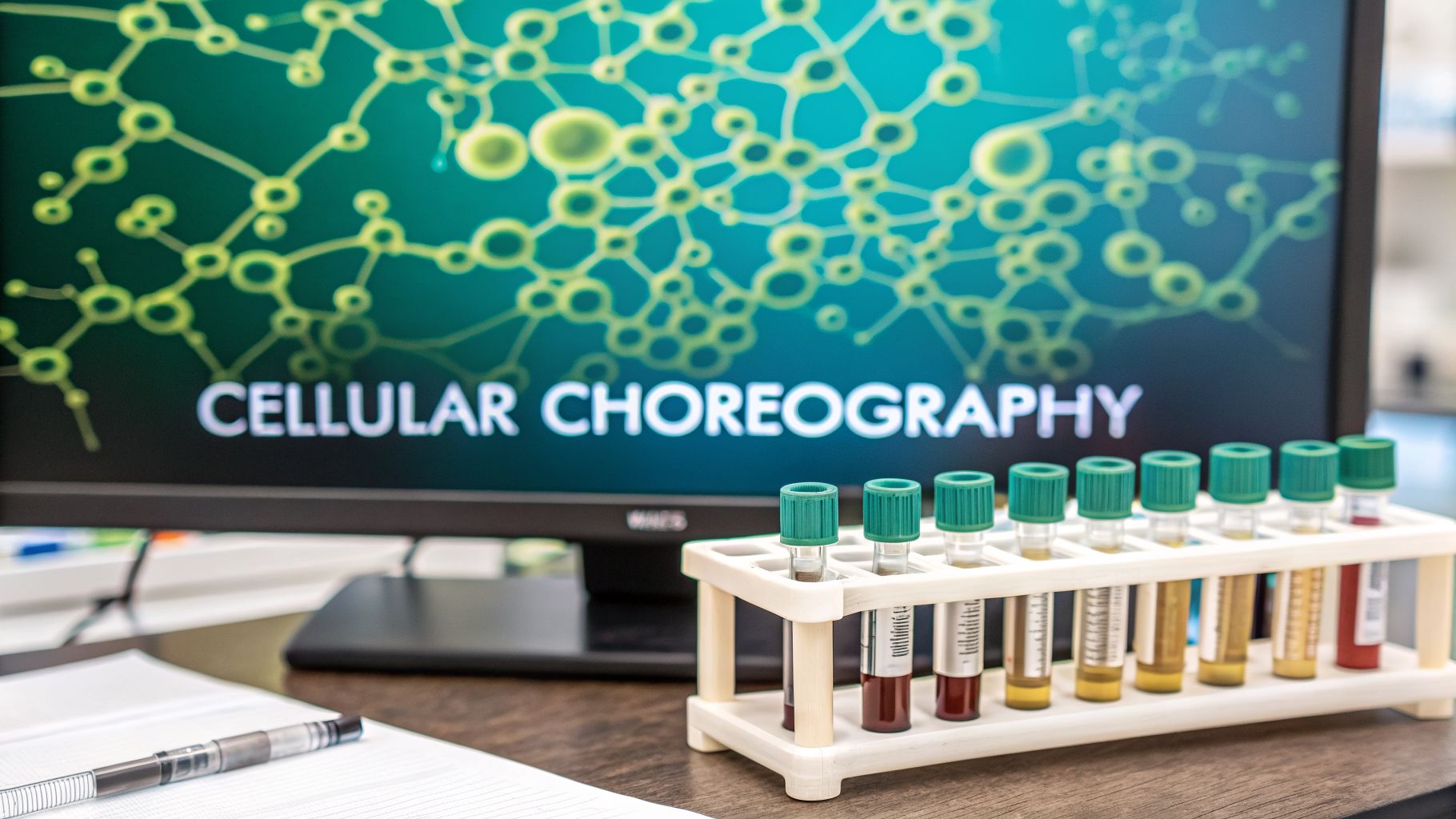
It's important to remember these cells aren't working independently. A cascade of specific growth factors, such as Vascular Endothelial Growth Factor (VEGF), Platelet-Derived Growth Factor (PDGF), and Fibroblast Growth Factor (FGF), acts as a sophisticated communication system, directing this cellular traffic and telling each cell exactly where to go and what to build. This cellular choreography is what makes healing possible.
From Temporary Scaffolding to a Permanent Fix
In the beginning, the scaffold that fibroblasts build is made mostly of a flimsy, flexible material called Type III collagen. This is perfect for the early days of healing because it's laid down quickly and allows cells to move around easily and get into position. However, it lacks significant tensile strength and is not suitable for a long-term fix.
As healing progresses and the proliferative phase transitions into the maturation (or remodeling) phase, the fibroblasts get to work remodeling this temporary structure. They gradually replace the weaker Type III collagen with a much tougher, more durable version known as Type I collagen. This is like a construction crew swapping out temporary wooden supports for permanent steel beams. It's this critical transition that gives the final scar tissue the strength it needs to hold up over time, although it typically only reaches about 80% of the original tissue's strength.
In a typical, uncomplicated acute wound, this process runs on a surprisingly predictable schedule. Granulation tissue usually hits its peak thickness around day 7. Of course, in more complex or chronic wounds, this can be delayed to day 15 or even later. Research has shown that key vascular markers, like vascular endothelial growth factor A (VEGF-A), spike right during this phase of peak formation, which directly correlates with how fast a wound heals. You can review the full research on healing timelines to see the data on these molecular events.
How to Assess Granulation Tissue at the Bedside
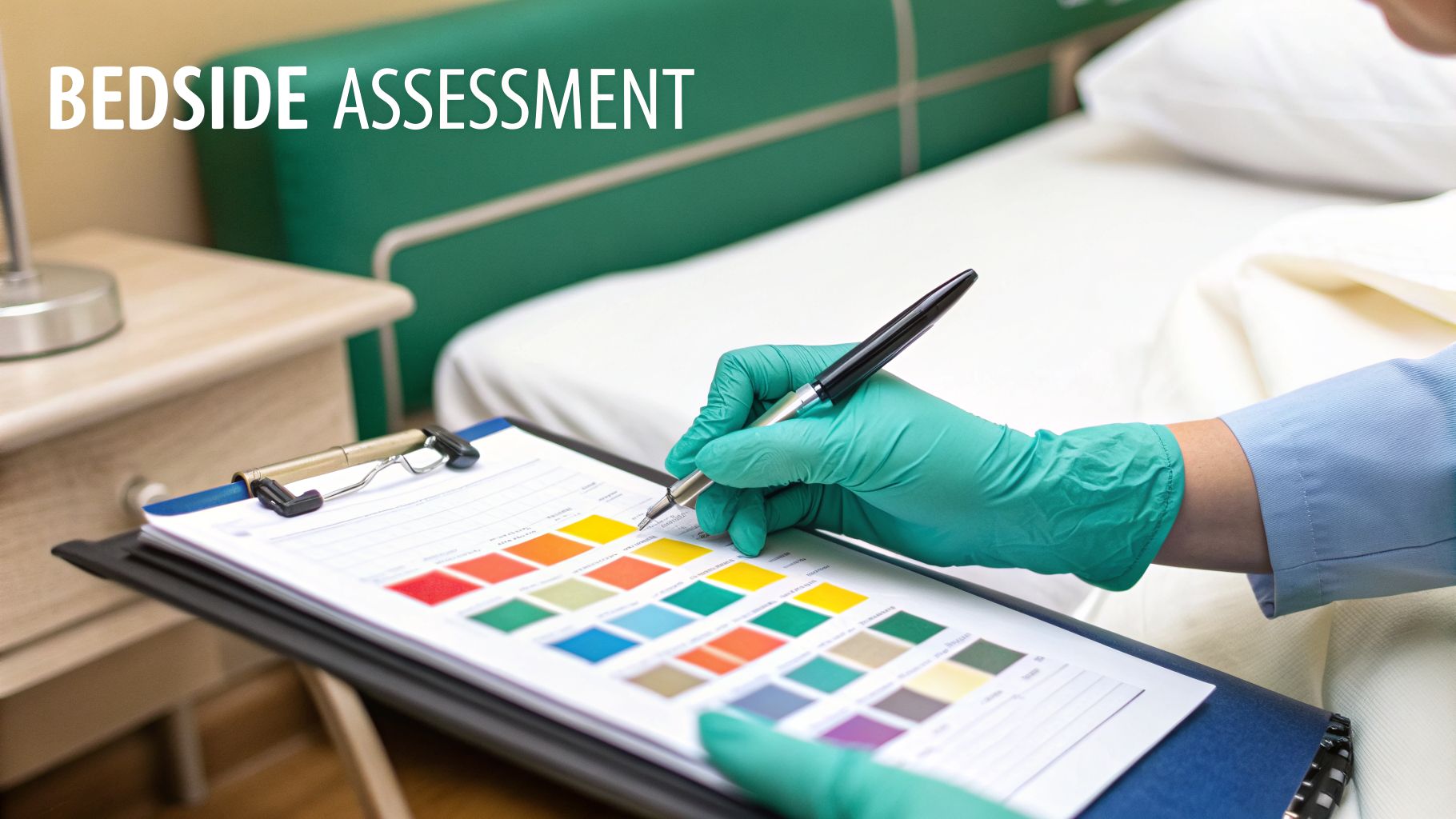
As clinicians, our eyes are one of our most powerful diagnostic tools, especially in wound care. Being able to confidently identify and assess granulation tissue at the bedside isn't just an academic exercise—it directly informs your care plan, helps predict healing trajectories, and tells you if the wound is on the right track. Let’s walk through what to look for and how to avoid common pitfalls in assessment.
When you see healthy granulation tissue, you'll know it. Think "beefy red." That classic, vibrant color is your first and best clue, signaling that a rich network of new blood vessels is forming and perfusing the new tissue. It's a sign of life and vitality in the wound bed.
The second thing to check for is texture. Healthy granulation tissue should have a bumpy, almost pebbled surface. A great way to remember this is to picture the surface of a raspberry. Those tiny bumps are actually the tips of new capillary loops pushing their way up to feed the healing wound. A smooth or glassy surface, by contrast, suggests poor vascularization and should be a cause for concern. It's a beautiful sign of progress when you see that healthy, bumpy texture.
Differentiating Granulation From Other Tissues
Of course, a wound bed is rarely that simple or uniform. You'll often find a mix of tissues, and your job is to tell them apart. Getting this right is critical, as mistaking non-viable tissue for healthy growth can stall healing and lead to ineffective treatment plans. Here’s a quick guide to telling friend from foe in the wound bed:
- Slough: This is the tricky one and a common source of confusion. Slough is non-viable tissue, a byproduct of the inflammatory response, composed of dead cells, fibrin, and exudate. It often shows up as a yellow, tan, or even whitish film. Unlike the firm, bumpy feel of granulation, slough is soft, stringy, or gelatinous. It's often loosely attached and can sometimes be gently wiped away, but may also be adherent.
- Eschar: You can’t miss this. Eschar is hard, dry, dead tissue—typically black or brown and leathery or scab-like. While it acts as a sort of natural "cover," it's a significant barrier to healing and must eventually be debrided to allow granulation and epithelialization to occur.
- Epithelium: This is the new skin advancing from the wound's edges. It has a very different look—smooth, shiny, and usually pale pink or almost pearly white. It contrasts sharply with the deep red, bumpy granulation tissue it’s trying to cover. Seeing this "leading edge" of new skin is a fantastic sign that the wound is moving toward closure.
Accurately identifying and quantifying these different tissues is the foundation of good wound documentation and treatment. Noting the presence, percentage, color, and texture of granulation tissue tells a powerful story about the wound's healing trajectory. For a deeper dive into what different shades can mean, our visual guide to granulation tissue color is an excellent resource.
Common Roadblocks to Healthy Tissue Growth
Even when a wound enters the proliferative phase, the path to full closure isn't guaranteed. A number of clinical hurdles can slow down or completely stall granulation tissue formation, turning what should be a healing wound into a chronic, frustrating problem. Knowing what these roadblocks are, both systemic and local, is the key to troubleshooting them and getting your patient's recovery back on track. A clear granulation tissue definition also includes understanding what inhibits its formation.
Many of these challenges are systemic, impacting the patient's entire body and its ability to heal. For instance, uncontrolled diabetes is a major barrier; monitoring Hba1c levels is critical for gauging how well blood sugar is being managed. Hyperglycemia damages small blood vessels (microangiopathy), impairs leukocyte function, and reduces the production of essential growth factors, effectively starving the wound of the oxygen and nutrients it needs for angiogenesis.
Peripheral vascular disease (PVD), whether arterial or venous, has a similar effect by directly compromising blood flow. This often results in pale, fragile, and poorly forming granulation tissue that simply won't granulate. Without adequate perfusion, healing is impossible.
The Impact of Aging and Comorbidities
Age itself plays a huge role in the efficiency of wound healing. You'll often see that elderly patients form granulation tissue much more slowly than younger individuals. This is a natural consequence of "immunosenescence," reduced cellular activity, a slower inflammatory response, and a less vigorous immune system, which can slow down the healing process by 20-30%.
Comorbidities make this delay even worse. Conditions like diabetes, cardiovascular disease, renal failure, and autoimmune disorders have been shown to throw another wrench in the works for older patients, making it significantly harder for the body to mount an effective repair. These conditions compound the age-related delays. You can explore the complete findings on healing in elderly patients for a deeper look.
These systemic issues can create a perfect storm. The wound bed simply lacks the basic resources—oxygen, nutrients, growth factors, and a strong cellular workforce—to build the new tissue it desperately needs. Addressing these underlying health problems is just as important as the local wound care itself.
Local Factors That Halt Progress
Beyond systemic problems, issues right in the wound bed can stop granulation cold. The most common local barriers we see at the bedside are often within our direct control as clinicians and include:
- Infection and Biofilm: Bacteria compete for vital nutrients and release toxins that are cytotoxic to new cells like fibroblasts. Biofilm is especially problematic; this slimy, bacterial shield is notoriously stubborn, resistant to antibiotics, and can make a wound completely resistant to healing by creating a state of chronic inflammation.
- Poor Nutrition: A patient's diet is a direct building block for wound repair. If they're deficient in protein (for collagen synthesis), key vitamins (especially C and A), or minerals like zinc and iron, tissue formation will grind to a halt. A nutritional assessment should be part of every chronic wound workup.
- Repetitive Trauma or Pressure: Constant pressure, particularly in pressure injuries, will crush the delicate new capillaries that are trying to form, leading to localized ischemia. This physically prevents tissue from growing and is a primary reason why offloading is non-negotiable in treating these wounds.
Successfully identifying and managing these roadblocks—whether they're systemic or local—is fundamental to promoting a healthy wound bed environment and finally achieving wound closure.
Strategies to Promote Healthy Granulation
Once you've figured out what's stalling a wound, your focus has to shift from assessment to action—actively encouraging new growth. This involves creating the ideal environment for the body’s own construction crew—the fibroblasts and endothelial cells—to get to work. The principle is simple: give the wound everything it needs to thrive and remove anything getting in its way. This is where the art and science of wound care truly intersect.
The foundation of any effective strategy is meticulous wound bed preparation. Think of it as clearing a construction site before you can pour the foundation. Any non-viable tissue, like yellow slough or black eschar, is a physical barrier that stops new capillaries from forming. It also serves as a breeding ground for bacteria, increasing the risk of infection and biofilm formation.
Preparing the Foundation for Growth
Debridement is the primary method for getting rid of those barriers. The objective is to expose the healthy, viable, bleeding tissue underneath because that’s the only surface new granulation tissue can actually grow on. Your choice of debridement method will depend entirely on the patient's condition, the wound characteristics, your scope of practice, and the clinical setting.
- Sharp Debridement: This is a quick and direct approach using sterile instruments like a scalpel, scissors, or curette. It's highly effective for getting rid of thick, adherent slough or hard eschar and is considered the gold standard for rapid removal of non-viable tissue.
- Autolytic Debridement: This is the body’s natural process, which we can enhance. By applying a moisture-retentive dressing (like a hydrocolloid or hydrogel), we let the body’s own enzymes gently dissolve the non-viable tissue. It's slower and more selective but much less invasive and painless for the patient.
- Enzymatic Debridement: This involves applying a prescription ointment containing enzymes, such as collagenase, to specifically break down and digest dead tissue (necrotic collagen), leaving the healthy tissue untouched. It's a good option when sharp debridement is not feasible.
The goal of debridement isn't just to clean the wound—it's to create a fully viable surface. A wound bed that is 100% covered in healthy, beefy red granulation tissue is the perfect launchpad for the final phase of healing: epithelialization.
Balancing Moisture and Protection
After the wound bed is clean, your next critical task is managing moisture. Granulation tissue is incredibly delicate. If it gets too dry, the new cells will desiccate and die, halting the healing process. If it gets too wet, the tissue can become macerated—mushy, fragile, and pale—which also impairs healing and raises the risk of infection as the periwound skin breaks down.
This is why dressing selection is an art. The ideal dressing protects this fragile new tissue from trauma, wicks away excess fluid (exudate) without drying out the wound bed, and maintains a warm, moist climate that cells love. Dressings like foams, alginates, and hydrofibers are excellent for this purpose.
For more challenging wounds that aren't responding to standard care, advanced therapies like negative pressure wound therapy (NPWT) can be a game-changer. NPWT uses a sealed dressing and a vacuum to apply gentle, controlled suction. This pulls excess fluid from the wound, reduces swelling (edema), increases perfusion, and actively stimulates the formation of new blood vessels (angiogenesis), effectively giving granulation a powerful jump-start.
Why Accurate Documentation of Tissue Matters
What you see and document at the bedside does more than just track a patient's healing journey. Those clinical notes are the critical link between your expert care, interdisciplinary communication, and your facility’s financial stability. In other words, how you describe the wound bed—applying the correct granulation tissue definition—directly influences medical coding, billing, and whether your organization gets paid for the work you do.
Think of it this way: describing a wound as "70% granulation with 30% slough" isn't just a clinical update. For a payer or an auditor, that specific detail paints a picture of medical necessity, justifying the skilled care and treatments you're providing, such as debridement. It’s the evidence that turns your clinical assessment into a language that billing and compliance departments can understand and use.
From Clinical Note to Financial Reimbursement
Your documentation is the legal and financial proof that backs up your coding choices. Specific ICD-10 codes depend on an accurate description of the wound's etiology, location, depth, and the types of tissue present. A note that clearly outlines the state of the granulation tissue—or the lack thereof—is what validates procedural codes (CPT codes) for services like debridement or the application of advanced therapies.
If your notes are vague, incomplete, or don't capture the wound's true complexity, payers are likely to question why your services were necessary. A note saying "dressing changed" is insufficient. A note that says "Sharp debridement of 4 sq cm of adherent slough from sacral pressure injury, revealing 100% granulation tissue" is defensible. Vague documentation is a fast track to claim denials, frustrating payment delays, and a mountain of administrative work for your practice.
This is why keeping the clinical granulation tissue definition in mind during documentation is so important. Strong, specific notes justify the need for ongoing advanced wound care, proving the patient requires more than just a simple dressing change. For clinicians aiming to make their documentation ironclad, using a structured wound care documentation template can be a game-changer for capturing every essential detail consistently and efficiently.
Ultimately, your precise descriptions do two jobs at once. They steer good clinical decisions and ensure continuity of care, while also ensuring your facility gets properly reimbursed for the expert care it delivers. It's how you protect your practice's financial health so you can continue providing that excellent care to every patient who needs it.
Frequently Asked Questions About Granulation Tissue
Even with a solid understanding of granulation tissue, a few tricky questions always seem to pop up at the bedside or during team meetings. Let's walk through some of the most common ones that clinicians run into, providing clear, actionable answers.
What Should I Do If Granulation Tissue Is Pale or Bleeds Easily?
Seeing pale (dusky pink) or friable granulation tissue—the kind that bleeds at the slightest touch—is a significant red flag. It’s the wound’s way of telling you something is wrong. This appearance often points to an underlying issue like poor perfusion (ischemia), a high bacterial load or infection brewing under the surface (biofilm), or significant nutritional deficits that are preventing healthy tissue formation.
Your job is to play detective. Start by assessing the wound and the surrounding tissue for other signs of infection (increased exudate, odor, periwound erythema). Then, zoom out and look at the whole patient. Re-evaluate their vascular status—are pulses palpable? Is there edema? What's their nutritional status really like? Answering these questions will help you adjust the care plan to address the root cause, whether that means consulting vascular surgery, initiating antimicrobial dressings, or involving a dietitian.
Is It Possible to Have Too Much Granulation Tissue?
Absolutely. We call this hypergranulation, or more informally, "proud flesh." This happens when the granulation tissue keeps growing unchecked and mounds up above the surface of the surrounding skin. It creates a bumpy, often friable, and wet-looking mass.
Think of it like a roadblock. The overgrowth creates a physical barrier, making it impossible for new epithelial cells to migrate across the wound bed and finish the job of closing it. The epithelial cells can't climb "uphill." This stalls the healing process completely and often requires intervention—such as the application of silver nitrate for chemical cauterization, topical steroids to reduce inflammation, or increased pressure from a foam dressing—to get things back on track and level with the skin.
How Can I Differentiate Granulation Tissue From a Blood Clot?
At a quick glance in a freshly debrided or traumatized wound, they can both look red, but the key difference is in their texture, adherence, and structure. Healthy granulation tissue has that classic bumpy, beefy-red appearance and is firmly attached to the wound bed. It's an integral part of the new tissue matrix and will not wash away.
A blood clot, on the other hand, is usually a darker, more gelatinous or jelly-like mass. It has a soft, unstructured feel and will often rinse away easily when you're cleansing the wound with saline. Granulation tissue is sturdy and organized; a clot is friable and temporary. A simple, gentle cleanse is often the easiest way to differentiate the two.
At Ekagra Health AI, we know the challenges of wound care documentation firsthand. That's why we built a platform that uses AI to turn your spoken clinical notes into accurate, structured documentation and clean claims. It helps you cut down on charting time by up to 70%, giving you more time to focus on what matters—your patients.
