The color of granulation tissue is one of the most immediate and telling signs of how a wound is healing. When you see that classic beefy red or a vibrant pink, it’s a great sign—it tells you there's healthy blood flow and the wound is on the right track. This healthy, new tissue is the foundation upon which complete healing is built, providing the necessary structure for epithelial cells to migrate across and close the defect.
But when you start seeing other colors—pale pink, a dusky or purplish-red, yellow, or even black—those are red flags. Each one points to a specific problem like poor circulation, a looming infection, or dead tissue that needs to be removed. Ignoring these visual cues can lead to stalled healing, complications, and poor patient outcomes. Understanding the language of granulation tissue color is not just a clinical nicety; it is an essential skill for any healthcare professional involved in wound management.

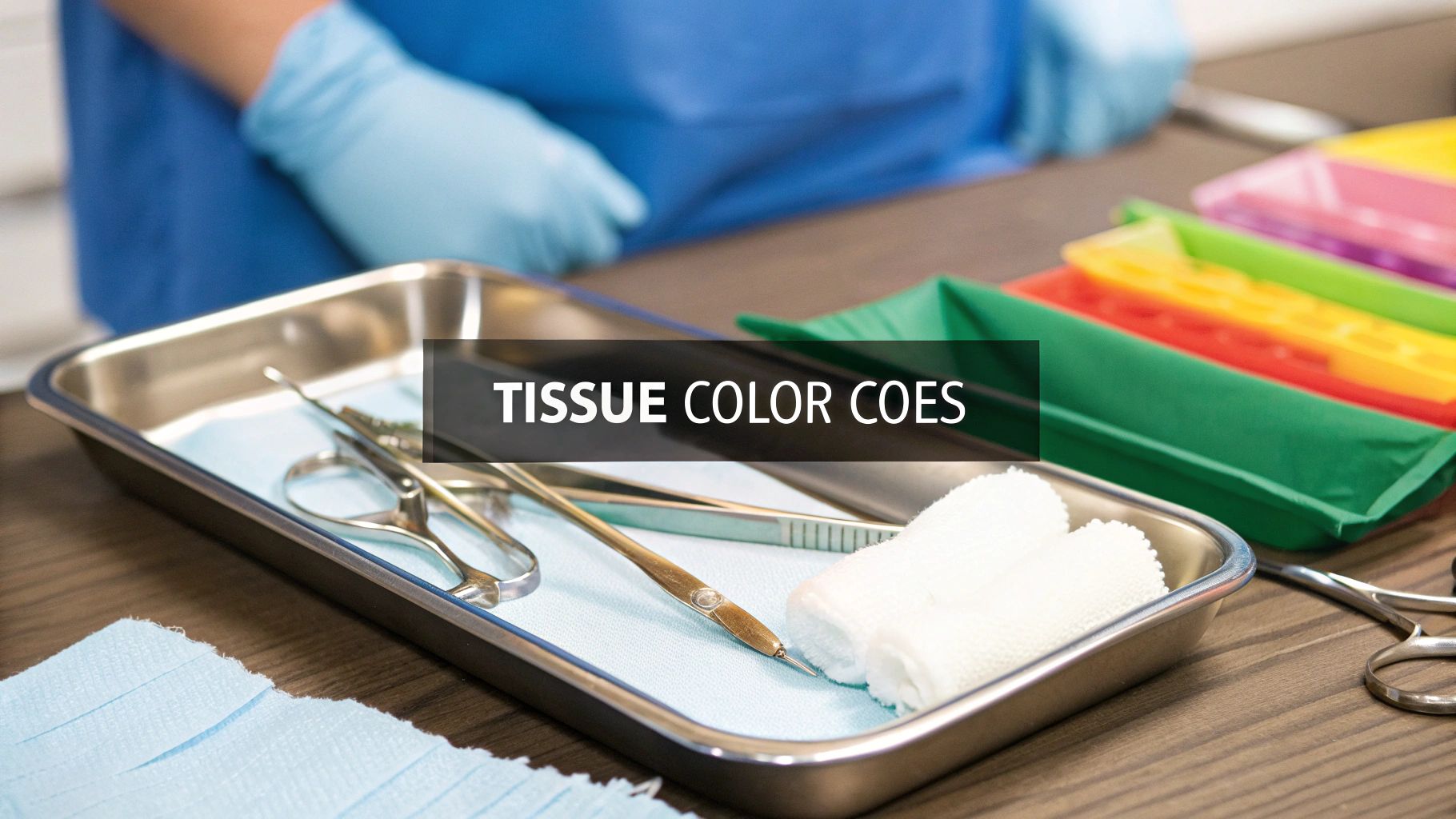
What Different Granulation Tissue Colors Mean
Think of the wound bed as a real-time report card for the healing process. Every color and texture tells a story about what’s happening beneath the surface, from the successful growth of new blood vessels (angiogenesis) to the presence of non-viable tissue that's holding back recovery. The cellular and vascular activity within the wound directly manifests as a specific color, providing a direct window into the wound's microenvironment.
For a busy clinician, being able to quickly read the granulation tissue color isn't just a nice-to-have skill; it's a core diagnostic tool. It helps you make smart, fast decisions right at the bedside. Mastering this "color language" is absolutely fundamental to good wound care, enabling you to anticipate problems and intervene before they escalate. It transforms a subjective observation into an actionable clinical insight.
This quick visual check helps you answer critical questions on the spot:
- Is the wound getting the oxygen and nutrients it needs to build new tissue?
- Is an infection brewing that I need to get ahead of?
- Do I need to debride this wound to clear the way for healthy tissue to grow?
- Is the current treatment plan effective, or does it need to be modified based on the tissue's response?
A Quick Guide to Tissue Colors
Knowing what each color means allows you to be proactive with your wound care, not just reactive. To dive deeper into the full picture of wound assessment, including factors like wound edge characteristics and exudate levels, check out our guide on comprehensive wound bed descriptions. For now, let’s focus on the colors, which are your direct signals for what to do next.
The appearance of granulation tissue isn't just a superficial detail; it's a direct reflection of the wound's microenvironment. A vibrant red color confirms that angiogenesis is successful and the foundational scaffolding for closure is being built effectively. Conversely, a pale or dusky hue is a clear indicator of compromised physiology that requires immediate investigation.
To get started, here’s a quick-reference table that connects the most common tissue colors to what they mean clinically and your initial next steps. Think of it as a handy cheat sheet before we dig into the details of each color.
Granulation Tissue Color At a Glance
| Color | Appearance | Clinical Meaning | Next Steps |
|---|---|---|---|
| Beefy Red | Moist, granular, bumpy | Healthy, well-vascularized | Protect tissue, maintain moisture |
| Pale Pink | Smooth, pale, anemic | Poor perfusion, ischemia | Assess circulation, nutrition |
| Dusky Red | Dark, purplish-red | Venous congestion, deep pressure | Elevate limb, assess for pressure |
| Yellow Slough | Stringy, wet, adherent | Non-viable tissue, bioburden | Debridement, moisture management |
| Black Eschar | Hard, dry, leathery | Necrotic, dead tissue | Assess for debridement, check perfusion |
This table gives you a solid starting point. Now, we'll break down each of these colors one by one to give you the context you need in your daily practice.
Reading the Signs of Healthy Wound Healing
When you're looking at a healing wound, what you want to see is a wound bed that looks vibrant and full of life. This is where granulation tissue color becomes a vital sign, with one particular shade acting as the gold standard for healthy healing: a bright, beefy red. This is the visual confirmation that the proliferative phase of wound healing is proceeding as expected.
That classic red appearance isn't just cosmetic—it's a direct visual report on the complex biological machinery humming along just as it should. Think of a healthy wound bed as a fertile garden, perfectly prepped to support new growth. Just like rich soil gives seedlings everything they need, healthy granulation tissue provides the ideal foundation for new skin to close the wound. It is composed of a matrix of new capillaries, fibroblasts, collagen, and inflammatory cells, all working in concert.

What Makes Granulation Tissue 'Beefy Red'
So, where does that deep, beefy red color come from? It's the result of angiogenesis, which is the body’s incredible process of building brand-new blood vessels. These tiny capillaries, budding from existing vessels, weave together to form a dense network that rushes oxygen and critical nutrients straight to the healing site. This neovascularization is the lifeline of the healing wound.
This robust blood supply is the engine driving two other crucial steps. First, it fuels fibroblast proliferation, where specialized cells arrive to lay down a new structural framework for the tissue. These fibroblasts are the construction workers of the wound. Second, it supports collagen deposition, the process of creating the strong protein fibers that give the new tissue its durability and tensile strength. When you see that bright red, you're looking at a wound that's successfully rebuilding itself from the inside out, a testament to a well-orchestrated physiological response.
Texture and Other Visual Cues of Healthy Tissue
Color is just one piece of the puzzle. The texture of healthy granulation tissue gives you even more confirmation that healing is on track. A healthy wound bed should have a bumpy, granular appearance, almost like the surface of a raspberry. This texture is a physical sign that all those new capillary loops are forming just right and pushing up towards the surface.
A moist, glistening surface is another hallmark of a healing wound. This indicates a well-hydrated environment where cells can easily migrate and perform their functions. The tissue should also be firm and resilient to gentle probing, not mushy or fragile, which could suggest excessive edema or underlying issues.
To make sure you’re seeing all the right signs, keep an eye out for these key indicators:
- Vibrant Color: A consistent beefy or salmon-red hue across the entire wound bed. There should be no significant patches of paler or darker tissue.
- Granular Texture: A bumpy, "berry-like" surface that signifies new blood vessel growth. This indicates a robust and healthy capillary network.
- Moisture Balance: A glistening, moist appearance without being overly wet or causing maceration at the wound edges. This reflects a proper balance of wound exudate.
- Firm Adherence: The tissue should be firmly attached to the wound base and shouldn't come apart easily. This demonstrates a strong connection to the underlying structures.
Recognizing these signs gives you a solid baseline for your assessment, allowing you to confidently identify when healing is progressing normally. This fundamental understanding is the first step in the wider wound healing process, equipping you to spot problems early and step in effectively when things go off track.
When Healing Slows Down or Is at Risk
While that beefy red tissue signals a healing process in full swing, not all granulation tissue tells such a positive story. Sometimes, the granulation tissue color shifts into more subtle, concerning shades that are actually early warning signs. These colors tell us the healing fire is either running low on fuel or, worse, being actively smothered.
Think of healthy granulation tissue as a roaring campfire—it's bright, hot, and crackling with energy. But what happens when that fire starts to die down? The glowing red embers fade to a weaker, paler color. That's exactly what happens in a wound bed when healing gets compromised. Learning to spot these subtle shifts from vibrant red to pale pink or a dusky, purplish-red is a critical skill for intervening before it's too late. These are not merely color variations; they are physiological alarms.

Decoding Pale Pink Granulation Tissue
When you see granulation tissue that looks pale pink, washed-out, or anemic, your first thought should be poor perfusion. The rich blood supply that gives healthy tissue its vibrant red hue has been choked off, starving the wound of the oxygen and nutrients it desperately needs to rebuild. This is a sign of ischemia.
This is the wound's equivalent of a fading fire. It's a clear signal that the metabolic activity needed for healing is grinding to a halt. This "anemic" look points directly to an underlying problem that you have to address, as the cellular machinery for repair cannot function without adequate oxygenation.
A few common culprits can cause this stalled state:
- Ischemia: This is a localized lack of blood flow. It can be caused by anything from arterial insufficiency (e.g., peripheral artery disease) and excessive pressure on the wound to severe edema that's physically squeezing the blood vessels shut.
- Systemic Issues: Sometimes the problem isn't local. Broader health issues like anemia (low hemoglobin) or significant malnutrition (low albumin/protein) can torpedo the blood's oxygen-carrying capacity, affecting every tissue in the body, including the wound.
- Heavy Bioburden: Even without a full-blown infection, a high concentration of bacteria can steal resources and impair circulation right at the wound site, leading to that pale appearance. The bacteria compete with host cells for limited oxygen and nutrients.
A pale pink wound bed is a call to action. It’s telling you to look beyond the wound itself to find and fix the root cause of the poor blood supply before that tissue dies off completely. This is a critical juncture where timely intervention can prevent progression to necrosis.
Understanding Dusky or Purplish-Red Tissue
Now, a dusky, purplish, or deep maroon granulation tissue color points to a completely different circulatory problem: venous congestion. Instead of a lack of incoming arterial blood, this color suggests blood is pooling in the area and can't get back to the heart. The tissue is engorged with deoxygenated hemoglobin, which gives it this characteristic dark hue.
Think of it as a traffic jam in the veins. This backup causes pressure to build and forces oxygen-depleted blood to linger in the tissue, giving it that dark, bruised look. This venous hypertension can lead to increased capillary permeability, leakage of fluid into the interstitium (edema), and ultimately, impaired tissue health.
You'll often see this in patients with venous insufficiency, congestive heart failure, or in dependent limbs where edema is a constant battle. It can also be an early red flag for a developing deep tissue pressure injury, where sustained pressure has crushed the underlying circulation and venous outflow.
From Observation to Clinical Intervention
Spotting these off-colors is just the first step. The real work begins when you translate that observation into a targeted clinical action plan. Your assessment needs to immediately pivot to detective mode to find the "why."
If you see pale pink tissue, start investigating:
- Assess Arterial Flow: Get your hands on the patient. Check for peripheral pulses and test the capillary refill time. Consider an Ankle-Brachial Index (ABI) to quantify arterial insufficiency.
- Review Systemic Factors: Pull up the chart. Look at recent lab results for hemoglobin levels and nutritional markers like albumin. Is the patient getting the building blocks they need? A nutritional consult may be warranted.
- Evaluate Pressure: Double-check that any offloading strategies are actually working. Is the pressure truly being relieved from the wound area? Ensure proper use of support surfaces and offloading devices.
If you see dusky red tissue, shift your focus:
- Check for Edema: Assess the limb for swelling. Is there pitting edema? How severe is it? Measure limb circumference to track changes.
- Consider Patient Positioning: If the wound is on a leg or foot, think about elevation to help promote venous return and clear that "traffic jam." Compression therapy is often a key intervention here, if not contraindicated.
- Investigate Comorbidities: Dive back into the patient's history. Do they have known conditions like venous stasis or heart failure that could be driving this? Ensure their systemic conditions are optimally managed.
By connecting the granulation tissue color to these potential underlying causes, you can stop just documenting a problem and start creating an informed intervention that gets the healing process back on track.
Identifying Non-Viable Tissue and Infection Risks
When you see the color in a wound bed shift away from red and into shades of yellow or black, it's more than just a slowdown in healing—it's an urgent distress call. These colors are tell-tale signs of non-viable, or dead, tissue. This tissue isn't just sitting there; it's a physical barrier to healing and a perfect breeding ground for bacteria. Recognizing and dealing with it through debridement is one of the most important things we do in wound care.
Unlike the subtle warnings you get from pale pink or dusky red tissue, seeing yellow slough or black eschar is a definitive sign of a problem. This dead material has to go before any healthy granulation can take its place. A good way to think about it is like trying to plant a garden that's choked with dead, thorny vines—you have to clear out all that debris before anything new and healthy can grow. Non-viable tissue prolongs the inflammatory phase and prevents the proliferative phase from beginning.

Understanding Yellow Slough
Yellow slough is essentially a collection of hydrated, dead tissue. It’s a mix of old cells, fibrin, proteins, and other gunk that collects in the wound bed. What can be tricky is that it doesn't always look the same from one wound to the next. Its presence indicates that the wound's natural autolytic processes are insufficient to clear the debris.
It's crucial not to confuse slough with pus, which points to an active infection. While they can look similar, they mean different things. You can explore our guide on whether pus is always a sign of infection to get a better handle on telling them apart. That said, the presence of slough absolutely creates an environment where infection can easily take hold by providing a nutrient-rich medium for bacterial growth.
Clinically, you'll see slough in a few common forms:
- Thin and Stringy: This kind is usually loosely attached and looks like fine, wet threads stretched across the wound.
- Thick and Gelatinous: This is a heavier, mucus-like slough that can coat parts of the wound, hiding the healthy tissue you want to see.
- Adherent Slough: This is the toughest to deal with. It's stuck on tight to the wound bed, making it difficult to remove without hurting the viable tissue underneath.
No matter what it looks like, yellow slough acts as a constant source of inflammation and provides a rich buffet for bacteria. This dramatically increases the risk of both infection and stubborn biofilm formation.
Getting that slough out of there through debridement (e.g., sharp, enzymatic, autolytic) is a non-negotiable step. It cleans up the wound bed, knocks down the bacterial load, and finally gives the body's own healing mechanisms a chance to take over.
Confronting Black Eschar
Black eschar is the most serious type of non-viable tissue you'll encounter. It’s a hard, dry, leathery patch of necrotic tissue that forms when the local blood supply has been completely cut off. Eschar essentially puts a "cap" over the wound, and that's dangerous for a couple of key reasons. It is desiccated, dead dermis and/or epidermis.
First, it physically stops healing in its tracks. New granulation tissue can't grow under it, and the wound edges can't pull together to close. Second, and maybe more insidiously, it can hide a disaster brewing underneath. A severe infection, an abscess, or deep tissue damage could be developing completely unseen beneath what looks like a stable, black covering.
The clinical decision-making around eschar is where things get really nuanced.
- When to Debride: If the eschar feels loose or boggy, or if you see any signs of infection—like drainage seeping from the edges, a bad odor, or spreading redness (cellulitis)—it needs to be debrided immediately. You have to get it off to see and treat what's happening below.
- When to Keep Stable: Here's the exception. In a patient with very poor circulation, like on the heel of someone with severe peripheral artery disease, a dry and stable eschar might be best left alone. In this specific case, it acts as a natural, biological barrier. Removing it could create an open wound that the body simply can't heal, potentially leading to a far worse outcome, like amputation. This is often referred to as a "stable, dry gangrene" scenario.
Making the right call requires a sharp assessment of the patient's circulatory status. Getting it wrong with eschar can lead to devastating complications, sometimes even limb loss.
AI Can Sharpen Our Clinical Eye, Not Replace It
As clinicians, we spend years honing our ability to assess a wound just by looking at it. That initial visual check is a crucial skill, but it has one major, built-in flaw: it's subjective.
Let's be honest—two highly experienced wound care specialists can look at the same wound bed and walk away with two different opinions. One might chart "70% healthy granulation," while the other documents "50%." This isn't about who is right or wrong; it's just a natural limitation of the human eye. This variability can muddy the waters, making it tough to track healing progress consistently or create a clear treatment plan that everyone on the team can follow.
The Problem with "Good Enough" Estimates
This isn't just a minor inconvenience; it's a well-documented challenge in wound care. When you rely solely on visual estimation, you get a lot of variation and imprecision.
A revealing analysis of wound photograph evaluations found some pretty startling differences in what clinicians saw. For about 8.5% of the photos (17 in total), the range of estimates for granulation tissue was a full 100%. That means one provider saw zero granulation, while another saw a wound bed that was completely granulated. You can dig into the specifics of this study on wound assessment variability at JAMA Network Open.
When assessments can vary that wildly, how can we confidently say a wound is getting better or worse? It makes it incredibly difficult to objectively measure the real impact of our treatment plans. This subjectivity undermines evidence-based practice and can complicate communication between care providers.
Bringing Objective Data into the Picture
This is exactly where AI-powered digital wound analysis comes in. Think of it as a tool that gives our clinical judgment a data-driven boost. Instead of relying on a subjective glance, these platforms use sophisticated color analysis to give us objective, measurable data about what’s really going on in the wound bed.
Tools like Ekagra Health AI can analyze a simple wound photo and turn it into hard numbers. For instance, it can generate a Granulation Red Index (GRI), which is a specific metric that quantifies the amount and quality of red granulation tissue. It can also precisely calculate the percentage of slough, eschar, and healthy tissue, removing the guesswork entirely.
Suddenly, a subjective note like "it looks pretty red" becomes a concrete data point that you can track accurately over time. This transition from qualitative to quantitative assessment is a game-changer for wound management.
This isn't about replacing clinical skill—it's about enhancing it. By pairing our hard-earned expertise with consistent, unbiased measurements, we can reduce documentation errors and standardize how healing is tracked across the entire care team. It allows us to make more confident, evidence-based decisions that ultimately lead to better outcomes for our patients.
Connecting Accurate Assessment to Better Reimbursement
It’s easy to think of a precise clinical observation as just good patient care, but its impact goes far beyond the bedside. It hits your practice right where it counts: the bottom line. When you document the color and percentages of granulation tissue with detail and objectivity, you're not just following best practices—you're building an ironclad case for your treatment choices and securing proper reimbursement.
Think about it from a payer's perspective. Vague documentation invites skepticism and can lead them to question the medical necessity of a procedure or an advanced wound care product. But when your notes paint a clear, quantitative picture, you create an undeniable record of the wound's status and the logic behind your care plan. That detail dramatically cuts down the risk of claim denials.
From Bedside Observation to Billable Service
Your wound notes are the evidence that backs up every single line on your claim. An entry like "wound improving" is subjective and nearly impossible to defend in an audit. Specific documentation, on the other hand, tells a clear, defensible story of the patient’s progress and your clinical decision-making.
A well-documented wound assessment tells a complete story. It not only tracks the patient's healing journey but also validates the resources, treatments, and clinician time required to achieve positive outcomes. It is the narrative that justifies medical necessity.
This is especially critical for accurate coding. The right ICD-10 codes often hinge on the wound's severity and complexity, which is something you can only demonstrate by documenting the percentages of viable versus non-viable tissue. For example, documenting the need for debridement is much stronger when it's supported by precise percentages of slough or eschar.
What Effective Documentation Looks Like
The best documentation connects what you see directly to what you do. It turns your visual assessment into measurable data that justifies every action you take.
Here’s what that looks like in practice:
- Example 1: "Wound bed is 70% beefy red granulation with 30% adherent yellow slough along the superior margin. Performed sharp debridement to remove non-viable tissue and promote further granulation." This clearly justifies the debridement procedure.
- Example 2: "Wound shows 90% pale pink granulation, indicating poor perfusion. Initiated consultation with vascular surgery and applied a dressing to increase local circulation." This note validates the vascular consult and the choice of a specialized dressing.
This kind of objective language leaves no room for interpretation. Anyone who picks up that chart—another clinician, a specialist, or a billing auditor—will immediately grasp the wound's condition and understand why you chose that specific intervention.
Making Great Documentation Effortless
Let's be honest: capturing this level of detail by hand is a drain on your time and adds to the mountain of administrative work. This is where tools designed for the job, like the AI-powered platform from Ekagra Health AI, can make a real difference. It takes the entire workflow—capturing high-res images, analyzing tissue types, and generating structured notes—and makes it seamless.
A quick photo becomes objective, actionable data. The platform helps you make the right coding decisions and builds a robust, audit-proof patient record automatically. This doesn't just solve an administrative headache; it helps minimize claim denials, speeds up the revenue cycle, and frees you up to spend less time on paperwork and more time where it matters most: with your patients.
Your Top Questions About Granulation Tissue, Answered
Even with a solid grasp of the basics, some practical questions always pop up when you're in the trenches, looking at a wound bed. Let's tackle some of the most common ones I hear from fellow clinicians.
How Often Should I Be Documenting Color Changes in Granulation Tissue?
This really comes down to your care setting and the wound itself. If you're managing a high-risk or acute wound, daily checks are often the standard. For a stable outpatient wound, documenting at each weekly visit is usually enough.
The golden rule is this: document any significant change the moment you see it. A shift in color is a direct signal that something has changed in the wound's healing journey, for better or worse. Consistent documentation frequency is key for establishing a trend, whether positive or negative.
Some of the newer AI-powered documentation tools are a huge help here, as they create a consistent visual timeline that makes spotting those subtle color shifts much easier over time.
Can Granulation Tissue Ever Be a Bad Thing?
Absolutely. The classic example is hypergranulation, which you might have heard called "proud flesh." This is when granulation tissue goes into overdrive, growing up and over the surface of the skin. It often looks like a bumpy, fragile, reddish mass that bleeds at the slightest touch. It is often a sign of a prolonged inflammatory stimulus, such as bacterial bioburden or friction.
The problem here is that this overgrowth creates a physical barrier, literally stopping the new skin cells (epithelium) from migrating across and closing the wound. This process, called epibole, is halted when epithelial cells meet the raised wall of hypergranular tissue.
To manage it, we often turn to topical corticosteroids or even silver nitrate to carefully reduce that excess tissue and get the healing process back on track. The goal is to level the tissue back to the height of the surrounding skin to allow for re-epithelialization.
What's the Protocol When a Wound Has Multiple Colors?
Seeing a mix of tissue types in one wound bed is incredibly common—in fact, it's more the rule than the exception. Your documentation needs to reflect this complexity.
The best practice is to estimate the percentage of each tissue type you see. For example, you’d note something like, "60% red granulation, 40% yellow slough." This is often called the "Red-Yellow-Black" system of wound classification.
This detailed breakdown is what drives your treatment plan. It tells you to focus your efforts on debriding the non-viable tissue (the slough) while protecting the healthy, new granulation tissue. You always want to tackle the biggest barrier to healing first. Your dressing selection will also be guided by this assessment; for instance, using a product that can absorb exudate from the sloughy areas while maintaining moisture over the healthy granulation.
Streamline your wound documentation from voice to claim in minutes. With Ekagra Health AI, you can capture objective wound data, generate structured notes automatically, and accelerate your reimbursement cycle. Learn more at the Ekagra Health AI website.
