
The Difference Between Eschar and Scab: What Actually Matters at the Bedside
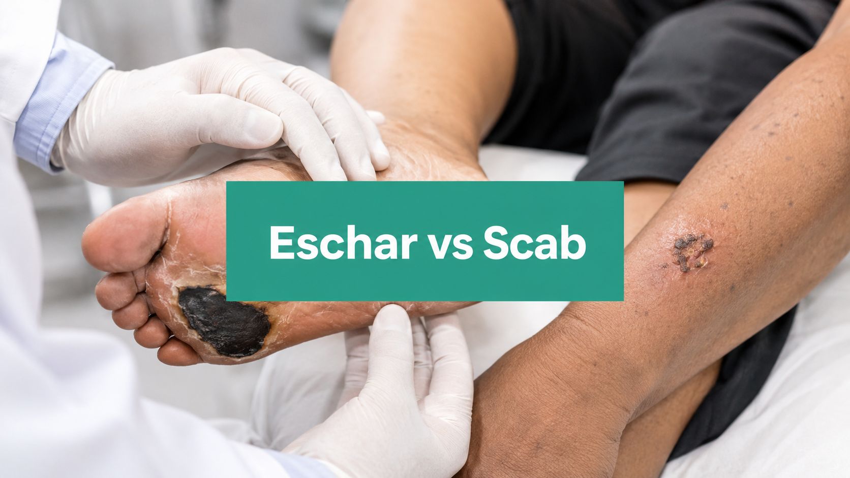
The most common mistake I see in wound charts is calling a black plaque on a diabetic heel a scab. This single word choice can derail a claim, delay appropriate treatment, and misrepresent what is happening in the tissue.
Eschar is devitalized tissue — often leathery, hard, and signaling deeper ischemia. A scab is a superficial crust of dried blood and serum sitting over intact epidermis that is actively healing. They are not interchangeable, and treating them as such creates clinical and financial problems.
Consider a common clinical scenario. A 68-year-old patient with diabetes presents with a black plaque over the calcaneus. A clinician documents it as a "scab" and bills a straightforward wound visit. The payer denies the debridement claim because the documentation used the wrong terminology and skipped the required measurements and staging. That revenue hit started with a simple failure of observation and terminology.
Clinical Definitions Worth Knowing
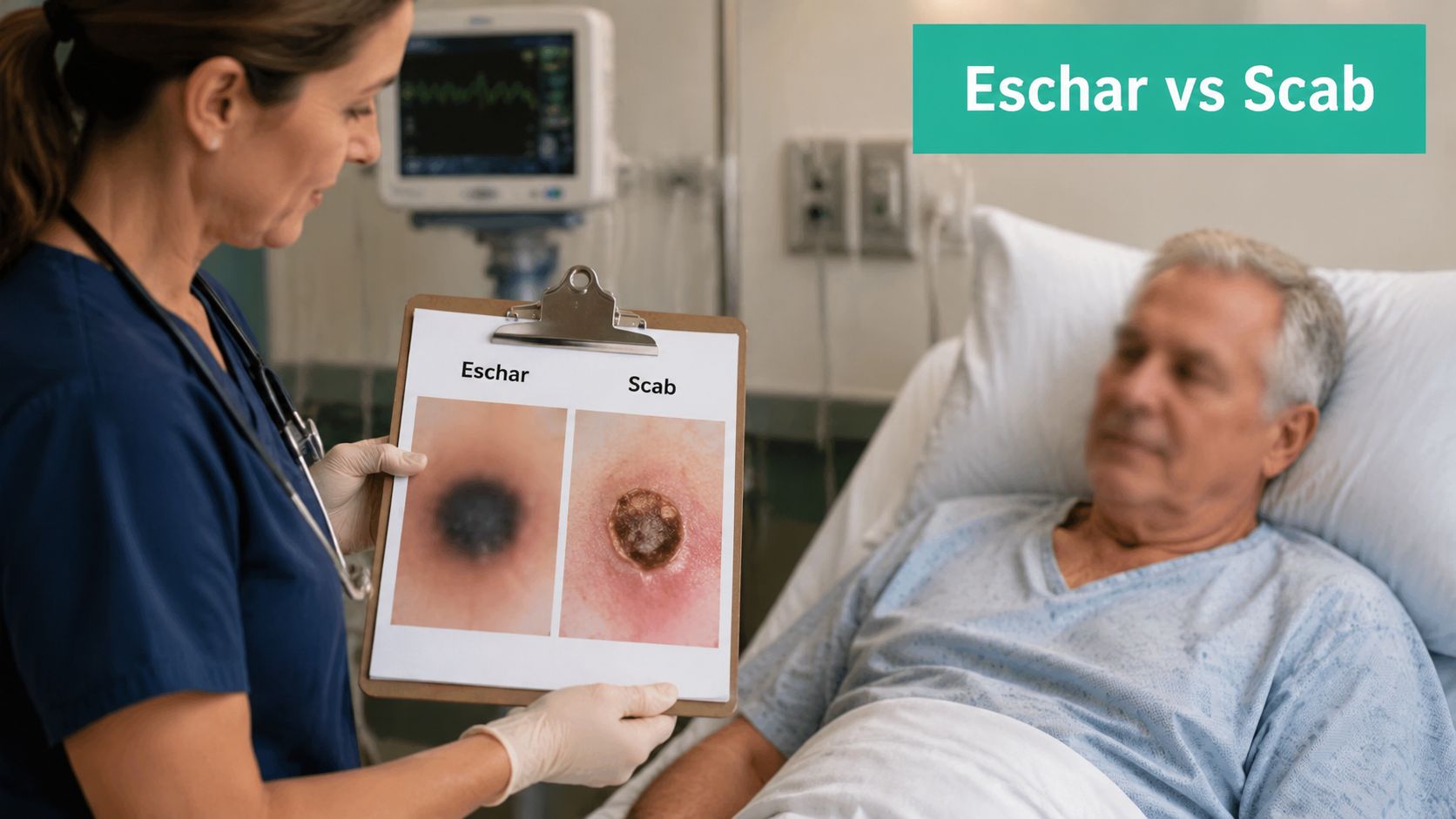
Eschar is necrotic tissue. It can be dry or moist, tan to black, and adherent or sloughing. The texture varies—sometimes leathery and hard, other times friable and boggy if infection has set in. Its clinical significance lies in what it represents: loss of perfusion, full-thickness tissue death, or both. Eschar often hides undermining, sinus tracts, or deeper injury that is not visible until the necrotic layer is addressed.
A scab, by comparison, is a protective crust. It is composed of dried blood, serum, fibrin, and cellular debris that forms within approximately 24 hours after an epidermal breach. It generally overlies viable tissue that is already working toward epithelialization. Lifting a scab typically reveals pink, healthy tissue underneath.
How to Tell Them Apart at the Bedside
I treat a lesion as eschar when it feels hard or inelastic, or when separating it reveals deeper tissue exposure. I lean toward scab when the covering is thin, lifts easily to show intact epidermis underneath, and the patient does not have significant ischemic risk factors.
Beyond visual inspection, document the objective data that supports your clinical reasoning. Record perfusion status—capillary refill, Doppler pulses, ankle-brachial index (ABI). Take precise wound measurements. Note probe-to-bone results when indicated. This practice is not just good medicine; it is what keeps your documentation defensible during a payer review.
Here's a note that holds up under review: "Dry, adherent, black necrotic plaque consistent with eschar; pulses Dopplerable; ABI pending; wound area 4.2 cm²." That level of specificity tells a clinical story that "scab present" never will.
Eschar vs. Scab at a Glance
The table below breaks down the core distinctions. Use it as a quick reference when making an initial assessment.
| Feature | Eschar | Scab |
|---|---|---|
| Tissue type | Necrotic devitalized tissue | Dried blood/serum crust |
| Depth | Often full-thickness or hides deeper injury | Epidermal or superficial |
| Color | Tan to black, variable | Brown to dark red |
| Texture | Leathery, hard, or boggy if infected | Thin, flaky, brittle |
| Acuity | May be chronic, ischemic, or pressure-related | Acute post-injury healing |
| Typical setting | Diabetic foot ulcer, pressure injury, burn | Minor abrasion, superficial laceration |
The texture and depth difference usually points in the right direction, but the clinical context—patient history, perfusion status, wound location—confirms the assessment.
What I Actually Document at the Bedside
When I evaluate a wound with necrotic tissue or crusting, here is what goes into my note:
- Measure and photograph — always with a scale and color card in frame. Record the area in square centimeters.
- Assess perfusion — document ABI results, pulse findings, and capillary refill. Probe-to-bone when the wound depth warrants it.
- Characterize the wound bed — describe the percentage of eschar versus slough, and note the exudate type (serous, sanguineous, purulent) and amount.
- Use precise language — vague terminology invites denials. Specific, clinically accurate descriptions support both patient care and appropriate reimbursement.
For a deeper dive into wound bed terminology and documentation strategies, read our guide on wound bed descriptions and how to document them.
Why the Distinction Changes Healing and Risk
Eschar signals nonviable tissue with impaired perfusion that physically blocks granulation and re-epithelialization. That distinction is critical.
Dry, adherent eschar almost always represents full-thickness necrosis over ischemic tissue. The collagen and capillary networks underneath are either gone or severely compromised, meaning the wound bed cannot support healthy granulation without revascularization or debridement.
Scabs, on the other hand, form within roughly 24 hours after a superficial epidermal injury. They are a protective crust of dried blood, serum, and fibrin, and they typically fall off on their own once re-epithelialization is complete.
A black heel on a patient with diabetes and peripheral arterial disease is far more likely to be eschar than a scab. In that context, the lesion falls into Wagner grade 1–2 territory if there is superficial ulceration beneath the necrosis—or when probing reveals deeper involvement. That finding shifts management immediately. Vascular assessment becomes urgent, and offloading is non-negotiable.
Clinical Implications for Immediate Action
Before reaching for a scalpel, get the perfusion data. Document ABI, toe pressures, or Doppler waveform prior to any definitive debridement. Poor perfusion fundamentally changes the risk–benefit calculation for sharp debridement and directly affects which CPT code is selected and how the ICD-10-CM diagnosis is documented.
For diabetic heel eschar, start with offloading and pressure redistribution while arranging vascular evaluation. This step can slow or prevent progression and limits the window for secondary infection.
Infection risk under a black plaque is a serious concern. Eschar can conceal boggy pockets, tunneling, or soft tissue infection that is not visible on the surface. Fluctuance, malodor, increased exudate, or periwound warmth are findings that push toward urgent debridement and possible systemic therapy.
A black heel is more than a color description—it's a signal to document perfusion and consider Wagner grading before choosing to leave an eschar in place.
Timing and Pathophysiology Differences
Eschar forms from ischemia or full-thickness injury and may persist chronically, particularly over the calcaneus where soft tissue is thin. Scabs indicate an active healing surface, and their separation timing, typically days to two weeks, corresponds with epithelial migration underneath.
When eschar softens, becomes boggy, or begins separating, that is a warning sign. The risk of underlying infection and hidden depth increases, and the clinician should reassess whether debridement is now necessary.
Practical Documentation Points
Your chart note should paint a clear picture. Describe tissue type and texture with precision—"dry adherent black necrotic plaque" versus "thin brown dry crust" tells a very different clinical story. Record perfusion objectively: ABI 0.55, biphasic Doppler waveform, capillary refill greater than four seconds. These numbers speak louder than adjectives.
If debridement is performed, document the surface area in cm², the depth, and the percentage of eschar versus slough present. This level of detail justifies CPT codes 11042–11047 and supports medical necessity.
Short case example: A patient with diabetes presented with an adherent black plaque over the calcaneus. Dorsalis pedis was palpable, ABI measured 0.6. The team offloaded the heel, ordered a vascular consult, and deferred sharp debridement pending revascularization. That charted reasoning—documenting the vascular status, the offloading decision, and the rationale for waiting—prevented an audit finding of inappropriate debridement.
The distinction between eschar and scab is not academic. It drives urgency, coding accuracy, and clinical safety. Your label should reflect tissue viability and perfusion data, not just color, because the label dictates the action that follows.
Sharp Debridement Versus Conservative Care
Always start with perfusion. Document pulses, ABI, toe pressures, or Doppler waveform before deciding between sharp debridement and conservative care. That step reshapes the risk assessment, determines which CPT code fits, and indicates whether to proceed now or await revascularization.
For dry, firmly adherent, nonfluctuant eschar on a non–weight-bearing heel, conservative care is often the smarter call. Offloading, serial observation, and protective dressings are appropriate while you coordinate a vascular workup.
When eschar becomes wet, boggy, malodorous, or separates from the edges, it indicates trapped bioburden and likely infection. These cases usually require removal. Sharp debridement reduces necrotic load and exposes the true wound bed. When you perform a debridement under CPT codes 11042–11047, record the surface area in cm², depth, and the percentage of eschar versus slough present.
When to Use Conservative Methods
Autolytic debridement works well for stable eschar when perfusion is sufficient or revascularization is planned. Moisture-retentive dressings allow endogenous enzymes to work; document the dressing choice and check the wound weekly. For patients who cannot tolerate an operative approach, enzymatic debridement is a reasonable fallback; note the agent, frequency, and tissue response. Mechanical options like pulsed lavage have their place but demand precise documentation of the rationale and patient tolerance.
- Autolytic: Fits stable, dry eschar with adequate perfusion; document dressing type and duration.
- Enzymatic: For patients with contraindications to sharp debridement; chart the product, frequency, and tissue response.
- Mechanical: Reserve for heavily contaminated wounds; note irrigation volume and clinical effect.
For a dry, adherent calcaneal eschar with no drainage or foul odor, conservative care with pressure redistribution and documented vascular assessment is defensible to surveyors.
Sharp Debridement Practical Rules
| Step | Action | Documentation Required |
|---|---|---|
| 1 | Assess perfusion and record objective measures | ABI, toe pressure, or Doppler waveform |
| 2 | If eschar is loose, boggy, foul-smelling, or tracking | Proceed to sharp debridement; culture as indicated |
| 3 | Document exact area debrided | Surface area in cm², percent eschar versus slough |
| 4 | Record procedural details | Hemostasis, instruments used, anesthesia level |
These details matter. They support your procedural note during audits and establish clinical necessity.
Burn Eschar Considerations
For full-thickness burns that cross joints, early removal of eschar preserves function. Delaying escharotomy costs range of motion. Document the location, size, and measurable effect on motion to justify the surgical decision.
Case example 1: A patient with diabetes and an ABI of 0.58 presents with a black, adherent heel plaque and no drainage. The team offloads, orders a vascular consult, and documents the conservative plan. This approach prevents inappropriate immediate sharp debridement and builds a defensible coding record.
Case example 2: Another patient presents with boggy, malodorous eschar, increasing exudate, and periwound erythema. Sharp debridement is performed, the wound is photographed with a scale, and CPT 11044 is documented with area and depth. This creates a record grounded in clinical necessity.
Learn more about wound debridement in our article on Wound Debridement Documentation and Workflow.
CPT, ICD-10-CM, and the Documentation That Survives Review
The line between what you document and what you can bill is thin. The story you tell in the chart determines whether a claim gets paid or denied.
The workhorses for debridement are CPT codes 11042–11047, which are tiered by tissue depth and surface area.
- CPT 11042 is for the first 20 cm² of debridement down to, but not including, muscle. This includes subcutaneous tissue or fascia.
- CPT 11043 and 11044 are for deeper debridement, reaching muscle or bone. Your note must state that you debrided devitalized muscle or bone and specify the area.
- CPT 11045–11047 are add-on codes for each additional 20 cm² (or part thereof) at the corresponding depth.
If your documentation does not explicitly state the depth and the exact surface area in cm², your claim is at risk.
Debridement CPT Codes and When to Use Them
This table provides a quick reference for selecting the correct CPT code based on the depth of tissue removed. Your clinical note must precisely match the depth described.
| CPT Code | Tissue Depth | Add-On Units | Typical Use |
|---|---|---|---|
| 11042 | Debridement of subcutaneous tissue (includes epidermis, dermis, subcutaneous tissue) | +11045 for each additional 20 cm² | Removing necrotic fat and fascia from a pressure ulcer or diabetic foot ulcer. |
| 11043 | Debridement of muscle and/or fascia (includes subcutaneous tissue) | +11046 for each additional 20 cm² | Debriding a deep wound where non-viable muscle is present. |
| 11044 | Debridement of bone (includes subcutaneous tissue, fascia, and/or muscle) | +11047 for each additional 20 cm² | Osteomyelitis cases or deep sacral ulcers where bone is exposed and debrided. |
Accurate coding hinges on linking the procedure to a diagnosis that justifies it. For instance, billing CPT 11044 for a simple abrasion (an S-code) is a guaranteed denial. The ICD-10-CM code must reflect the wound's severity, such as L89.- for a pressure ulcer or an L97.- code for a non-pressure chronic ulcer with necrosis.
Documentation Essentials That Win Audits
Vague notes get claims denied. Your documentation needs to paint a clear, measurable picture.
Always include these five elements:
- Exact surface area in cm²: Specify the measurement method (e.g., acetate tracing, digital imaging).
- Tissue depth: Use precise terms like epidermis, dermis, subcutaneous fat, fascia, muscle, or bone.
- Wound bed composition: Give a percentage breakdown (e.g., "70% black, adherent eschar and 30% yellow, fibrinous slough").
- Structural details: Note any undermining or tunneling, with probe-to-resistance measurements in centimeters.
- Vascular status: Document perfusion data like ABI, toe pressures, or Doppler waveforms, especially for lower extremity wounds.
A Note That Works: "Sharp debridement was performed on the left heel ulcer, removing 8.5 cm² of full-thickness necrotic tissue down to healthy, bleeding subcutaneous tissue. The wound bed was composed of 80% dry eschar and 20% slough. Hemostasis was achieved with direct pressure. This note supports CPT 11042."
Why Claims Get Denied
I see the same documentation mistakes sink otherwise valid claims. These are the most common denial triggers:
- Depth Mismatch: Billing a muscle debridement (11043) when the note only says "debrided to subcutaneous tissue."
- Missing Measurements: Failing to document the surface area in cm² or simply writing "wound debrided."
- Vague Language: Using generic phrases instead of specific descriptions of tissue type and appearance.
- No Perfusion Data: Not documenting vascular status for stable, dry eschar on an ischemic limb, which is crucial for justifying medical necessity.
Practical Charting Example
Here is how to put it all together in a clear, defensible progress note:
- Location and Size: "Left plantar heel ulcer measuring 4.2 cm x 2.0 cm for a total surface area of 8.4 cm² (measured via acetate tracing)."
- Wound Bed: "Wound bed is covered by 80% dry, black, adherent eschar with a rim of 20% yellow slough. No fluctuance or erythema noted."
- Depth: "Debridement was carried down to viable subcutaneous fascia; no muscle or bone was exposed."
- Perfusion: "Dorsalis pedis pulse is palpable. Ankle-Brachial Index (ABI) is 0.62. Awaiting vascular consult."
- Procedure: "Under local anesthesia, 8.4 cm² of necrotic tissue was sharply debrided. CPT 11042 will be billed. Pre- and post-debridement photos with a measurement scale were saved to the patient's chart."
When sharing documentation with payers or for legal reviews, protecting patient privacy is critical. Knowing how to redact sensitive data from PDFs is a vital skill for maintaining HIPAA compliance.
Wound Staging, Nursing Language, and Case Examples
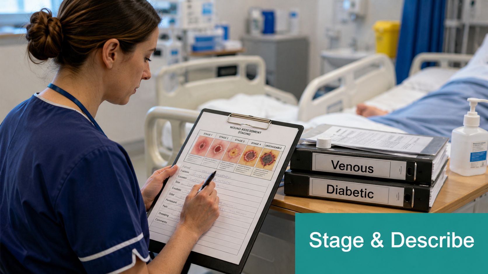
Start with language that holds up under review. For eschar, write "dry, adherent, black necrotic plaque consistent with eschar." For a scab, use "thin, brown, dry crust consistent with scab." These objective phrases set up accurate staging and coding, reducing queries from reviewers.
Clinical Translation to Staging and Grading
When eschar covers a pressure site and hides the wound bed, NPUAP classifies it as an Unstageable Pressure Injury until debridement exposes the depth. After removal, restage according to standard NPUAP criteria.
For diabetic foot ulcers, Wagner grading is used. Eschar masking a superficial ulcer often maps to Wagner 1 or 2, depending on probe-to-bone results and post-debridement depth. Always document probe-to-bone findings and vascular data alongside any staging change.
- Document perfusion with ABI, toe pressures, and Doppler waveform.
- Record probe-to-bone findings and take photographic scale images.
- Note tissue composition: for example, 70% eschar, 30% slough.
Labeling a lesion correctly directs urgency, coding, and the next clinical step.
Case Example: Venous Leg Ulcer With Scab
A 72-year-old patient with venous insufficiency has a lateral gaiter-area ulcer with a thin crust along the margin. The chart reads: "Left medial lower leg ulcer 3.0 x 1.8 cm; wound bed pink with 95% granulation; 5% thin brown dry crust consistent with scab at superior margin."
Staging is NPUAP Stage 2 if there is partial-thickness skin loss; it is not unstageable. Management is straightforward: moist wound healing, compression bandaging, and weekly reassessment. Use an L97.- code for a chronic ulcer. No debridement CPT code applies when only a scab is present.
Practical points:
- Photograph the crust with a scale for the record.
- Do not use sharp debridement on an intact scab over viable epidermis.
- Schedule follow-up at one week to check dressing tolerance and compression.
Case Example: Diabetic Heel With Stable Eschar
A 65-year-old patient with diabetes and PAD presents with a black, adherent plaque over the calcaneus. The chart entry: "Right plantar heel 4.2 cm²; dry, adherent black necrotic plaque consistent with eschar; dorsalis pedis palpable; ABI 0.58."
Staging is Unstageable Pressure Injury because the depth is obscured. Management focuses on offloading the heel, urgent vascular referral, and serial monitoring. Debridement is deferred until revascularization occurs or the eschar becomes boggy or malodorous.
Action steps and coding considerations:
- Document vascular status and the clinical rationale for deferring sharp debridement.
- If debridement happens later, record the area in cm², depth, and percent breakdown of eschar versus slough to support CPT 11042–11047 selection.
- Follow-up cadence: vascular consult within 48–72 hours; wound checks every 3–7 days depending on perfusion and exudate levels.
Nursing Charting Phrases That Hold Up
Short, objective templates work best.
- "Dry adherent black necrotic plaque consistent with eschar; no drainage; ABI 0.58."
- "Thin brown dry crust consistent with scab; underlying tissue pink and epithelializing."
A numbered checklist for the bedside keeps documentation consistent:
- Capture a photo with scale and color card.
- Measure and calculate cm².
- Record perfusion objectively.
- State staging and the rationale behind the debridement decision.
These steps connect assessment to staging, guide the clinical plan, and create documentation that supports appropriate ICD-10 and CPT claims.
Practical Takeaways for Clinicians, Coders, and Surveyors
Document the lesion with precision. For necrosis, write "dry, adherent black necrotic plaque consistent with eschar." For superficial crusts, use "thin brown dry crust consistent with scab." This specific language holds up under payer and surveyor review.
Always capture objective perfusion data—ABI, toe pressures, Doppler waveform—and record wound area in cm². These numbers directly drive CPT selection and medical necessity determinations.
When to Call It Eschar
- Hard, leathery, or nonelastic covering over suspected full-thickness injury
- Obscures the wound bed or probes to deeper tissue
- Associated with ischemia, pressure, or chronic ulceration
When to Call It Scab
- Thin crust that lifts to reveal intact or pink epidermis
- Forms within 24–72 hours of superficial injury
- No undermining, purulent drainage, or systemic signs of infection
When to Debride Versus Wait
- Debride now if eschar is boggy, malodorous, shows fluctuance, or has increasing exudate.
- Defer sharp debridement and offload if a dry, adherent heel eschar with poor perfusion is present; document your rationale and revascularization plan.
Checklist for Point-of-Care Team
- Photo with scale and color card
- Measure length and width, then compute cm²
- Note tissue percentages (eschar vs. slough) and exudate type
- ABI/toe pressure and probe-to-bone if indicated
Checklist for Revenue Cycle
- Match CPT 11042–11047 to documented depth and area
- Include an ICD-10-CM code reflecting necrosis or ulcer severity (L97.-, L89.-)
- Avoid vague terms; supply measurements, percentages, and vascular data
Bottom line: Choose chart words that reflect tissue viability and perfusion. They determine treatment urgency and reimbursement.
Understanding the nuanced differences between eschar and scab is fundamental; see DocsBot in healthcare for a concise reference.
Frequently Asked Questions About Eschar vs. Scab
Can a Scab Form Over an Eschar?
Yes. A thin crust can sometimes sit on top of devitalized tissue, but the underlying problem remains eschar and must be documented and staged as such. Note the covering's appearance, but prioritize describing the tissue beneath, perfusion status, and probe findings. Record surface area in cm² and percent eschar versus slough to show clinical reasoning and support coding.
Do You Ever Debride a Scab?
No. Scabs that sit on intact epidermis serve a protective function, and debridement is not medically necessary for CPT codes 11042–11047. Removing a scab over viable tissue disrupts epithelial migration and raises infection risk. Document the scab as a "thin brown dry crust consistent with scab" and avoid procedural language that suggests necrotic tissue removal.
Is Stable Heel Eschar Ever Left in Place?
Yes. Stable, dry, adherent heel eschar on a non–weight-bearing calcaneus with no fluctuance or malodor can be managed conservatively. Always obtain objective perfusion data (ABI, toe pressures, Doppler waveform) and document the vascular plan and offloading strategy. Surveyors expect serial notes showing reassessment and the rationale for deferring sharp debridement.
What Wording Reduces Payer Denials for Debridement?
Payers deny claims when documentation is vague. Use measurable, specific language: tissue type, depth, surface area in cm², and percent eschar versus slough. Note method (sharp, enzymatic, autolytic), anesthesia level, and hemostasis. For example: "Sharp debridement of left heel removing 8.4 cm² of dry adherent necrotic tissue down to bleeding subcutaneous tissue; wound bed 80% eschar, 20% slough; ABI 0.62; CPT 11042." This level of detail links clinical necessity to the CPT/ICD-10-CM choice and withstands an audit.
Precise measurements and perfusion data are the chart elements reviewers look for first. Words like "scab" or "debridement of necrotic tissue" without numbers invite denials.
