
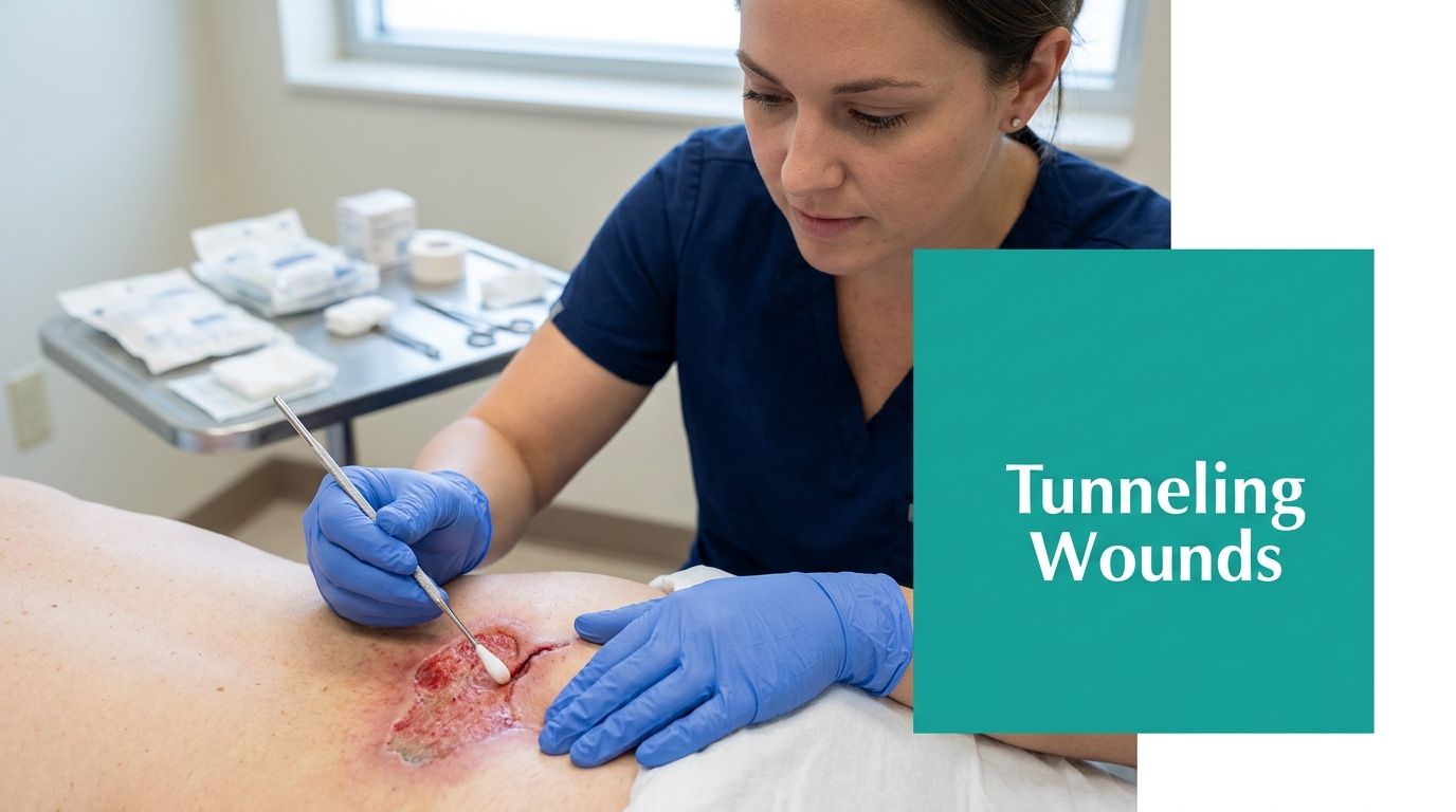
The first thing I look at is the opening that looks too small for the problem underneath it. A 1 cm sacral opening that probes into a 6 cm tract at the 4 o'clock position is not a tiny wound. It's a dead-space problem, a documentation problem, and often an escalation problem all at once.
A tunneling wound behaves like a hidden cavity. The surface can look calm while the tract keeps a pocket open under intact skin, which is why bedside probing, serial measurement, and honest charting matter more than the surface footprint alone. That's true in pressure injuries, post-surgical dehiscence, pilonidal disease, hidradenitis suppurativa, and any wound where drainage keeps finding somewhere to go instead of closing from the base up.
What Tunneling Actually Looks Like at the Bedside
A wound that “doesn't look that bad” can still be a mess under the skin. I've seen a shallow-appearing sacral defect that accepted a cotton-tip applicator far beyond what anyone expected, and I've seen groin wounds with a tiny mouth but a long, branching tract underneath. The surface opening misleads people. The probe doesn't.
Tunneling and undermining are not the same thing
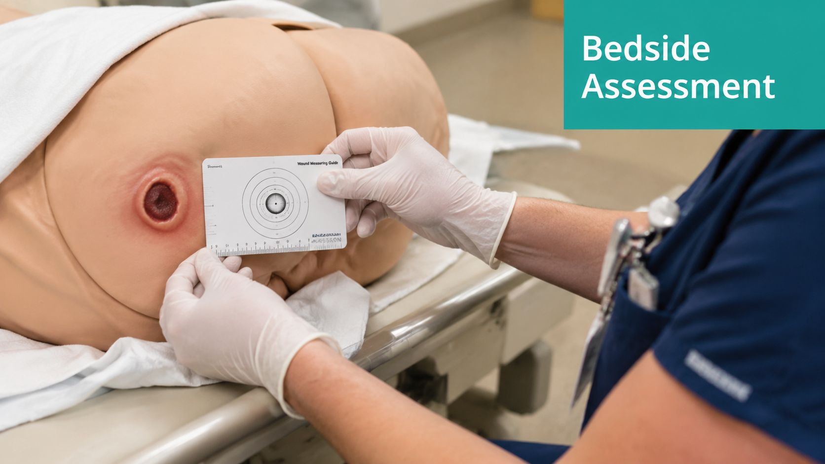
Tunneling is a narrow tract under the wound base that extends away from the visible opening. It's documented by direction and length, not just by the wound's surface dimensions, using the clock-face method with a soft probe or cotton-tip applicator (wound measurement references). Undermining is different. That's tissue destruction that extends under the wound edge at the perimeter, so you measure the depth of that shelf at the margin rather than the length of a tract.
That distinction matters in the chart and at the bedside. A payer, a surveyor, or a colleague reviewing the note will read those fields separately, because a wound with undermining is not the same clinical shape as a wound with a tunnel. If you only write “tunneling present,” you've told me almost nothing useful.
Practical rule: If the wound bed hides dead space, the note has to show where it is and how far it runs.
Dead space is the real enemy
The reason tunneling gets underestimated is simple. The visible opening can be small while the dead space underneath remains large enough to hold drainage, bacteria, and nonviable tissue. Clinical wound teaching materials emphasize that tunnels may need irrigation, packing, reassessment, and debridement because the problem is not cosmetic, it's structural (wound assessment and management reference).
That's why a 1 cm opening with a 6 cm tunnel is not a minor wound. It behaves like a much larger defect, especially if the drainage is purulent, the periwound is indurated, or the base isn't granulating. The chart has to reflect what the probe finds, not just what the eye sees.
If you need a quick visual reminder of how to separate measurements cleanly, I keep a simple wound measurement example reference handy for newer staff and float nurses.
Measuring a Tunnel Correctly
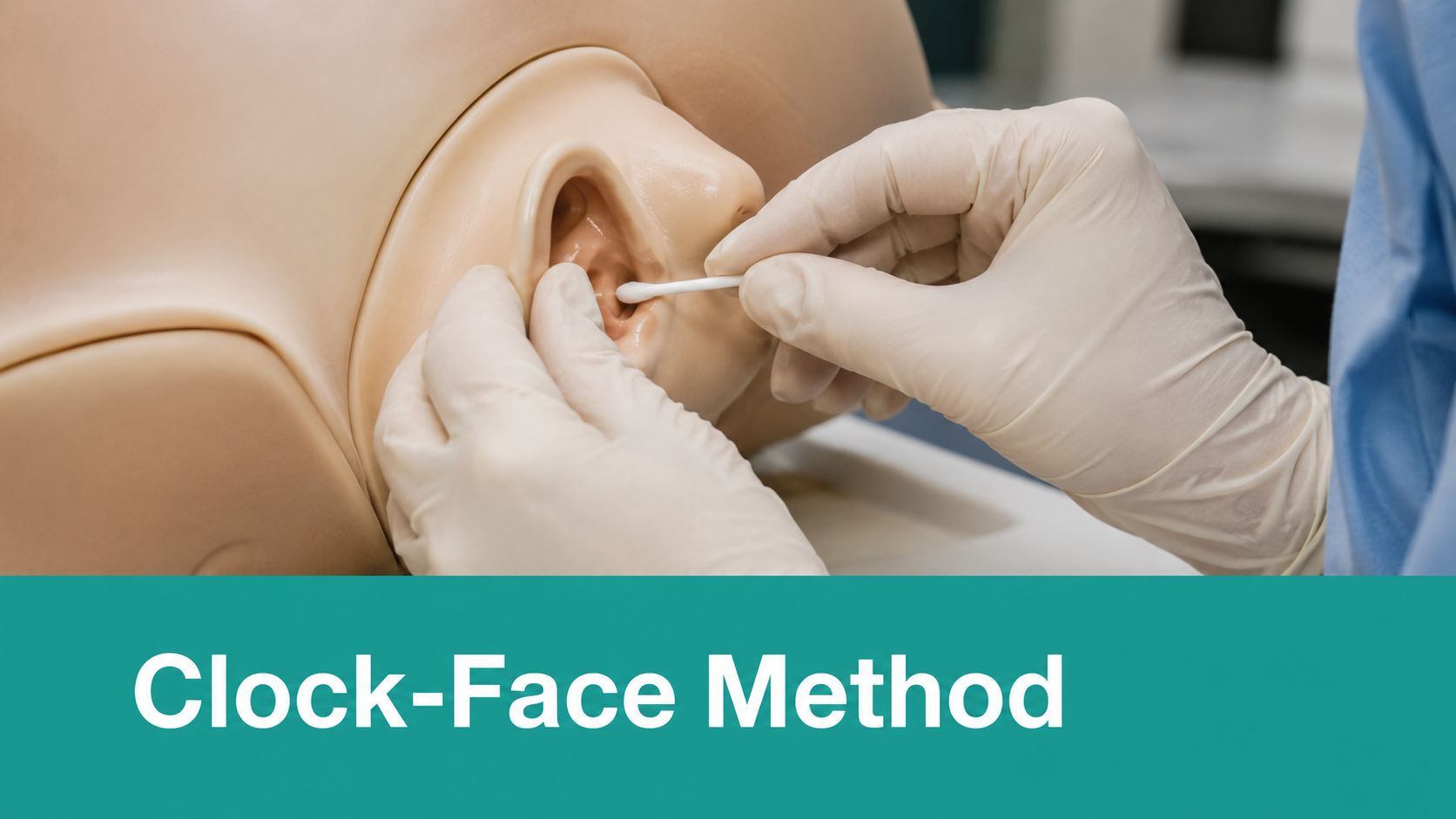
A tunnel should be measured the same way every time, and the note should start before the probe enters the tract. Clean the wound enough to see the opening, position the patient so the opening does not collapse on itself, and keep the clock face consistent from visit to visit. 12 o'clock points toward the patient's head. Once the team uses that convention every time, the chart is easier to defend and easier to compare.
The actual probing sequence
Use a sterile cotton-tipped applicator or similar soft probe, not a rigid instrument that can create a false passage. Advance it gently along the tract until you reach the full depth, then mark where the applicator meets the skin surface and measure that length in centimeters. Wound-measurement references describe tunneling as a narrow tract under the wound base that should be documented by direction and length, not just surface size, using the clock-face method.
Write the number. Write the clock position. Write both at every visit. “Tunneling present” without a baseline depth is a billing problem and a continuity problem, not documentation. A tract that cannot be compared over time cannot be called improving, worsening, or unchanged with confidence.
What should be in the note every visit
At minimum, I want the following in the record:
- Clock-face location, such as 4 o'clock or 9 o'clock.
- Length of the tunnel in cm.
- Undermining depth, if present, recorded separately.
- Exudate amount and character, for example scant serous, moderate serosanguineous, or purulent.
- Periwound condition, including maceration, induration, erythema, warmth, or intact skin.
- Pain, ideally tied to the patient's report during probing and care.
- What was placed into the tract, including the dressing or packing material.
Documentation habit that pays off: Put the baseline probe length in the first note, then compare every follow-up to that number. Audits go better when progress is measurable, not implied.
If a nurse writes “small tunnel” or “improving” without a number, that note is weak on appeal. If the wound is billed under a debridement family, the measurement record has to match the procedure narrative. That is where the work stands or falls.
A quick visual reference can help newer staff keep the measurements clean and consistent, especially when the tract direction is hard to document on a busy shift. See this measurement technique reference for a plain example of how to separate location, length, and surrounding wound dimensions.
Why Tunnels Form and Why They Persist
A tunnel usually forms because the wound bed never gets a clean path to closure. Pressure, shear, moisture, infection, necrosis, and poor offloading keep tissue from closing in a straight line, so the surface can look smaller while the deeper tract stays open. In practice, the common settings are pressure injuries, diabetic foot ulcers, post-surgical wounds that break down, and chronic inflammatory disease like hidradenitis suppurativa.
The tract usually tells you what is still active
A wound that keeps tunneling usually still has the driver in place. A pressure injury with a long tract often means pressure relief is incomplete. A diabetic foot ulcer that tracks deeper may be dealing with persistent mechanical stress, retained necrosis, or infection. A dehisced surgical wound with dead space often needs source control, not another dressing change.
Hidradenitis suppurativa is a clean example of why tunnel-prone disease deserves respect. A review reports a global prevalence of 0.40% with a 95% confidence interval of 0.26% to 0.63%, and the same review describes a minimally invasive tunnel protocol with daily antibiofilm flushing that achieved a 93% tunnel resolution rate by day 28 and an average healing time of 12 days (hidradenitis suppurativa review). That does not mean every tunnel behaves like HS. It does mean tunnels are measurable, and in the right disease state they can respond when the driver is named. For the biofilm piece, the biofilm on skin reference is the right conceptual backdrop for why these wounds stall and recur.
Hidden pathology is the part people miss
A persistent tract can signal occult infection, pressure-related dead space, retained necrosis, or a wound that has never been offloaded. That is why a tunnel should not be treated as a dressing nuisance. It is a marker of deeper pathology until proven otherwise.
I use that mindset when a wound keeps cycling through packing and dressing changes without meaningful change. If the tract is not shortening, the problem is not just the material in the cavity. The biology underneath is still wrong.
A tunnel that is not changing, or is changing in the wrong direction, should make you rethink source control, pressure relief, and whether imaging is needed to look for deeper extension. In the record, that same pattern should prompt tighter documentation, clock-face location, measured depth, and a cleaner tie between the tract and the procedure code set, especially when debridement levels under CPT 11042–11047 are being considered and infection coding such as ICD-10 L03.90 is part of the claim.
Packing Technique and Dressing Choices
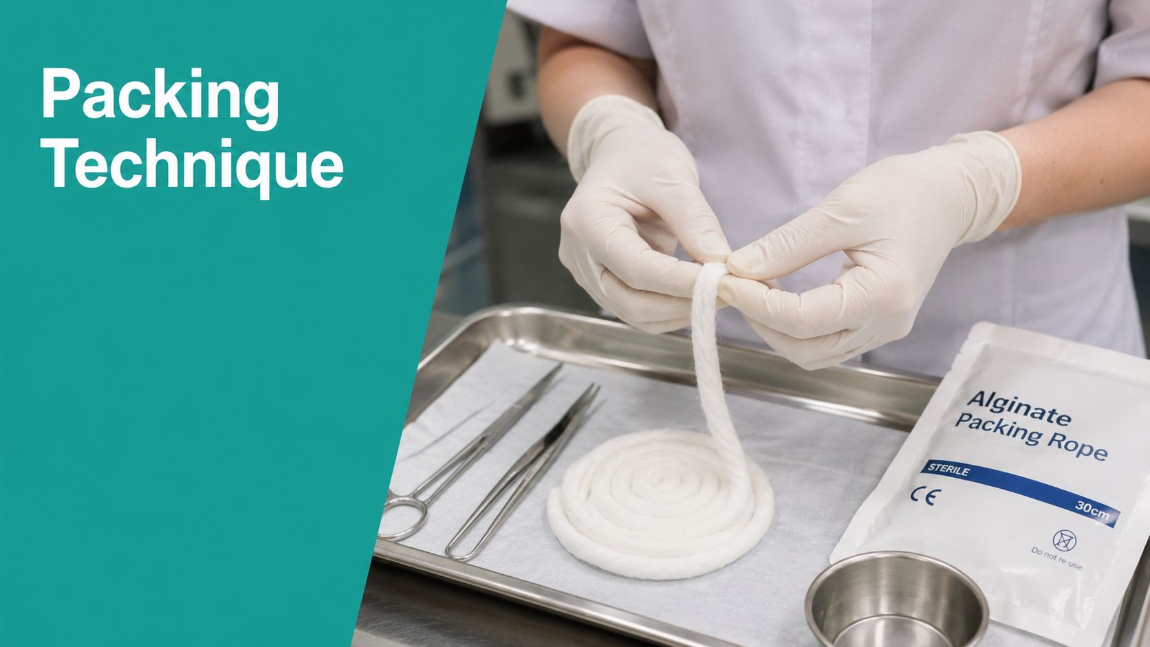
The old habit of stuffing a tract until it feels “filled” is how people create new problems. A tunnel needs gentle cleansing, soft irrigation, and loose packing. Fill the dead space without creating pressure on the tract wall, because pressure can compromise perfusion and force premature closure at the surface while the deeper space remains open.
Loose fill, not tight pack
Clinical teaching materials describe tunnel management as cleansing, gentle irrigation, and loose packing with alginate rope, hydrogel-impregnated gauze, medicated rope, iodine-based dressings, collagen dressings, or negative pressure wound therapy when wound characteristics support it, and they warn against overpacking (tunnel treatment reference).
That warning is real. Overpacking creates outward pressure, can damage granulating walls, and can close the entrance before the tract is healed. The result is the kind of wound that looks smaller while the drainage problem keeps going.
Practical rule: If the packing resists going in, it's too much material. A tunnel should be lightly filled, not wedged.
Match the dressing to the biology
I match the dressing to drainage and bioburden. A wetter tract usually needs something more absorbent, like alginate rope. A drier but still open tract may do better with a moisture-supporting option like hydrogel-impregnated gauze. If the bed supports it and the seal is achievable, NPWT can be useful, but not when the tunnel is actively purulent or when the wound geometry makes the seal unreliable.
Debridement matters too. Sharp bedside debridement is for conservative, serial removal of nonviable tissue when the wound is otherwise manageable. Surgical debridement belongs in the OR when the tract is deep, extensive, branching, or needs source control you can't get at the bedside. A tunnel with dead tissue and ongoing drainage is not a place to be timid.
If you want a practical way to line up dressing choice with wound features, keep a wound dressing selection chart available for the team so the choice isn't guessed from memory at the end of a long clinic day.
Infection, Antibiotics, and Red Flags
A tunneled wound can be colonized without being infected, and that distinction saves a lot of bad prescribing. Contamination is expected. Colonization is common. Infection changes the plan. I care most about the shift in drainage, odor, pain, tissue quality, and periwound appearance.
What should make you change course
When a tract starts producing more purulent drainage, new or worsening odor, expanding induration, spreading erythema, or stalled granulation, I assume the wound biology has changed. If the patient develops systemic signs, the wound isn't a dressing problem anymore. It's a source-control problem.
Antibiotics do not fill dead space. They treat spreading infection. If the tract is still open and the base is still necrotic or biofilm-laden, you'll often need debridement, irrigation, and better local management before antibiotics can do much. That's why a surface swab usually doesn't settle the question when the wound is not improving. The tract itself needs attention.
When to escalate urgently
These are the scenarios that should push fast referral or surgical input:
- Tunneling into a joint or near a joint capsule.
- Bone exposure or a probed-to-bone finding.
- Necrotizing soft tissue signs, including rapidly worsening pain or tissue change.
- Rapidly expanding tunnel despite care.
- New sepsis physiology or systemic decline.
Expert wound-care sources note that persistent tunneling may require CT or MRI when bedside inspection cannot define the full tract, and that regular cleansing plus loose packing with alginate rope or antimicrobial gauze helps reduce bacterial burden and prevent abscess formation (wound care assessment and treatment reference). That's the point where bedside confidence stops being enough.
A Decision Framework for Escalation
A tunnel that's shrinking, cleaner, less painful, and less exudative can stay on a conservative path. A tunnel that's not changing, or is changing in the wrong direction, needs a different move. The practical minimum is weekly reassessment, because you need enough cadence to know whether the tract is shorter, the same, or longer, and whether the drainage, pain, and periwound changes are trending the right way.
Three paths clinicians actually use
If the tract is clearly improving, keep bedside care going and document the trend. If the tract is deep, branching, anatomically unclear, or raises concern for a fistula, move to CT or MRI. Practitioner-focused wound sources note that visualization may be insufficient for tunneled wounds and that CT or MRI are often necessary for complete assessment, which is the inflection point between continued conservative care and surgical exploration.
If there's abscess, necrotizing concern, bone involvement, or stalled progress after optimized bedside care, get a surgical consult. That's where unroofing or operative debridement enters the conversation. Pressure injuries, diabetic foot ulcers, post-surgical wounds, and hidradenitis suppurativa all push differently, but the decision logic is the same. Measure response, then escalate when the tract stops behaving.
Practical rule: If the wound is not shorter, cleaner, and less inflamed after optimized care, assume the tract needs a new workup.
A good home health or clinic workflow should make that call easy. The wound nurse should be able to compare the current tunnel length, clock-face direction, and drainage pattern to the last visit without hunting through a disorganized chart.
Documentation and ICD/CPT Tagging That Survives Audit
A strong note links the wound shape, the treatment, and the code family without making the reviewer work for it. I'd rather see a concise, specific note than a long one that says very little. For a sacral pressure injury, a clean line might read: Stage 4 pressure injury of the sacrum, L89.154, 4 o'clock tunnel measuring 6 cm, undermining 1 cm at 2 to 3 o'clock, moderate serosanguineous drainage, periwound intact with mild maceration, pain 4/10, wound gently irrigated and loosely packed with alginate rope.
The coding logic has to match the note
For debridement, the CPT 11042–11047 family is chosen by depth and surface area, so the note has to support what tissue was removed and how extensive the work was. Tunneling doesn't automatically force a higher code, but it does support the medical necessity for a more intensive assessment and treatment plan when the tract is deep, chronic, or repeatedly reassessed.
If infection is part of the picture, the diagnosis has to fit the story. L03.90 is a common cellulitis code, but only if the clinical record supports cellulitis. Don't hang a procedure on a diagnosis that isn't documented. That's a denial waiting to happen.
Common audit pitfalls
| Documentation Field | What to Write | Audit Pitfall to Avoid |
|---|---|---|
| Wound type and etiology | Stage, location, and cause, such as sacral pressure injury or post-surgical dehiscence | Generic “open wound” language |
| Tunnel location | Clock-face direction, such as 4 o'clock | “Tunneling present” with no direction |
| Tunnel length | Measured length in cm | “Small tunnel” or no baseline |
| Undermining | Separate depth and location | Mixing undermining into tunneling |
| Exudate | Amount and character, such as moderate serosanguineous | No drainage description |
| Periwound | Maceration, induration, erythema, intact skin | “Looks okay” |
| Treatment | Irrigation, loose packing, dressing type, debridement | Missing link to the intervention |
| Code support | CPT 11042–11047 and diagnosis linkage | Procedure with no diagnosis rationale |
Revenue-cycle teams get hit when the narrative is vague, the tunnel isn't measured, or the treatment description doesn't match the code. Surveyors notice the same thing. A wound with dead space is only defensible when the chart shows you understood the dead space.
Weekly Cadence and Escalation Reminders
Tunneled wounds reward discipline, not heroics. Reassess every week, compare tunnel length, direction, exudate, and periwound condition to the prior note, and don't pretend a stable-looking surface means the tract is safe. If the tunnel stops improving despite optimized packing, or if bone, joint, or systemic infection becomes a concern, move to imaging or surgical review.
The bedside checklist is short:
- Measure the tunnel with clock-face location and centimeters.
- Compare today's length with the last visit.
- Inspect exudate, odor, and periwound change.
- Confirm the dressing is loosely filling dead space.
- Link the diagnosis, procedure, and debridement code in the note.
If you keep those fields tight, the care gets better and the claim gets stronger. That's the part people miss when they treat a tunnel like a dressing issue. It's really a longitudinal assessment problem with billing consequences attached.
If your team is still documenting tunnel length in free text, or struggling to keep the note, code, and wound image in sync, EkagraHealth AI can help build that documentation backbone at the point of care. Visit EkagraHealth AI to see how it supports wound note drafting, measurement capture, and cleaner coding workflows for tunneled wounds.
