
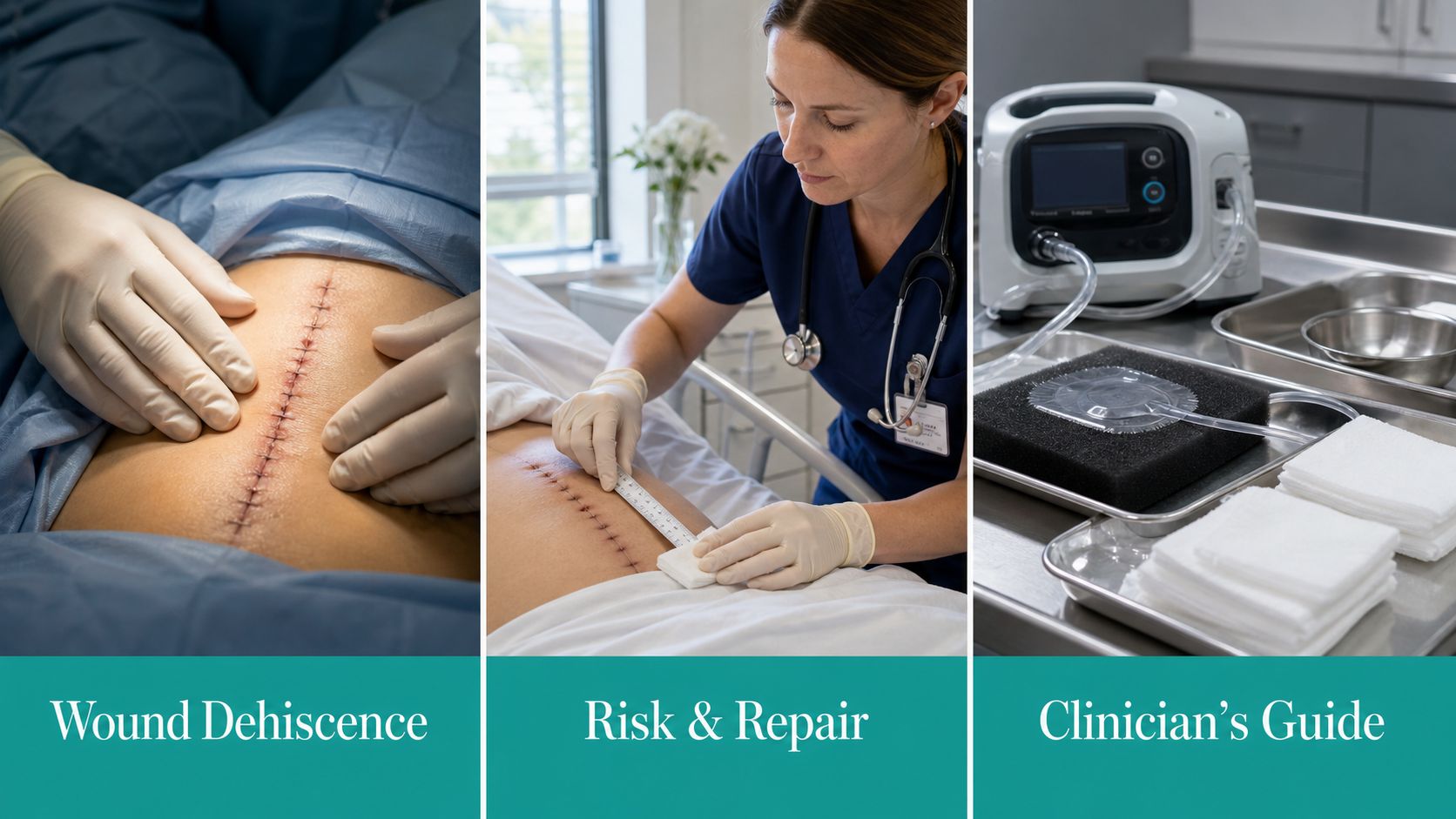
The call comes late. A post-op nurse says the midline incision on a diabetic patient is draining more than yesterday, the skin edges look a little wider, and the patient coughs every time the dressing is changed. That's the moment a lot of teams start thinking in terms of dressings, but the core question is sharper, which layer failed, how far it's gone, and whether you're looking at a wound-care problem or a surgical one.
Wound dehiscence is the separation of a previously closed incision, and the clinically useful part is not the word itself, it's the depth. A skin line that gaps is one thing. Fascial failure is something else entirely, because the management, urgency, and documentation all change once the load-bearing layer is involved. Timing matters too. Dehiscence often shows up in the early post-op window, commonly around days 4 to 14 after surgery, when tensile strength is still weak and the incision hasn't earned much mechanical forgiveness yet (PubMed review).

A good bedside mental model is simple. The incision didn't just “open.” The wound lost tensile strength, and something in the biology, the mechanics, or both, gave way. That's why the fresh laparotomy with new drainage, the midline in an obese patient, or the contaminated emergency case deserves tight follow-up rather than casual reassurance.
See also the basic distinction between an open wound and a separated closed incision in what are open wounds.
What Wound Dehiscence Actually Means at the Bedside
When the dressing strikes you as heavier than yesterday and the patient winces when they move, start with the incision line itself. If the edges are separating, you're not looking at a minor cosmetic issue, you're looking at a closure that's failing under load. That failure may be superficial, or it may involve fascia, and that difference decides whether you're packing a wound or calling the surgeon.
The layer that failed matters more than the skin looks
A superficial gap can look dramatic and still be manageable with local care if the deeper layer holds. A fascial defect is a different beast. Once fascia gives way, abdominal contents are no longer safely contained, and the situation can move quickly from a wound visit to an operative problem.
This is why “the incision opened” is too vague for clinical work. A more useful statement is, “partial superficial separation with serosanguinous drainage,” or “complete fascial dehiscence with exposed deeper tissue.” That language tells the next clinician what happened and tells the coder what was documented.
Practical rule: if the wound is under tension, draining, or separating at all, treat it as a healing failure until proven otherwise.
The biology also explains the timing. Early healing wounds are mechanically weak, so a cough, a bout of vomiting, or abdominal distension can turn a tenuous closure into a failure point. That's why the same incision may look fine on round one and clearly worse on round two. A wound can deteriorate fast when the patient is stressed, swollen, infected, or both.
For a concise overview of partial separation concepts, the partial-thickness wound discussion is a useful companion when you're writing up a superficial case.
Partial vs Complete and Superficial vs Fascial
Partial dehiscence means some of the closure has separated, but not all of it. Complete dehiscence means the wound has fully separated along the involved segment. The difference sounds academic until you're standing at the bedside, because the exam dictates the next move, not the label alone.
Superficial separation can stay in the wound clinic lane
If the skin and subcutis have separated but fascia is intact, the wound may heal by secondary intention with local care. That usually means cleaning, protection from contamination, and a dressing plan that matches the amount of exudate and the amount of dead space. A small, clean, shallow gap is not the same as a deep, draining tract with undermining.
Fascial failure is a surgical problem
Once fascia is open, you're in urgent territory. The issue is not just drainage, it's containment. In abdominal wounds, fascial separation can progress to burst abdomen, and that's the kind of failure that needs immediate surgical escalation. Exposed viscera, or anything approaching it, changes the tone of the entire encounter.
Evisceration is not just dehiscence with flair. It means abdominal contents are outside the wound, and the patient needs urgent surgical response. A clean superficial gap may be followed. A fascial gap may need negative pressure therapy or return to theatre. Full evisceration gets the escalation pathway moving now.
For a practical shorthand, think in three tiers, superficial and partial, deep or fascial, then catastrophic with evisceration. That's the vocabulary that survives a handoff, a wound note, and an audit.
Why Dehiscence Happens Patient, Wound, and Perioperative Drivers
The risk factors cluster in predictable ways, and they rarely sit in one bucket. I sort them into patient factors, wound factors, and perioperative factors because that is how you find the failure point and decide what to fix.
Patient factors create the baseline vulnerability
High BMI and diabetes keep showing up across procedure types in risk-factor work, so they belong in the risk stratification, not as a casual note in the assessment. Other recurring contributors include older age, male sex, hypoalbuminemia, anemia, jaundice, malignancy, chronic steroid exposure, smoking, and poor tissue quality (systematic review, StatPearls review). Those factors matter because the wound starts healing in a host that may already be short on reserve.
In diabetic patients, inflammation control and glycemic control pull in the same direction. If the metabolic side is uncontrolled, the incision often pays for it at the edge of the wound. For a readable patient-facing explainer you can share when education is needed, managing inflammation in diabetes is a practical adjunct.
Wound and perioperative stress finish the job
Contamination, tissue ischemia, emergency surgery, and closure under tension push a wound toward failure. Coughing, vomiting, and abdominal distension add force before the incision has enough tensile strength to tolerate it. Poor closure technique can do the same thing, especially if the fascia was closed under excessive strain.
A red, draining, tight incision on a malnourished patient deserves respect on first glance. I do not treat that as routine drainage until the exam proves the fascia is intact and the wound is stable.
There is also a reason dehiscence stays familiar in surgical practice. A 2006 review noted that the rate had not meaningfully dropped in recent years and sat around 1% to 3%, despite modern sutures and protocols (PubMed review). The basics still matter because the biology has not changed.

Bedside Assessment That Actually Holds Up in the Chart
Good notes start with measurements, not adjectives. “Looks worse” won't survive a payer review, and it won't help the surgeon decide whether the fascia is involved. I want a wound assessment that someone else can reproduce tomorrow, not a narrative that only makes sense to the person who wrote it.
Document the wound like you expect an audit
Record length, width, depth, undermining, tunneling, edema, warmth, pain, exudate type and amount, odor, and periwound skin condition. Then state whether fascia, mesh, hardware, or viscera is exposed. If purulent drainage is present, write it plainly. Purulence is treated as evidence of infection in common wound-assessment pathways, and that changes both the clinical plan and the downstream documentation.
The chart should also show reassessment at every dressing change. A dated, reproducible measurement set is the backbone of medical necessity. If the wound is deteriorating, the note should show exactly how. If it's improving, the note should prove it. Surveyors look for that trend, not just a single snapshot.
Use language that links assessment to action
A strong note doesn't just describe a wound. It connects findings to a decision. For example, “full-thickness superficial separation with moderate serosanguinous drainage, no fascial exposure, periwound intact, continue packing and monitor for increased depth,” is far better than “open area noted.” One is clinical. The other is a liability.
Charting tip: if you wouldn't be comfortable defending the wording in front of a medical director, it probably isn't specific enough.
Photography helps when your workflow allows it, but measurement and narrative still matter. Photos don't replace a good wound exam, and they don't substitute for an assessment of tissue depth or infection. The note has to stand on its own.
Management From Packing to OR Escalation
Minor superficial dehiscence can often be managed with cleaning, protection, and packing to support healing by secondary intention. That's the route when the wound is shallow, the fascia is intact, and the tissue looks viable. If the exudate is modest and the bed is clean, simple local care may be enough to let the wound granulate in a controlled way.
Match the dressing to the biology
Heavy exudate, deeper dead space, or a wound that needs more structured granulation support often pushes you toward negative pressure wound therapy. NPWT is not a reflex, and it's not a substitute for source control, but it's useful when the wound needs fluid management and tissue approximation support. If necrotic tissue is present, debridement comes first.
Debridement coding follows tissue depth. 11042 fits subcutaneous tissue, 11043 muscle and/or fascia, and 11044 bone. Add-on codes 11045 and 11046 cover additional 20 sq cm increments, and 11047 is the add-on for deeper tissue beyond the first 20 sq cm when the work extends into muscle, fascia, or bone. The code has to match what was removed, not what was present on the wound bed.
When the wound is full-thickness or fascial
A complete fascial dehiscence is an OR problem. Practical surgical principles include opening the incision enough to inspect the fascia, releasing inadequately closed fascia, and re-closing with large interrupted retention sutures when needed (TeachMeSurgery). Exposed viscera should be covered with saline-soaked gauze during transport, broad-spectrum IV antibiotics should start before theatre, and the patient needs an A-to-E assessment on the way to definitive management.
Older literature also shows why nobody should brush this off. In one laparotomy trial, dehiscence occurred in 56 of 1,958 patients, 2.9%, and mortality among those who dehisced was 25% (PubMed review). Severe cases have been reported with mortality as high as 45% in older reviews, which is why the threshold for escalation should stay low when fascia is involved (PubMed review).
Closing the Loop Prevention Strategies That Actually Work
Prevention starts before the incision. Nutritional optimization, glycemic control, smoking cessation, and a hard look at steroid exposure all belong in the pre-op workup when a patient is headed toward a high-risk closure. If the wound is going to be under tension, the OR plan should say so out loud, not pretend otherwise.
Tight closure, less tension, fewer surprises
Intraoperatively, layered fascial closure and appropriate suture technique matter more than marketing around materials. Retention sutures belong in selected high-tension closures, especially when the abdomen is at risk of strain. Antibiotic prophylaxis, when indicated, is part of the same safety net.
Postoperatively, the job is to protect the repair. Abdominal binders, early mobilization without excess load, prompt control of surgical site infection, and clear patient education on warning signs all reduce the odds of a bad surprise in clinic. The wound team should also feed outcomes back to the surgeon. If a pattern is showing up, it needs to be visible upstream.
For discharge education, especially when language barriers get in the way of instructions about drainage, odor, or wound support, prevent medication errors with translation is a useful reminder that communication failures can become wound failures too.
Documentation, Coding, and the Denials That Follow
A decent clinical note can still get rejected if dehiscence coding lacks specificity, as vague language is the fastest route to a denial. If you document only “wound dehiscence” without depth, encounter type, or a clean tie to what you did, the claim often looks unfinished.
Code the diagnosis to the depth you actually assessed
ICD-10-CM dehiscence codes live in the T81.30 through T81.33x range, with the 6th and 7th characters carrying encounter type and depth detail. That means the note has to tell the story clearly enough for the code to be defensible. If the wound is superficial, say so. If fascia is involved, say that too. If this is an initial encounter versus follow-up care, the chart should not make the coder guess.
The procedure code has to line up with the wound bed. If you debride subcutaneous tissue, don't bill a deeper code. If the work extends beyond the initial 20 sq cm, the add-on codes must be documented with actual surface area. Payers deny these claims when the record doesn't say what tissue was removed, how much was removed, and why debridement was medically necessary.
Denials usually come from missing proof, not missing work
The common failure points are predictable, unspecified depth, missing surface area, lack of medical necessity for the level billed, and no diagnosis-to-procedure linkage. Reviewers also look for a dated, reproducible wound assessment at each visit, plus a clear reason the chosen treatment remains appropriate. If the wound isn't changing, the note needs to show why the plan still makes sense.
A structured workflow can help. EkagraHealth AI is one option that drafts SOAP notes, maps CPT and ICD-10-CM codes, and supports point-of-care documentation so the record is closer to claim-ready before it leaves the room. For a deeper look at the documentation standard payers expect, the medical necessity documentation resource is worth keeping handy.
For surgical site infection context that often overlaps with dehiscence documentation, the insights from BacteriaFAQ about SSIs can help frame how infection language should be written when drainage, odor, or purulence are part of the note.
Putting It All Together at the Point of Care
Classify it first, superficial or fascial, partial or complete. Measure it the same way every time. Then choose the path, pack and observe, NPWT, debridement, or straight to the OR. If the note supports the decision and the diagnosis matches the work, the wound is more likely to heal and the claim is more likely to pay.
If your team is tired of rewriting dehiscence notes, chasing down missing depth, and getting claims kicked back for avoidable documentation gaps, EkagraHealth AI can help. It captures the visit, drafts the wound note, and maps the coding so the clinical record and the claim match the care you delivered. Visit EkagraHealth AI to see how it supports wound documentation from bedside assessment through billing.
