
A debrided diabetic foot ulcer with an exposed Achilles tendon will teach a new clinician this lesson fast. If you pack that wound with the wrong foam, the next dressing change becomes a fight. The tissue is angry, the patient remembers the pain, and your note won't explain why you made that choice in the first place.
That's where wound vac white foam earns its place. It isn't the default foam. It's the protective foam. When the wound bed is fragile, tunneled, undermined, or sitting over something you cannot afford to traumatize, foam selection stops being a supply choice and becomes a clinical decision that has to be defensible at the bedside and on the claim.
When Black Foam Is the Wrong Tool for the Job
The classic setup is a Wagner Grade 3 or 4 foot wound after sharp debridement. You've opened the cavity, controlled obvious nonviable tissue, and now you're staring at exposed tendon, deep undermining, and a patient who already has enough reasons not to trust the next dressing change.
Black foam is often too aggressive in that moment.
A wound bed that needs protection
White foam belongs in wounds where aggressive tissue ingrowth is not the goal. The material is denser, less adherent, and more resistant to tearing in undermining. That matters when you're laying foam over tendon, around a graft, near a flap, or into a sinus tract where fragment retention would become tomorrow's problem.
In practice, the wrong foam choice usually shows up later as one of three failures:
- Pain at dressing change when tissue has grown too aggressively into the dressing
- Trauma to exposed structures that should have been protected
- Poor documentation that says only “NPWT applied,” which invites payer scrutiny when the wound is clearly high acuity
White foam is rarely about convenience. It's about control.
The decision has to match the wound
If the wound bed is broad, healthy, and you want fast granulation across an open surface, black foam may still be appropriate. But if the wound has a narrow opening with a deeper cavity, exposed critical structures, or a painful bed where atraumatic removal matters, white foam is often the better call.
That's the standard I teach new hires. Don't ask which foam you use most. Ask what the wound can tolerate, what the next dressing change will look like, and whether your chart explains the reasoning well enough for a medical director or surveyor reading it cold.
White Foam vs Black Foam A Material Science Breakdown
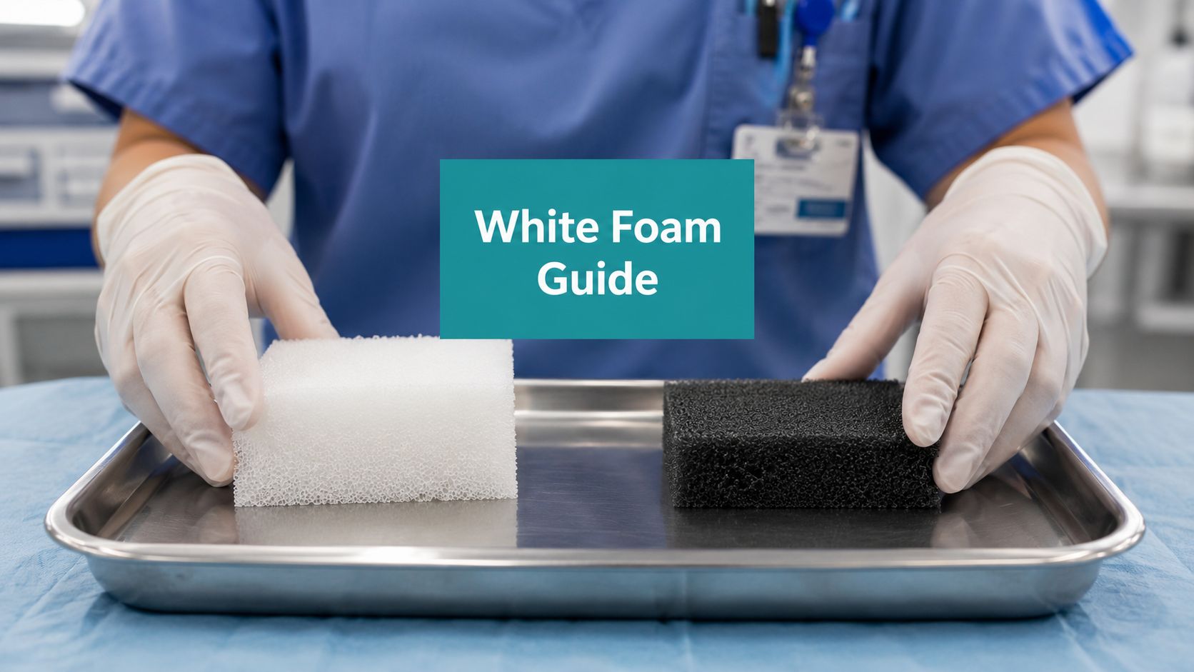
Set two pieces of foam on the tray and they may look like a simple color choice. They are not. The material changes how the dressing transmits negative pressure, how much the wound bed grows into it, how removal feels three days later, and how clearly you can defend the choice in your note.
White foam is polyvinyl alcohol (PVA). Black foam is polyurethane. PVA is denser, less porous, and less likely to let tissue grow far into the dressing. Polyurethane is more open and tends to create stronger mechanical interaction with the wound surface. As noted earlier in the article, clinical guidance supports white foam when the treatment goal includes protection of vulnerable tissue and less traumatic removal.
Clinical Comparison of NPWT Foams
| Characteristic | White Foam (PVA) | Black Foam (Polyurethane) |
|---|---|---|
| Material behavior | Higher density | More open and aggressive in wound contact |
| Tissue ingrowth | Restricts ingrowth | Allows more ingrowth |
| Dressing change experience | Typically less painful to remove | Often more adherent to the wound bed |
| Use over exposed structures | Better suited when protection matters | Less ideal when delicate structures are exposed |
| Tunnels and undermining | More resistant to tearing during removal | Greater concern for fragmentation in difficult cavities |
| Clinical role | Protective interface and cavity filler in select wounds | Common choice when stronger granulation stimulus is desired |
At the bedside, that difference shows up fast. White foam usually gives you more control in wounds where you cannot afford a dressing that grabs the tissue too aggressively. Black foam usually gives you more surface contact and faster granulation response across a broad, open wound bed.
Density matters for another reason. White foam holds its shape better in narrow undermining and small cavities, so it is less likely to shear or leave a fragment behind during removal. That does not remove the need to count pieces and document placement, but it lowers one avoidable risk in wounds with awkward geometry.
Pore structure matters too. Black foam's open structure helps distribute suction through a larger open surface, which is useful when building granulation is the main objective. White foam sacrifices some of that aggressiveness to reduce adherence. That is the trade-off. You get less tissue ingrowth and often an easier dressing change, but you should not expect it to behave exactly like black foam in a large shallow wound.
That trade-off should appear in the chart. If you select white foam, document the wound feature that justified a protective interface: exposed tendon, painful wound bed, graft or flap coverage, narrow tunnel, undermining, or concern for atraumatic removal. Payers do not reimburse material preference. They reimburse a medically necessary treatment choice tied to wound characteristics and risk.
For teams reviewing broader dressing categories, this guide to wound healing dressings helps place NPWT foam selection in the larger wound care plan.
Practical rule: Choose white foam when protection, controlled contact, and safer removal matter more than aggressive granulation stimulation. Choose black foam when the wound can tolerate stronger ingrowth and that response is the goal.
Clinical Indications When to Choose White Foam

A common bedside scenario is a dehisced foot wound with a short tunnel, exposed tendon, and a patient who already dreads the next dressing change. That is a white foam case until the wound proves otherwise.
Exposed tendon bone mesh or graft material
White foam belongs in wounds where the contact surface needs to protect tissue, not grip it. Use it over exposed tendon, bone, hardware, mesh, vascular structures, graft material, or flap tissue when direct black foam contact would raise the risk of ingrowth and traumatic removal.
Chart that decision clearly. "NPWT applied" does not support medical necessity. "White PVA foam placed over exposed Achilles tendon to limit adherence and protect structure during dressing changes" does. That sentence ties the material choice to a wound feature, which is what reviewers look for.
Tunnels sinus tracts and undermining
White foam is often the safer choice in narrow tunnels, sinus tracts, and undermined pockets because it holds together better during removal from confined spaces. That matters in real practice. The harder the geometry, the more disciplined the foam selection and piece counting need to be.
The placement has to maintain contact with the portion of the wound you intend to treat. If a tunnel is being dressed with foam, leave enough material visible at the opening to confirm retrieval and communication with suction. Teams that need a refresher on technique should review this guide on wound vac placement for tunneled and undermined wounds.
Painful wounds and atraumatic dressing changes
Some wounds technically can accept black foam but clinically do better with white because the patient cannot tolerate repeated traumatic removals. I make that switch in painful post-op wounds, diabetic foot wounds after debridement, and wounds sitting over sensitive structures where every change risks setting the patient back.
That choice should also be documented in plain terms. State the pain, the friability, or the high-risk surface. Then state why a less adherent interface was selected. If the chart only says provider preference, payment gets harder to defend.
Grafts flaps and wounds you are trying not to disturb
White foam is useful when the treatment goal includes protecting a graft, supporting a flap, or managing drainage without repeated disruption of a delicate wound bed. In these cases, the trade-off is straightforward. You may give up some of the faster granulation response seen with black foam in exchange for a dressing that is less likely to traumatize the surface at the next change.
That is a sound clinical choice if the wound needs protection more than aggressive tissue ingrowth.
A short bedside checklist
Choose white foam when the wound has one or more of these features:
- Exposed critical structures that need a protective contact layer
- Tunnels, sinus tracts, or undermining where safe retrieval is a concern
- Painful or friable wound beds where less adherent foam improves tolerance
- Fresh grafts or flap coverage where minimizing trauma is part of the plan
- Complex diabetic foot wounds where cavity management and atraumatic removal both matter
If you choose white foam, document three things every time: the wound feature that justified it, where each piece was placed, and why that choice was safer than black foam for this wound on this date. That is good care, and it is how you support the claim.
Application Protocols Pressure and Dressing Changes
A common bedside failure looks like this. The wound is packed correctly, the drape holds, the pump is running, and two days later the tunnel still looks stagnant and the patient says the dressing never really pulled down. In many of those cases, the problem is not the machine. The white foam was treated like black foam, and the settings and packing technique did not match the material.
Set pressure for the foam you chose
White foam is denser and less compressible. It often needs more negative pressure to transmit therapy through the dressing and into the wound bed. If the pressure is set too low, the dressing may not contract well, exudate control can be inconsistent, and tunneled areas may not communicate the way you expect.
Start with the ordered pressure that fits the wound goal and patient tolerance. If white foam is being used in a deep cavity, tunnel, or undermined space, verify that the foam is drawing down after the seal is achieved. If it is not, reassess the fill, the bridge, and the pressure setting instead of assuming the wound is slow to respond.
For clinicians reviewing placement mechanics, this guide to proper wound vac placement is a useful refresher.
Change the dressing on a schedule, then adjust to the wound in front of you
For a stable, monitored wound, dressing changes usually fall in the 48 to 72 hour range. In practice, the wound decides whether that interval holds.
Change sooner if drainage is heavy, the seal keeps failing, the periwound is macerating, pain rises, odor increases, or the patient develops new signs that raise concern for infection. White foam around a delicate surface can buy you a safer removal, but it does not give you permission to leave a struggling wound untouched.
A protocol that holds up clinically and on audit
Use a repeatable process:
- Prepare the wound bed first. White foam will not compensate for untreated necrotic tissue, uncontrolled bioburden, or a cavity that has not been properly assessed. If surgical debridement is performed, document the tissue level removed and code the service accurately.
- Measure before you cut. Record length, width, depth, tunnel depth and direction, undermining by clock face, exposed structures, drainage amount and character, odor, and periwound condition.
- Cut one purposeful piece whenever possible. In tunnels or narrow undermined areas, avoid multiple disconnected fragments. If a second piece is necessary, document the piece count and where each piece was placed.
- Pack to contact, not to fill every millimeter. Overpacking blocks pressure transmission and increases pain at removal. Underpacking leaves dead space and weakens therapy.
- Confirm function after the drape is down. Look for visible contraction of the dressing, listen for leaks, and ask about pain once suction is applied.
One charting habit prevents a lot of denials. Document why white foam was selected for this wound on this date, how many pieces were inserted, where they were placed, the ordered pressure, the therapy mode, and the planned change interval. If that rationale is missing, a reviewer can argue that the material choice was preference rather than medical necessity.
Know when to continue and when to stop
NPWT should show progress. That may be better granulation, less undermining, improved exudate control, or a wound bed that is ready for the next step in closure. If the wound becomes dusky, pain is out of proportion, drainage shifts in a concerning direction, or the wound stalls despite correct application, reassess the whole plan.
Do not let the device run on autopilot. White foam is a good tool, but only when the pressure, packing, change frequency, and documentation all match the wound you are treating.
Troubleshooting Retained Foam and Other Complications
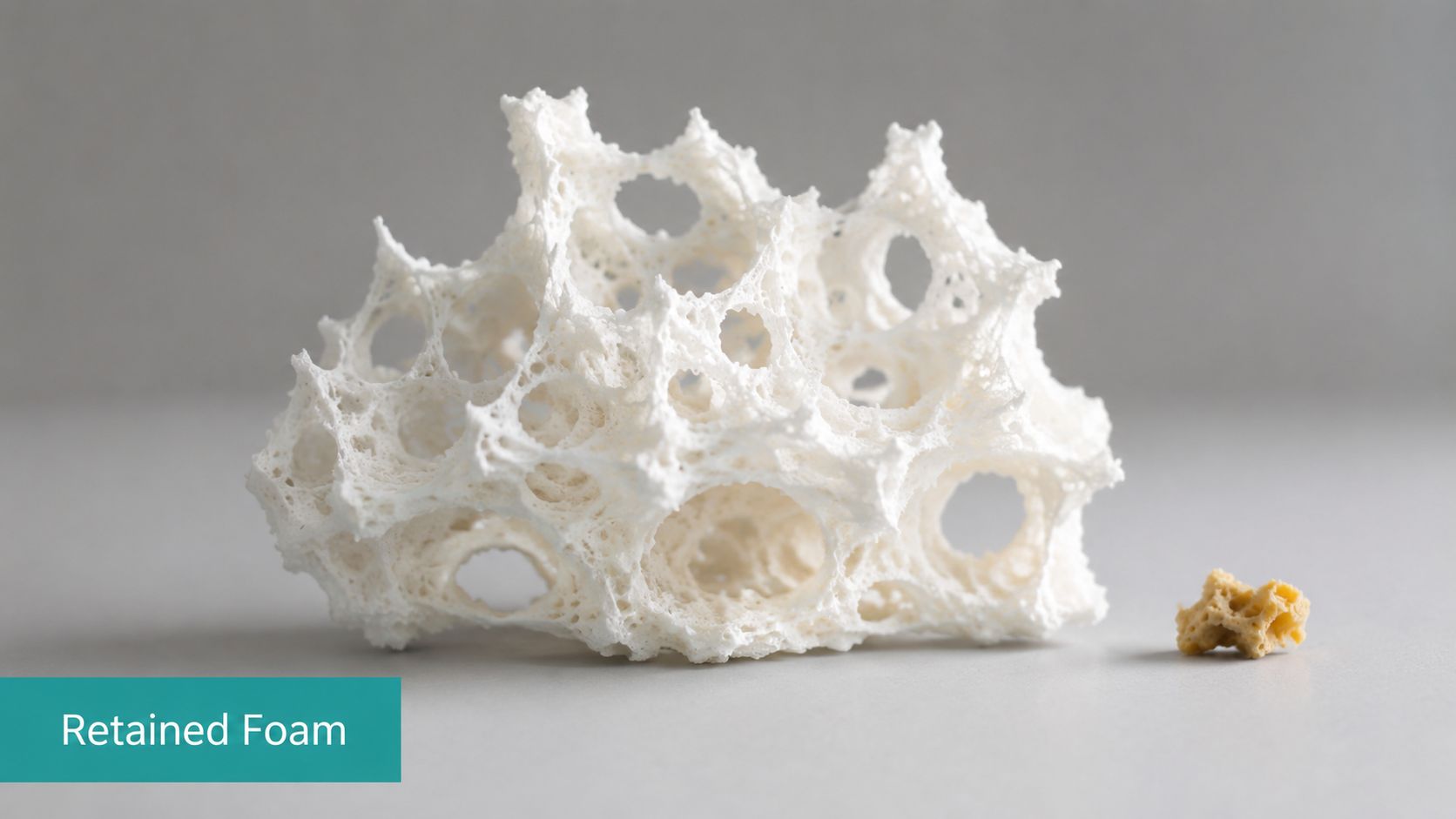
Retained foam is the complication people mention discreetly after it happens to them once. It should be discussed a lot louder.
Why retained foam matters
A retained white foam fragment acts like a foreign body. It can hold bioburden, stall granulation, and turn a straightforward dressing visit into an infection workup. Data cited in the literature notes that 18% of NPWT-related infections in SNFs are directly linked to retained dressing material, and the same discussion points out that post-removal inspection techniques are often not spelled out clearly in guidelines, as described in this retained dressing material analysis.
That's a patient safety problem. It's also a documentation problem. If your note doesn't describe removal integrity and wound bed inspection, you've left a hole in the record.
How to lower the risk
Retained foam usually follows one of three patterns:
- The foam was cut into multiple small pieces and one piece was forgotten in undermining.
- The tunnel was packed without a visible tail or clear communication to the wound opening.
- Removal was rushed and nobody verified that the extracted piece matched what was inserted.
Use one piece whenever the wound geometry allows it. If you must shape the dressing for a tunnel or undermined cavity, document the number of pieces placed and removed. Counting matters.
What to look for after removal
The visual cues are often subtle. A persistent dark pocket that doesn't granulate normally, a focal area that stays unusually moist or inflamed, or a cavity that keeps producing unexpected exudate despite otherwise decent progress should raise concern.
After removal, inspect and probe carefully.
- Use saline-moistened swabs to gently explore tunnels and undermined areas
- Compare removed foam to the inserted configuration before discarding it
- Document cavity inspection instead of writing the vague phrase “dressing removed intact”
If you can't describe how you confirmed the wound was free of retained material, your chart is weak and your process probably is too.
Other trouble signs worth acting on
Retained foam gets the attention, but don't miss the basics:
| Complication clue | What it may mean |
|---|---|
| Dusky wound bed | Inadequate perfusion or poorly tolerated therapy |
| Persistent malodor | Infection, retained material, or inadequate debridement |
| Rising pain at therapy site | Pressure intolerance, seal problem, infection, or contact trauma |
| Increasing periwound maceration | Excess exudate, poor drape control, or delayed dressing changes |
Documentation and Coding for White Foam NPWT

Clinicians lose payment on white foam cases for one reason more than any other. The chart says what was done, but not why that specific dressing was medically necessary.
What payers and auditors look for
They want the wound characteristics that justify your foam choice. They want depth, tissue type, exposed structures, tunnel measurements, exudate description, infection status, and prior treatment failure or treatment rationale. If white foam was chosen because the wound was painful or because tendon was exposed, say that plainly.
Surveyors and medical reviewers also look for internal consistency:
- Debridement depth must match the code. If you bill 11042 to 11047, the note has to identify the deepest tissue debrided.
- NPWT rationale must match wound findings. White foam over exposed tendon makes sense. White foam with no documented reason looks arbitrary.
- Ongoing need must be visible. Serial measurements, exudate trends, granulation progression, and periwound response should support continued therapy.
Language that holds up better
These phrases are stronger than “NPWT continued”:
PVA foam selected to protect exposed Achilles tendon and reduce adherent granulation tissue at dressing changes.
White foam placed within undermined lateral cavity due to need for atraumatic removal and reduced fragmentation risk.
Dressing change frequency increased due to heavy serous exudate and concern for bioburden accumulation.
Those aren't fancy phrases. They're clinically specific. That's what protects the claim.
For teams aligning diagnosis coding with wound documentation, this page on wound vac ICD-10 coding is a useful reference.
A note template that works
A defensible white foam NPWT note should include:
- Wound etiology and severity such as diabetic foot ulcer, post-surgical dehiscence, or pressure injury, plus Wagner grading when relevant
- Exact measurements including length, width, depth, tunnel depth and direction, and undermining by clock position
- Wound bed description with granulation, slough, exposed tendon, fascia, bone, or hardware if present
- Exudate and periwound assessment such as serous, serosanguineous, or purulent drainage, plus maceration, erythema, or callus
- Foam selection rationale stating why white foam was chosen instead of a more adherent option
- Removal integrity including whether all foam was removed and how the cavity was inspected
- Plan with pressure setting, change frequency, offloading, debridement plan, and stop criteria
Weak documentation gets denied as not medically necessary, not specific enough, or inconsistent with billed services. Strong documentation supports care, coding, and continuity all at once.
The Right Tool for the Right Job
A common handoff problem looks like this. The wound is deep, tender, and tracks under the edge. The prior note says NPWT was continued, but it does not explain why white foam was packed into the tunnel, what pressure was used, or how removal was verified. That creates two problems at once. The next clinician has to guess, and the payer can say the dressing choice was not supported.
White foam earns its place when the wound needs a gentler interface and more controlled placement than black foam can provide. That decision should be tied to what is before you. Exposed tendon. A narrow sinus tract. Fragile granulation. A painful cavity where aggressive tissue ingrowth into the foam will make the next change harder than it needs to be.
Good clinicians make that call at the bedside. Strong teams also make it legible in the chart.
The endpoint is not only 'continue wound vac.' The endpoint is measurable progress with a safe wound bed and a documented reason to keep going. If the wound is filling in, granulating, and tolerating therapy, continue with a clear plan. If the wound stops progressing, develops signs of infection, or shows tissue compromise, reassess the whole setup, including whether NPWT is still appropriate and whether the contact layer or filler should change.
That is the job. Choose the filler that fits the wound. Apply it in a way that avoids preventable harm. Write the note so another clinician can repeat your work safely and a reviewer can see why the treatment was reasonable.
Foam choice is a clinical decision with billing consequences. If white foam was selected to protect delicate structures, reduce pain with removal, or lower the risk of fragmentation in a difficult cavity, say that plainly. That single sentence often makes the difference between a clean claim and a denial tied to vague documentation.
EkagraHealth AI helps wound care teams turn bedside decisions into clean documentation and cleaner claims. If your clinicians are spending too much time writing NPWT notes, matching debridement codes, chasing ICD-10 specificity, or fixing denials after the fact, EkagraHealth AI gives you a faster way to document, code, and bill wound care without losing the clinical detail that gets you paid.
