
In modern wound care, the difference between a successful claim and a costly denial often comes down to the quality of your documentation. Incomplete, inconsistent, or subjective notes are a primary driver of claim rejections, delaying reimbursement and creating significant administrative burdens for providers across all settings. Flawless documentation is not just a best practice; it is a fundamental requirement for financial viability and regulatory compliance.
This article cuts straight to the solution, providing seven detailed wound documentation examples designed to be immediately applicable. We will break down the exact templates and protocols that top-performing wound care teams use to support medical necessity, justify treatment choices, and ensure they are paid appropriately for the critical care they provide. Each example is accompanied by strategic analysis and actionable takeaways you can implement today.
Readers will learn how to:
- Construct audit-proof narrative notes that clearly articulate the patient's condition.
- Utilize structured tools like the PUSH Tool and Bates-Jensen Wound Assessment Tool (BWAT) effectively.
- Document measurements, exudate, and bioburden with precision to track progress and justify interventions.
- Link care plans directly to evidence-based standards.
Whether you're managing complex cases in a hospital wound team, providing care in an outpatient clinic, or visiting patients in a home health setting, these examples serve as a definitive guide. This comprehensive resource is built to help your team achieve documentation excellence, streamline workflows, and secure the reimbursement you've earned. Let's dive into the examples.
1. PUSH Tool (Pressure Ulcer Scale for Healing) Documentation Entry
The PUSH Tool, or Pressure Ulcer Scale for Healing, is a standardized instrument developed by the National Pressure Ulcer Advisory Panel (NPUAP) to provide an objective, quantifiable method for monitoring pressure injury healing over time. It evaluates three key wound characteristics: surface area (length × width), exudate amount, and tissue type. Each parameter is assigned a sub-score, which are then totaled to produce a single composite score ranging from 0 (healed) to 17 (worst possible state). This numerical output provides a clear, at-a-glance trajectory of the wound's progress, making it an invaluable tool for clinical decision-making.
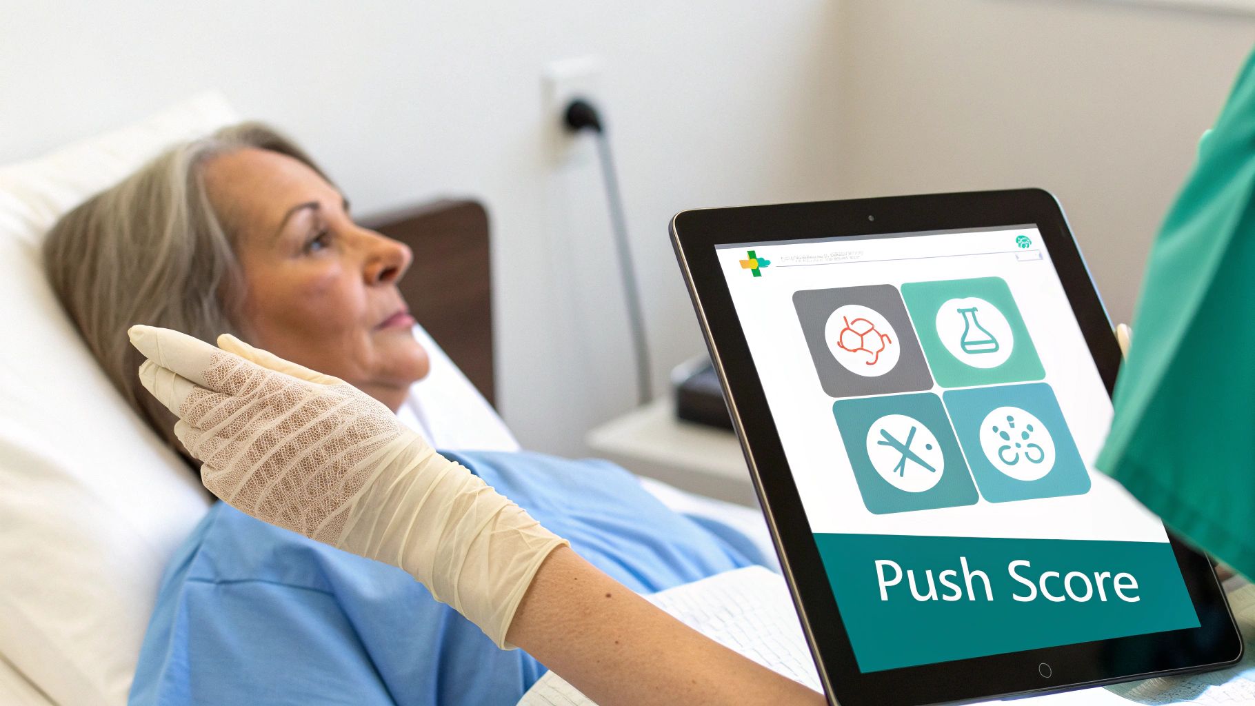
This method shines in settings requiring consistent, interdisciplinary communication and for justifying ongoing treatment to payers. A decreasing PUSH score over several weeks provides powerful, data-driven evidence that the current care plan is effective. Conversely, a stalled or increasing score signals the need for prompt re-evaluation and intervention. It’s a classic example of how structured data transforms subjective observations into actionable clinical intelligence.
Example PUSH Tool Documentation Entry
This example demonstrates how a nurse in a skilled nursing facility (SNF) might document a weekly assessment for a Stage III pressure injury on a resident's sacrum.
Patient: John Doe
Date: 10/26/2023
Wound Location: Sacrum
PUSH Tool Assessment:
- Wound Size (Length x Width): 4.2 cm x 3.5 cm = 14.7 cm²
- Sub-score: 8 (for surface area between 12.1 and 16.0 cm²)
- Exudate Amount: Moderate (Wound tissue is moist; dressing is 25% to 75% saturated)
- Sub-score: 2
- Tissue Type: 75% Granulation Tissue, 25% Slough
- Sub-score: 2 (as Slough is the worst tissue type present)
Total PUSH Score: 12
Narrative Note: "Weekly PUSH assessment performed on sacral pressure injury. Wound bed shows a 10% increase in granulation tissue since last week's assessment (10/19/2023, PUSH Score: 13). Exudate remains moderate, serosanguinous, and non-odorous. Periwound skin intact. Resident tolerates dressing change with minimal discomfort. Will continue current treatment with silver alginate and cover foam dressing, changed daily and PRN. Will reassess PUSH score in 7 days to monitor healing trajectory."
Strategic Analysis & Actionable Takeaways
- Objective Trajectory: The documentation clearly shows a one-point decrease in the PUSH score from 13 to 12. This simple number provides a defensible, objective measure of improvement that supports medical necessity for continued care and satisfies payer requirements.
- Component-Specific Insight: By breaking down the sub-scores, a clinician can pinpoint why the score changed. In this case, the improvement came from better tissue quality, even though the size and exudate levels remained in the same sub-score categories.
- Narrative Synergy: The narrative note complements the PUSH score, adding crucial context that the numbers alone cannot convey (e.g., periwound condition, patient tolerance, and specific dressing types). This combination is a best practice for robust wound documentation examples.
When to Use This Approach
The PUSH Tool is ideal for tracking the healing of any open pressure injury, particularly in long-term care, home health, and hospital inpatient settings where multiple providers interact with the same patient. Its standardized format reduces variability in assessment between different clinicians. It is one of the most effective wound assessment tools for nurses aiming to bring consistency and objectivity to their documentation practices. Implement PUSH scoring at regular intervals (e.g., weekly) to create a clear trend line for every pressure injury you manage.
2. Bates-Jensen Wound Assessment Tool (BWAT) Documentation
The Bates-Jensen Wound Assessment Tool (BWAT), sometimes known as the Pressure Sore Status Tool (PSST), is a comprehensive instrument used to evaluate the status of a wound as it heals. It assesses 13 distinct characteristics, including size, depth, edges, undermining, necrotic tissue type, exudate type and amount, and surrounding skin condition. Each item is rated on a 1-5 scale, generating a total score from 13 (healed) to 65 (most severe). Unlike the more focused PUSH Tool, the BWAT captures a more granular and holistic view of wound complexity, making it exceptionally valuable for multifaceted wounds.

This tool is a powerhouse in settings like hospital multidisciplinary wound teams and home health agencies where complex wounds, such as diabetic foot ulcers or non-healing surgical sites, are common. The BWAT's detailed parameters allow clinicians to document subtle but clinically significant changes, such as the quality of wound edges or the health of the periwound skin, which might otherwise be missed. A declining score over time provides a robust, evidence-based justification for the care plan, while a static or increasing score is a clear indicator that a change in strategy is required.
Example BWAT Documentation Entry
This example illustrates how a home health nurse might document a complex diabetic foot ulcer on a patient's left heel using the BWAT.
Patient: Jane Smith
Date: 10/27/2023
Wound Location: Left heel, plantar aspect
Bates-Jensen Wound Assessment Tool (BWAT) Assessment:
- 1. Size: 3.8 cm x 2.9 cm (Score: 3)
- 2. Depth: 0.8 cm, wound base visible (Score: 3)
- 3. Edges: Indistinct, attached (Score: 2)
- 4. Undermining: 1.5 cm from 12:00 to 3:00 (Score: 3)
- 5. Necrotic Tissue Type: Yellow slough (Score: 4)
- 6. Necrotic Tissue Amount: 25% of wound bed (Score: 3)
- 7. Exudate Type: Serosanguinous (Score: 3)
- 8. Exudate Amount: Moderate (Score: 3)
- 9. Skin Color Surrounding: Redness extending <2 cm (Score: 2)
- 10. Peripheral Tissue Edema: Firm, pitting edema <4 cm (Score: 3)
- 11. Peripheral Tissue Induration: Moderate firmness (Score: 3)
- 12. Granulation Tissue: <25% of wound bed (Score: 4)
- 13. Epithelialization: <25% of wound bed (Score: 4)
Total BWAT Score: 40
Narrative Note: "Initial BWAT assessment completed for left heel diabetic ulcer. Score of 40 reflects wound complexity with moderate slough, undermining, and periwound edema. Offloading boot fit confirmed and patient educated on consistent use. Plan includes enzymatic debridement ointment and calcium alginate dressing to manage exudate and promote autolysis. Will reassess weekly. Patient verbalizes understanding of plan of care."
Strategic Analysis & Actionable Takeaways
- Granular Clinical Detail: The BWAT score of 40 immediately communicates a severe, complex wound. The itemized scores pinpoint specific problem areas like undermining and poor granulation, guiding the clinician to select targeted interventions (e.g., specific dressings for undermining, debridement for slough).
- Holistic Assessment: The tool forces documentation beyond the wound bed itself. By scoring peripheral tissue edema and surrounding skin color, it creates a comprehensive clinical picture that is crucial for managing conditions like venous or diabetic ulcers, where surrounding tissue health is a key prognostic indicator.
- Data-Driven Care Planning: This detailed documentation provides a strong baseline. Future assessments can compare scores for each of the 13 items, allowing the team to see if the enzymatic debrider is reducing necrotic tissue (Item 6) or if compression is improving edema (Item 10). This level of detail makes it a superior choice for complex wound documentation examples.
When to Use This Approach
The BWAT is best suited for the initial and ongoing assessment of complex wounds where multiple factors contribute to non-healing. It is ideal for hospital-based wound care teams, outpatient clinics, and home health agencies managing surgical, diabetic, or vascular ulcers. While it can be used for pressure injuries, it is often paired with the PUSH Tool; the BWAT provides the initial comprehensive assessment, and the simpler PUSH tool is used for routine weekly tracking. Regular training is recommended to ensure inter-rater reliability among clinicians.
3. Wound Measurement and Photography Documentation Protocol
A Wound Measurement and Photography Documentation Protocol combines precise physical measurements with standardized serial photography to create a comprehensive, objective record of a wound's status over time. This method involves documenting length, width, and depth at each visit, often supplemented with tracings or digital planimetry, and capturing high-quality images from consistent angles, distances, and lighting conditions. This dual approach provides both quantitative data and qualitative visual evidence, which is essential for accurate clinical tracking, payer validation, and robust medico-legal protection.
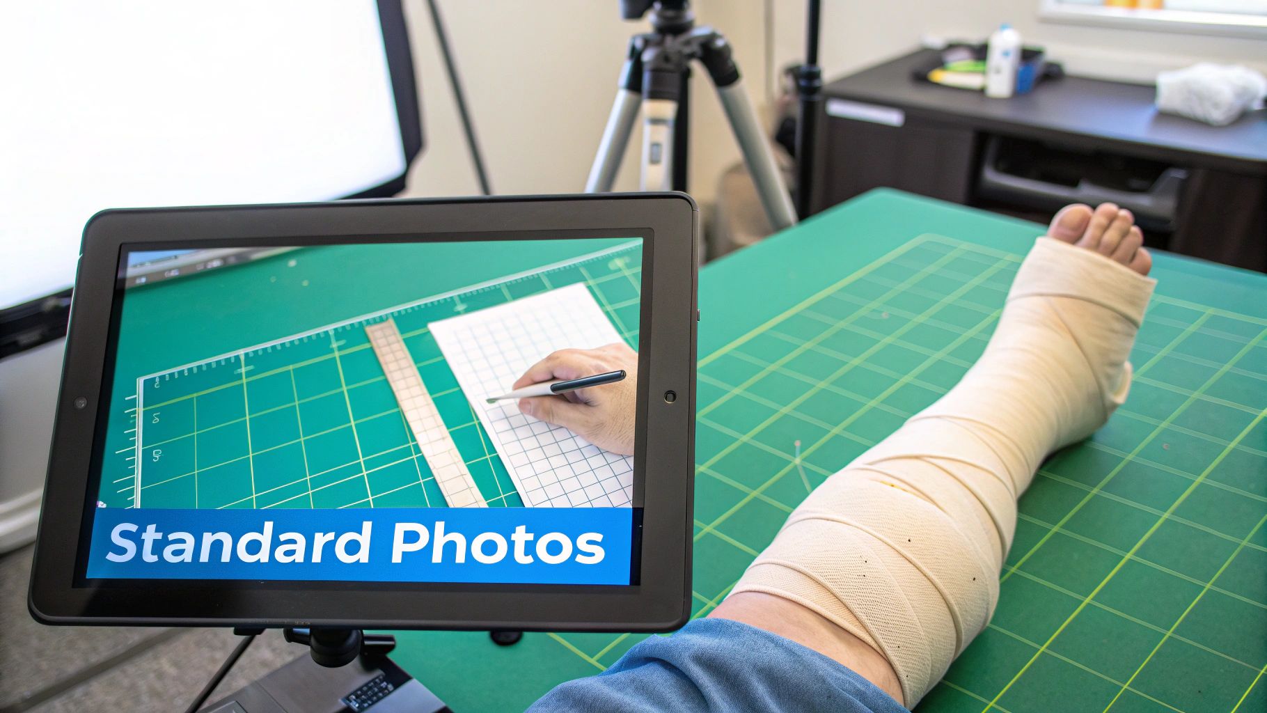
The power of this protocol lies in its ability to create an undeniable visual timeline of healing or decline. While measurements provide the raw data, serial photographs tell a story that numbers alone cannot, highlighting subtle changes in tissue type, periwound condition, and edge characteristics. In an era of telehealth and remote consultations, this visual evidence becomes even more critical. Modern AI-powered platforms can now automate surface area calculations from images, reducing manual errors and providing data-driven insights that support clinical decision-making.
Example Measurement & Photography Documentation Entry
This example shows how a home health nurse might document a follow-up visit for a patient with a diabetic foot ulcer on their great toe, integrating measurements and photo documentation into the electronic health record (EHR).
Patient: Jane Smith
Date: 11/02/2023
Wound Location: Plantar aspect, left great toe
Wound Assessment:
- Measurements: 1.5 cm (L) x 1.2 cm (W) x 0.2 cm (D)
- Surface Area: 1.8 cm²
- Wound Bed: 90% granulation tissue, 10% fibrin slough at wound margins. No exposed bone or tendon.
- Exudate: Scant, serous.
- Periwound: Skin warm, dry, and intact. No erythema or maceration.
Photo Documentation Note: "Standardized digital photograph (Image ID: JS_110223_LGT) taken from 30 cm distance with disposable ruler for scale, per agency protocol. Image uploaded to patient's secure media tab in EHR. Compared to photo from last visit (10/26/2023), there is a visible increase in granulation and reduction of slough at the periphery."
Narrative Note: "Wound dimensions show a 10% reduction in surface area from 2.0 cm² last week. Patient remains compliant with offloading footwear. Continued treatment with hydrogel and dry sterile dressing daily. Will continue weekly measurements and photography to monitor progress. Shared latest image with supervising physician via secure message for review."
Strategic Analysis & Actionable Takeaways
- Evidence-Based Justification: The combination of a 10% reduction in measured surface area and a supporting photograph provides powerful, multi-modal evidence to justify continued skilled nursing care to payers. This objective data is difficult to dispute.
- Enhanced Clinical Collaboration: Securely sharing standardized images facilitates effective remote collaboration between the home health nurse, the supervising physician, and the wound specialist, enabling timely adjustments to the care plan without requiring an in-person visit.
- Medico-Legal Protection: A consistent, dated photographic record serves as a definitive legal document of the wound's condition and the care provided over time, protecting the clinician and agency from potential liability claims.
When to Use This Approach
This protocol is a best practice for virtually all wound types but is especially critical for chronic wounds like diabetic foot ulcers, venous leg ulcers, and arterial ulcers. It is the standard of care in outpatient wound clinics, home health, and telehealth settings. Implementing a strict photography and measurement protocol is one of the most effective wound documentation examples for creating a defensible, data-rich, and visually compelling patient record that supports optimal clinical and financial outcomes.
4. Wound Care Plan and Intervention Documentation with Evidence Mapping
Effective wound documentation goes beyond simply describing the wound; it must articulate a clear, evidence-based plan of care. This method involves explicitly linking specific interventions-such as dressings, debridement, or offloading-directly to the wound assessment findings and measurable clinical goals. This creates a defensible narrative that demonstrates medical necessity, connecting each treatment to an expected outcome, like a 10% reduction in size within two weeks or complete control of exudate.
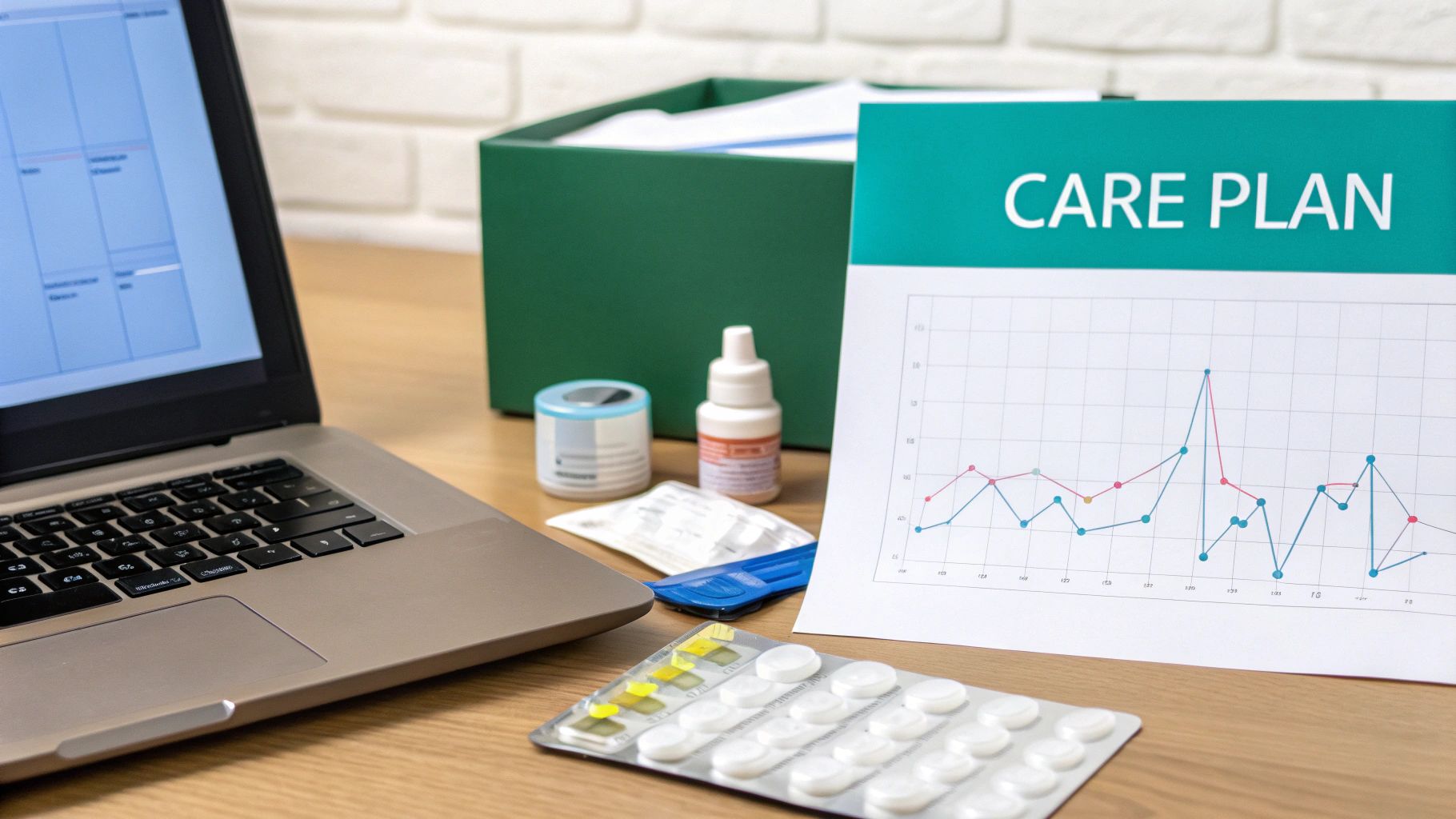
This approach transforms the care plan from a simple list of tasks into a strategic document. By mapping interventions to goals and evidence, clinicians can justify the chosen treatments to payers, auditors, and other team members. Advanced systems, like those powered by AI from providers like Ekagra Health, can even auto-populate evidence-based recommendations and link interventions to CPT codes, significantly reducing documentation gaps and claim denials. This proactive documentation is a cornerstone of modern, compliant wound care.
Example Wound Care Plan Documentation Entry
This example shows a wound care specialist in a diabetic foot clinic documenting a comprehensive plan for a new neuropathic ulcer.
Patient: Jane Smith
Date: 10/26/2023
Wound Location: Plantar aspect of the right great toe
Care Plan & Intervention Mapping:
- Problem #1: Excessive Plantar Pressure:
- Intervention: Application of a total contact cast (TCC) for offloading.
- Goal: Achieve >50% pressure reduction at the ulcer site to promote granulation.
- Metric: Wound size reduction of 15% within 14 days. Re-measure at next TCC change.
- Problem #2: Non-viable Tissue (10% Slough) Impeding Healing:
- Intervention: Sharp debridement performed at bedside today.
- Goal: Achieve a 100% granular wound bed within 7 days.
- Metric: Tissue type assessment at next visit.
- Problem #3: Moderate Serous Exudate:
- Intervention: Apply silver alginate dressing directly to the wound bed before TCC application.
- Goal: Maintain a moist wound environment and manage bioburden without maceration.
- Metric: Periwound skin integrity and exudate level assessment at next TCC change.
- Problem #4: Patient Knowledge Deficit:
- Intervention: Educated patient on signs of TCC complications (pain, swelling, foul odor) and importance of non-weight bearing.
- Goal: Patient will verbalize understanding and demonstrate safe ambulation with crutches.
- Metric: Patient teach-back confirmed.
Narrative Note: "Comprehensive care plan established for new neuropathic ulcer on the plantar R great toe. TCC applied to offload pressure, a primary etiological factor. Silver alginate used to manage moderate exudate and bioburden. Patient and spouse educated on TCC care and safety, with competency confirmed via teach-back. Plan is evidence-based per American Diabetes Association (ADA) guidelines for diabetic foot ulcers. Will follow up in 7 days for TCC change and wound reassessment."
Strategic Analysis & Actionable Takeaways
- Demonstrates Medical Necessity: Each intervention is explicitly tied to a clinical problem and a measurable goal. This "if-then" logic (if pressure is the problem, then TCC is the solution) creates an ironclad justification for the treatment plan that is easy for payers to approve.
- Proactive Goal Setting: The plan sets specific, time-bound goals (e.g., 15% size reduction in 14 days). This establishes a clear benchmark for success and triggers a re-evaluation if progress is not met, aligning with quality care standards.
- Holistic & Compliant: The documentation includes patient education, a critical but often overlooked component. Referencing clinical guidelines (like the ADA's) further strengthens the note by grounding the plan in nationally recognized standards of care, making these some of the most robust wound documentation examples.
When to Use This Approach
This problem-intervention-goal format is the gold standard for all wound care plans, especially for chronic, complex, or high-cost wounds. It is essential in outpatient wound clinics, home health, and any setting where justifying ongoing, specialized care is necessary. Use this structure when initiating a new plan of care or significantly modifying an existing one. It creates a clear roadmap for the entire care team and provides a powerful defense against payment denials.
5. Wound Exudate Assessment and Drainage Tracking Documentation
Wound exudate, or drainage, provides a real-time window into the wound's physiological state. Detailed documentation of its characteristics, including type, amount, color, and odor, is a fundamental clinical skill that directly informs treatment decisions. By consistently tracking these factors, clinicians can assess inflammation, detect early signs of infection, evaluate dressing performance, and justify the medical necessity of specific interventions.
This documentation method moves beyond vague terms like "some drainage." Instead, it uses a standardized vocabulary to describe the fluid, such as serous (clear, watery), sanguineous (bloody), seropurulent (thin, cloudy, yellow/tan), or purulent (thick, opaque, colored). Amount is quantified objectively by observing dressing saturation over a specific timeframe, linking the volume of exudate directly to the need for particular dressing types and change frequencies.
Example Exudate Documentation Entry
This example shows how a nurse in an outpatient venous ulcer clinic documents a significant change in wound drainage, justifying a change in the care plan.
Patient: Jane Smith
Date: 11/02/2023
Wound Location: Medial aspect, right lower leg
Exudate Assessment:
- Amount: Heavy. Primary foam dressing and secondary wrap were 100% saturated with visible strikethrough after 24 hours.
- Type: Seropurulent.
- Color: Cloudy, light tan.
- Odor: Mildly foul, sweetish odor noted upon dressing removal, which was not present at last week's visit.
Narrative Note: "Patient presents for weekly venous ulcer assessment. Reports increased drainage and discomfort over the past 48 hours. Upon dressing removal, heavy, cloudy, tan seropurulent exudate with a new, mildly foul odor was observed. Periwound skin is macerated along the inferior border due to moisture. Wound bed remains 90% granular. Given the change in exudate character and new odor, a wound culture was obtained to rule out infection. Care plan updated to increase dressing change frequency to daily and switch to a super-absorbent polymer dressing to manage high exudate volume and protect periwound skin. Will follow up via phone regarding culture results in 48-72 hours."
Strategic Analysis & Actionable Takeaways
- Justification for Action: The documentation clearly links the change in exudate (new odor, increased volume) to a specific clinical action (wound culture) and a change in treatment (upgrading to a super-absorbent dressing and increasing change frequency). This creates a strong, defensible record.
- Objective Volume Quantification: Stating "dressing was 100% saturated with visible strikethrough after 24 hours" is far more powerful than "heavy drainage." It provides objective evidence to payers justifying the use of a more expensive, high-performance dressing and the need for daily changes.
- Early Infection Indicator: The note correctly identifies the change in odor and fluid character as potential early signs of infection. This proactive documentation is a key part of risk management and demonstrates a high standard of care.
When to Use This Approach
This detailed level of exudate documentation is essential for all wound types but is particularly critical for chronic wounds (e.g., venous ulcers, diabetic foot ulcers), post-operative wounds, and any wound at high risk for infection. It should be a standard component of every single wound assessment. Consistently using standardized descriptors, as promoted by organizations like the Wound, Ostomy, and Continence Nurses Society (WOCN), ensures clear communication across the entire care team and provides the robust data needed to optimize wound care plans effectively.
6. Wound Bioburden and Infection Risk Assessment Documentation
Documenting wound bioburden and infection risk involves a structured assessment of clinical signs and symptoms that suggest microbial proliferation. This goes beyond simple observation by linking specific findings (e.g., erythema, purulent drainage, malodor) to established clinical criteria, such as the Cutting & Harding framework for local infections or IDSA guidelines for systemic issues. This methodical approach justifies clinical interventions like wound cultures, antimicrobial dressings, or systemic antibiotics, creating a clear and defensible record.
This type of documentation is critical for risk management and ensuring appropriate care escalation. By systematically recording signs of infection, clinicians create a data trail that supports medical necessity for higher-cost treatments and protects against liability. It transforms subjective concerns like "the wound looks worse" into objective, evidence-based assessments that drive timely and effective treatment decisions, which is a cornerstone of high-quality wound documentation examples.
Example Infection Risk Assessment Documentation Entry
This example shows a home health nurse documenting a concerning change in a patient's chronic venous leg ulcer, prompting an escalation of care.
Patient: Jane Smith
Date: 11/02/2023
Wound Location: Medial aspect, right lower leg
Infection Risk Assessment (Based on Cutting & Harding Criteria):
- Increased Pain: Yes, patient reports new-onset, persistent pain at the wound site, rated 5/10.
- Erythema: Yes, 2 cm area of erythema and induration noted at the superior wound margin.
- Local Warmth: Yes, periwound skin is warm to the touch compared to the contralateral limb.
- Edema: Moderate pitting edema (2+) noted, an increase from a trace edema last visit.
- Purulent Drainage: Yes, small amount of thick, yellow, malodorous exudate noted on dressing.
- Delayed Healing: Yes, wound size has increased by 0.5 cm² in the past week.
- Systemic Signs: No fever, chills, or altered mental status reported.
Narrative Note: "Patient's venous leg ulcer on the right medial malleolus now presents with three new classic signs of local wound infection: increased pain, localized warmth, and purulent, malodorous drainage. Periwound erythema has developed. These findings, combined with wound deterioration, indicate a high probability of critical colonization or local infection. Dr. Allen was notified via phone of these changes. New orders received and implemented: obtain wound culture via Levine's technique, start Cadexomer Iodine dressing daily, and follow up with the office in 48 hours. Patient educated on signs of spreading infection to report immediately."
Strategic Analysis & Actionable Takeaways
- Evidence-Based Justification: The documentation explicitly references clinical criteria for infection. This elevates the note from a simple description to a clinical assessment that justifies the need for a wound culture and a switch to a costly antimicrobial dressing, satisfying payer scrutiny.
- Clear Escalation Pathway: The note clearly outlines the communication and actions taken: physician notification, new orders received, and patient education. This creates a closed-loop communication record, which is vital for care coordination, especially in a home health setting.
- Differentiating Local vs. Systemic Infection: By noting the absence of systemic signs (fever), the nurse appropriately categorizes the issue as a probable local infection. This distinction is crucial for determining the right course of action, such as whether to start with topical antimicrobials versus systemic antibiotics. To better understand this distinction, it's helpful to know more about the signs that differentiate contamination from infection, such as understanding if pus is always a sign of infection.
When to Use This Approach
This structured infection assessment is essential for all wounds at every visit, but it becomes critically important when a wound is failing to progress, or new signs or symptoms appear. It is the standard of care in diabetic foot clinics, post-operative care, and for managing chronic wounds like pressure injuries or venous ulcers in any setting (hospital, home health, SNF). Implement a standardized checklist based on recognized criteria to ensure no signs are missed and documentation is consistent across all clinicians.
7. Patient Progress Note with Healing Trajectory and Functional Outcome Metrics
Effective wound documentation goes beyond just measuring size and describing tissue. A comprehensive patient progress note integrates objective wound data with patient-centered functional outcomes. This approach creates a holistic narrative that demonstrates not only that the wound is healing, but also that the patient's quality of life is improving. It links clinical metrics like PUSH/BWAT scores and measurements with tangible results, such as reduced pain, increased mobility, and a greater ability to perform Activities of Daily Living (ADLs).
This narrative format is powerful because it tells the full story of the patient's recovery. By documenting the healing trajectory alongside functional gains, clinicians can more effectively justify the medical necessity of ongoing, multidisciplinary care. It also supports patient engagement by clearly showing them how the treatment plan is impacting their daily life, which can significantly improve adherence to complex care regimens like compression therapy or offloading.
Example Progress Note Documentation Entry
This example shows how a visiting nurse might document a follow-up visit for a patient with a venous leg ulcer in a home health setting.
Patient: Jane Smith
Date: 11/15/2023
Wound Location: Medial aspect, left lower leg
Objective Wound Assessment:
- Measurements: 3.5 cm x 2.8 cm x 0.2 cm (Previously 4.1 cm x 3.2 cm x 0.3 cm on 10/25/2023)
- Wound Bed: 85% granulation, 15% slough. No signs of infection.
- Exudate: Scant serous.
- Periwound: Skin is intact, mild erythema, 1+ non-pitting edema noted from ankle to mid-calf.
Functional Outcome Metrics:
- Pain Level: Patient reports pain is now 3/10 with activity, down from 6/10 at the start of care.
- Mobility/ADLs: Patient states, "I can now walk to the mailbox without stopping for a rest." She is able to stand for 15 minutes to prepare meals, an increase from 5 minutes previously.
- Adherence: Compression wrap adherence is estimated at >90% based on patient report and wrap condition at visit.
Progress Note Summary:
"Venous leg ulcer on left lower leg is demonstrating a positive healing trajectory with a 25% reduction in surface area over the past three weeks. The wound bed has improved, with a significant increase in granulation tissue. Functionally, the patient reports a 50% decrease in pain and has regained the ability to perform light household ADLs and ambulate short distances without assistance. Progress is attributed to excellent adherence with the multi-layer compression wrap system. Will continue current plan of care, with dressing changes twice weekly. Estimated time to closure is 5-7 weeks if current healing velocity is maintained."
Strategic Analysis & Actionable Takeaways
- Connects Clinical Data to Patient Life: This note masterfully links the "25% reduction in surface area" to the patient's ability to "walk to the mailbox." This makes the data meaningful and justifies the cost and effort of care to payers and stakeholders.
- Quantifies Functional Improvement: Using specific numbers for pain (3/10 from 6/10) and function (standing for 15 mins vs. 5 mins) provides objective evidence of improvement that complements wound measurements.
- Highlights Patient Adherence: Documenting high adherence to the compression plan directly links the prescribed intervention to the positive outcome. This reinforces the value of the clinician's plan and the patient's effort, which is critical for understanding the overall wound healing process.
When to Use This Approach
This integrated documentation style is essential in any setting where demonstrating the value of care is crucial, particularly in home health, outpatient wound clinics, and long-term care. It is the gold standard for managing chronic wounds like venous ulcers, diabetic foot ulcers, and non-healing surgical wounds. Use this approach to build a strong case for continued care, showcase the effectiveness of your interventions, and keep patients motivated by tracking progress that matters most to them.
7-Point Wound Documentation Comparison
| Documentation Type | 🔄 Implementation Complexity | ⚡ Resource & Time Efficiency | ⭐ Effectiveness / Quality | 📊 Primary Outcomes / Impact | 💡 Ideal Use Cases / Key Tip |
|---|---|---|---|---|---|
| PUSH Tool (Pressure Ulcer Scale for Healing) | Low–Medium — 3-item standardized scale, minimal training | High — ≤5 min per assessment; easily automated | ⭐⭐⭐⭐ — Gold standard for pressure injuries; payer-recognized | Standardized numeric tracking (0–17); supports reimbursement and trend analysis | Best for routine pressure‑injury follow-ups — standardize measurement technique |
| Bates-Jensen Wound Assessment Tool (BWAT) | High — 13-item comprehensive instrument; training required | Moderate–Low — 10–15 min without automation; benefits from AI | ⭐⭐⭐⭐ — Very thorough for complex wounds; strong clinical credibility | Detailed severity scoring (13–65); captures tunneling, undermining, systemic factors | Use for initial/complex wound assessments; pair with PUSH for follow-ups |
| Wound Measurement & Photography Protocol | Medium–High — protocol + photography setup and training | Moderate — time for imaging/storage; efficient with AI auto-measure | ⭐⭐⭐⭐⭐ — Irrefutable visual/metric evidence; medico-legal/payer value | Objective metrics + serial photos; precise size/area trends and audit-ready records | Ideal for telehealth, payer audits, medico-legal needs — standardize distance/scale/consent |
| Wound Care Plan & Intervention with Evidence Mapping | High — links assessment to interventions, goals, and coding | Moderate — template/knowledge intensive; AI improves speed | ⭐⭐⭐⭐ — Strong for justifying medical necessity and billing | Maps interventions to measurable goals and CPT/ICD codes; reduces denials | Use when justifying high-cost therapies; employ structured templates and evidence links |
| Exudate Assessment & Drainage Tracking | Medium — requires standardized descriptors and staff training | Moderate — quick to record but subjective; photos help; AI can flag trends | ⭐⭐⭐⭐ — Important for dressing selection and infection surveillance | Tracks exudate type/volume/timeline; justifies dressing frequency/costs and alerts infection risk | Best for high‑exudate wounds (venous, postop); adopt consistent vocabulary and saturation timing |
| Wound Bioburden & Infection Risk Assessment | Medium–High — clinical criteria + potential culture interpretation | Moderate — may require labs/serial assessments; AI can flag risks | ⭐⭐⭐⭐ — Critical for timely infection management and stewardship | Documents infection signs, cultures, treatment rationale; supports antibiotic justification | Use when infection suspected; document objective metrics (e.g., erythema mm) and culture plans |
| Patient Progress Note with Healing Trajectory & Functional Metrics | High — integrates scores, measurements, QoL, and functional data | Low — time‑intensive data collection; AI can summarize trends | ⭐⭐⭐⭐ — Valuable for holistic outcomes, payer/value arguments, patient engagement | Demonstrates functional improvement and healing velocity; supports transitions and outcomes reporting | Best for multidisciplinary care, value‑based reporting — calculate healing velocity and record functional gains |
From Documentation to Dollars: Automating Your Path to Faster Reimbursement
Throughout this guide, we've dissected a comprehensive array of wound documentation examples, moving far beyond simple templates to uncover the strategic layers that underpin effective clinical and financial outcomes. We've seen how tools like the PUSH and Bates-Jensen Wound Assessment Tool (BWAT) provide a structured language for communicating healing progress, and how meticulous measurement protocols create an undeniable visual and metric-based narrative of patient care.
The core lesson is this: excellent wound documentation is not merely a record-keeping task; it is an active instrument of care coordination, compliance, and revenue generation. Each detailed note on exudate, bioburden, or intervention mapping serves a dual purpose. It guides the next clinical decision while simultaneously building an ironclad justification for the medical necessity of the services rendered. This creates a clear, auditable trail that directly supports the assigned CPT and ICD-10 codes, significantly reducing the risk of claim denials and payment delays.
The Bridge Between Best Practice and Daily Practice
The true challenge lies in consistently applying these best practices under the pressure of a demanding clinical schedule. Remembering every specific element for a debridement note or ensuring every photograph is perfectly calibrated can feel overwhelming. This is where the gap between knowing what to do and actually doing it every single time can widen, leaving practices vulnerable to compliance issues and lost revenue.
The most critical takeaways from the examples provided are:
- Objectivity is Paramount: Replace vague terms like "healing well" with specific data points: "Wound surface area decreased by 15% from 4.2 cm² to 3.57 cm² over 7 days."
- Connect Interventions to Outcomes: Explicitly link the actions you take (e.g., application of a silver alginate dressing) to the observed changes (e.g., "reduced malodor and moderate serosanguinous drainage").
- Consistency Builds the Narrative: Using the same standardized tools (like BWAT) and measurement techniques consistently across visits creates a powerful, data-driven story of the patient's healing trajectory, which is essential for justifying continued care.
Mastering the art of crafting these detailed narratives is the first crucial step. The next is building a system that makes this level of detail achievable and sustainable for every patient, every day. The future of efficient and profitable wound care isn't about working harder; it's about working smarter by leveraging technology that embeds these principles directly into your workflow.
From Meticulous Notes to Automated Revenue
The ultimate goal is to transform the principles seen in these wound documentation examples from a manual, time-intensive effort into a seamless, automated process. The transition from capturing clinical observations to submitting a clean, compliant claim should be as swift and error-free as possible. When documentation is consistently accurate, detailed, and properly coded from the moment of creation, the entire revenue cycle accelerates. Denials decrease, pre-authorization requests are stronger, and cash flow becomes more predictable and robust.
This is where the power of automation becomes a strategic imperative. Imagine a workflow where the ambient conversation during a patient encounter is instantly translated into a perfectly structured, coded, and compliant progress note. This system would automatically calculate PUSH scores, map interventions to evidence-based guidelines, and suggest the appropriate billing codes, all without requiring the clinician to spend hours typing after their shift. By adopting such a solution, you ensure the high standards demonstrated in this article are not just an occasional achievement but the consistent output of your practice, turning documentation from a necessary burden into a streamlined engine for clinical excellence and financial stability.
Ready to eliminate documentation bottlenecks and ensure every note meets the highest standard of compliance and reimbursement? Discover how Ekagra Health AI uses ambient voice technology to instantly generate the kind of detailed, structured, and coded wound documentation examples you've seen here, directly from your patient conversations. Visit Ekagra Health AI to see how you can reduce documentation time by 70% and accelerate your path from voice to claim.
