
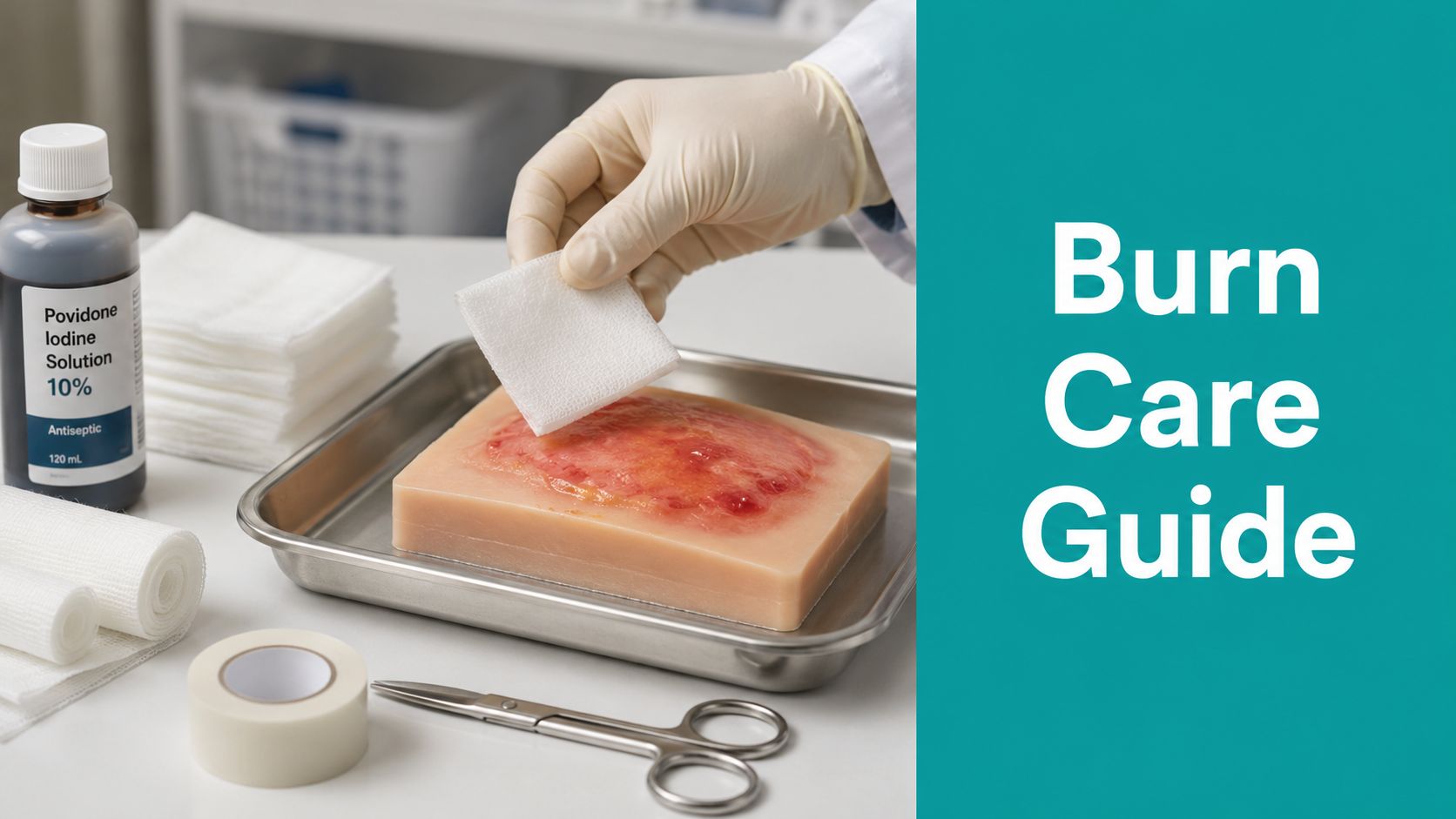
A patient arrives with a fresh scald across the dorsum of the hand. The skin is blistered, wet, painful, and already tightening under a watch band that stayed on too long. In that moment, treatment and reimbursement are tied together. If the first note says only “partial-thickness burn,” the clinical picture stays blurry, the follow-up plan gets weaker, and any later procedure or supply claim becomes harder to defend.
Second-degree burns create problems because they sit between urgent injury care and serial wound management. They can look similar on day one and behave very differently by day three. A wound that heals with simple coverage and close follow-up does not need the same cadence, supplies, or coding support as one that declares itself deeper after the initial visit. Payers notice that mismatch fast. If the exam is vague, medical necessity is easy to challenge.
Good burn care starts with disciplined observation and disciplined charting.
Teach newer staff to assess what the wound is doing, not just what it is called. Pain pattern, moisture, blister quality, refill, edema, and location all matter because they shape both the treatment plan and the expected healing course. That same reasoning applies across the broader wound healing process, where an early classification error changes dressing choice, visit frequency, and the story the chart tells over time.
Anatomy still drives the bedside decisions. If a clinician needs a refresher on skin layers and related structures, I'd point them to explore Natomy's anatomy recommendations, because burn depth assessment is anatomy applied under time pressure.
I have seen otherwise solid care lead to denied claims for one simple reason. The wound improved or worsened over several visits, but the original documentation never established a clear baseline to justify the changes.
Introduction
A patient arrives with a red, blistered, very painful burn from a kitchen spill. At first glance, it looks routine. Three days later, part of it is drying out, another part is still weeping, and the area over the joint is getting stiffer. If the first note only says “second-degree burn, cleansed and dressed,” both the care plan and the claim are already on weak ground.
Second-degree burns create problems because treatment and reimbursement rise or fall on the same question. How much viable dermis is left. The label alone does not tell you enough. Partial-thickness burns can heal quickly with simple coverage and close follow-up, or they can declare themselves deeper and need a different dressing plan, more visits, and sharper documentation to support medical necessity.
I teach newer clinicians to stop treating “second-degree burn” as a complete assessment. It is a starting category, not a plan. What matters in practice is whether the wound behaves more like a superficial partial-thickness injury or a deeper partial-thickness injury that will heal slowly, scar more aggressively, and justify a different level of care. That clinical reasoning should already be visible in the chart on day one.
A practical rule helps. If another clinician cannot read your note and understand why you chose that dressing, that visit interval, and that follow-up plan, a payer will have the same problem.
Good day-one practice is simple to describe and harder to do well. Record the location, size, blister status, wound bed appearance, moisture, pain response, and periwound condition. State what threatens function, especially on the hand, foot, face, perineum, or across a major joint. Then match the plan to what you found. “Burn cleansed and dressed” is not enough if you later bill debridement, advanced dressings, or repeated visits.
This is also where basic anatomy still matters. Depth assessment depends on knowing which skin structures are likely intact, which is why I still tell clinicians to explore Natomy's anatomy recommendations if they need a quick refresher on skin layers under clinical pressure. The same bedside logic applies across the broader stages of the wound healing process, because an early depth error changes treatment intensity, expected healing time, and the story the record tells over time.
The first note sets the baseline. If that baseline is thin, every later change in treatment is harder to defend.
The First 15 Minutes Assessment and Triage
Your first job is to decide whether this patient belongs in your setting at all. Your second job is to make the initial depth assessment as defensible as possible, knowing some burns declare themselves better over the next couple of days.

What to check before you even think about dressing choice
Look beyond color. Depth clues in partial-thickness burns come from bedside findings you can repeat and chart.
- Blanching response: A more superficial wound usually blanches briskly. Deeper partial-thickness areas may have sluggish or absent capillary refill.
- Pain pattern: Very painful burns often still have viable superficial dermal structures. Reduced sensation in a patchy area can signal deeper injury.
- Moisture and blister behavior: Tense, clear blisters over a pink wound often behave differently from flaccid blisters over pale, waxy, or mixed-depth skin.
- Periwound assessment: Document edema, advancing erythema, and whether the border is sharply demarcated or evolving.
- Functional location: Hand, foot, face, perineum, and burns crossing a major joint change the threshold for referral.
I teach newer clinicians to chart what they can see and provoke. Don't write “appears deep partial thickness” without describing why. Write the blanch response. Write whether the wound bed is pink, pale, or mottled. Write whether pain is preserved.
Immediate cooling and first aid
If the patient presents soon after injury, or if staff are fielding a call right after the burn happened, first aid still matters. Active cooling with running tap water between 8°C (46.4°F) and 25°C (77°F) for at least 20 minutes reduces burn depth, shortens healing time, and decreases grafting requirements, provided cooling begins within 30 minutes of injury per the AAFP review.
That's one of the few early interventions with a clear practical payoff. Not ice. Not random ointment first. Cooling.
Hard referral triggers
Some wounds don't belong in office-based management, even if the surface area looks manageable.
- Face involvement: Airway, vision, and cosmetic stakes are too high.
- Hands and feet: Small errors here become long functional problems.
- Perineum or genital region: High contamination risk and difficult aftercare.
- Crossing major joints: Contracture risk starts early.
- Mixed-depth concern or rapidly worsening exam: If the wound is declaring itself as deeper than first assumed, don't sit on it.
If you're hesitating because the wound “might be okay to follow,” but it involves a high-risk functional site, refer early and document why.
A quick note on coding mindset. Don't drag in unrelated chronic wound frameworks just because you use them elsewhere. I still see people loosely borrowing language from pressure injuries or diabetic foot workflows. Wagner grading belongs to diabetic foot ulcer severity, not burns. Burn documentation needs burn depth, anatomic site, laterality when relevant, encounter status, and procedure detail that matches what you performed.
Wound Prep and Debridement Choices
Blisters are where practice patterns diverge fast. Some clinicians leave nearly all of them intact. Others deroof aggressively. Neither extreme is reliable.
The blister question clinicians still argue about
The American Burn Association takes a position that surprises many general wound teams. It recommends sterile needle drainage of intact blisters with antibiotic coverage, but provides no data on infection rates, which leaves clinicians relying on judgment when deciding whether drainage helps or creates more risk as described by the American Burn Association guidance.
That matters clinically and financially. Once a blister becomes an open wound, your documentation burden changes. So can your coding options.
My practical approach is simple.
- Leave small, stable blisters alone if they aren't impairing motion, dressing contact, or visibility of the wound bed.
- Drain or open blisters selectively when size, tension, location, or imminent rupture makes intact management unrealistic.
- Deroof nonviable loose epidermis when it's contaminated, obscuring assessment, or trapping exudate against the wound.
I'm more aggressive on the plantar foot, over joints, and in patients who won't be able to protect the blister between visits. I'm more conservative when the blister roof is still acting like a useful biologic cover and I can offload friction.
A blister plan should answer two things in the note: why you intervened, or why you didn't.
Cleansing versus true debridement
Payers deny claims when the chart says one thing and the code says another. Cleansing, irrigation, and removal of loose surface debris are not automatically billable debridement. If all you did was wash the wound and place a dressing, don't force a debridement code.
When there is devitalized tissue, ruptured blister remnants, slough, or clearly nonviable epidermal tissue that you selectively remove, then you may be in CPT 97597 territory depending on tissue level and technique. If the wound has progressed and tissue-level debridement extends deeper, then the surgical debridement family 11042 to 11047 may become relevant. Your note must support the depth debrided, total area, instrument used, tissue removed, hemostasis, tolerance, and post-debridement measurements when appropriate.
For clinicians using enzymatic support after the wound declares itself more clearly, it helps to understand where collagenase-type strategies fit in the broader debridement plan. This overview on enzymatic debridement considerations for wounds is useful if you're trying to line up tissue condition with documentation language.
What payers usually push back on
A few denial patterns show up again and again:
- Missing tissue description: “Debridement performed” is not enough. Say what tissue was removed.
- No pre-procedure wound status: If there's no slough, necrotic debris, or nonviable tissue documented, the procedure looks unsupported.
- No size or surface area linkage: You need the wound measurements that justify the service billed.
- Confusing burn management with chronic ulcer templates: Burn wounds need burn-specific findings, not copied chronic wound phrasing.
If you're mentoring staff, audit this line in every note: “Why did this wound need debridement today?” If the chart can't answer that cleanly, the claim is vulnerable.
Selecting Dressings and Setting the Cadence
A dressing can protect a burn or sabotage it. The wrong one either dries the wound out, traps too much fluid, or forces more dressing changes than the patient can realistically tolerate.
Match the dressing to depth and exudate
For early shallow deep second-degree burns within 24 to 48 hours, foam or hydrocolloid dressings are preferred. Once wound depth stabilizes over 48 to 72 hours, enzymatic debridement may be appropriate. Silver-impregnated foam dressings are now standard in many clinics because they often require only one or two changes until reepithelialization, which lowers visit burden based on the outpatient burn dressing review.
That's the part many teams miss. Dressing choice isn't just about wound biology. It's also about cadence. If a dressing can hold the moisture balance and control exudate without constant disruption, you reduce pain, reduce handling, and make your schedule easier to manage.
Second-Degree Burn Dressing Selection Guide
| Dressing Category | Primary Use Case | Exudate Management | Typical Change Frequency |
|---|---|---|---|
| Foam | Early partial-thickness burns with moderate drainage | Good absorption while maintaining moisture balance | Often less frequent than simple gauze, based on strike-through and edge integrity |
| Hydrocolloid | Shallow burns with lower to moderate exudate where occlusion is helpful | Retains moisture well, less ideal for heavy drainage | Usually left in place until loosening, leakage, or clinical reassessment |
| Silver-impregnated foam | Outpatient burns where antimicrobial coverage and lower visit burden matter | Handles exudate while supporting a moist environment | Often one or two changes until reepithelialization in appropriate cases |
| Hydrogel | Drier wounds or later autolytic support after necrotic burden is reduced | Adds moisture rather than absorbing much drainage | More frequent reassessment if exudate rises or maceration develops |
What works in clinic and what usually doesn't
A few practical points save time and skin.
- Foam works well early when the wound is wet enough to need absorption but not so dry that you're sticking dressing material to a fragile surface.
- Hydrocolloid can be excellent on the right wound, but if exudate is high or the periwound is delicate, maceration can sneak up fast.
- Hydrogel has a place later when you're shifting toward autolytic support and the wound is no longer draining heavily.
- Simple dry gauze changed too often usually makes everybody miserable. More pain, more disruption, worse consistency.
I want exudate documented every visit in plain language. Serous. Serosanguineous. Purulent if that's what it is. Scant, moderate, or heavy, if that's how your workflow captures it. Dressing choice should make sense when read next to the exudate description.
Dressing selection should follow the wound, not the shelf.
Build a visit schedule the patient can actually keep
If you choose a plan that requires frequent dressing disruption, write down why. Sometimes that's appropriate. An unstable depth assessment, a contaminated wound, or concerning exudate justifies closer monitoring. But if the burn is behaving well, repeated unnecessary changes slow practice flow and often increase discomfort without helping healing.
Good wound care second degree burns management balances three variables at once: moisture, protection, and reassessment interval. When one of those is ignored, the wound usually tells you.
Managing Pain Infection and Complications
A burn can look inflamed and still be on track. It can also look only slightly worse and be heading in the wrong direction. You need a threshold-based approach, not guesswork.

Separate expected inflammation from infection
Expected burn inflammation includes warmth, pain, edema, and erythema near the injury. Infection changes the pattern. I worry when exudate character shifts, odor appears, erythema advances beyond the expected edge, pain escalates rather than settles, or the wound bed dulls and deepens.
Infection is the primary complication that stalls healing in second-degree burns, and the 2024 consensus calls for immediate local wound management, surgical excision, and systemic antimicrobial therapy for severely infected burns. Deep partial-thickness wounds may require four to six weeks to heal and carry a higher complication burden as summarized in the clinical guidance referenced by Shriners Children's.
That means you can't document “watching closely” forever. At some point you either escalate or you're delaying definitive care.
Pain control that doesn't interfere with assessment
Use multimodal pain control, but don't blunt the exam so heavily that you lose useful clinical signals. Systemic analgesics usually do more for functional pain management than repeated topical anesthetic use. Elevation, protection from friction, and fewer dressing disruptions often matter just as much as medication.
Some clinicians also look at comfort-supportive options around treatment sessions. For a patient education resource on topical comfort strategies, you can review Medistik for faster recovery. Keep those discussions grounded in the actual wound plan.
When I culture and when I escalate
I culture when the exam suggests infection, not because the wound is merely painful or draining. Burns drain. That alone isn't a culture indication. But purulence, a sudden exudate shift, or wound bed deterioration deserves action.
- Local decline: Worsening tissue quality, increasing slough, foul odor
- Border changes: Expanding erythema or spreading tenderness
- Systemic concern: Fever or a patient who looks unwell
- Functional risk: Hand and foot burns that are deteriorating need a lower threshold for escalation
Don't confuse delay with prudence. A worsening burn rarely rewards extra days of indecision.
Documentation and Billing for Burn Care
The claim lives or dies on the note. Burn care is one of those areas where treatment can be appropriate and still unpaid because the documentation is soft, copied forward, or too generic.
What has to be in the note
For partial-thickness burns, I want the record to answer five basic questions fast:
- Where is the burn and how extensive is it
- What depth category are you treating
- What does the wound bed and periwound look like today
- What exactly did you do
- Why was that medically necessary
If you bill procedures, add procedural detail that would make sense to an auditor who never saw the patient. That includes measurements, tissue type removed, level of debridement, instrument, tolerance, and dressing applied. If conservative treatment failed and that failure justifies escalation, say so plainly.
Coding details that people still miss
In the US setting, don't get casual with ICD-10-CM burn coding. The site, laterality when relevant, degree, and 7th character for initial, subsequent, or sequela encounter all matter. If the patient returns for ongoing wound management, make sure the encounter character matches the stage of care.
For CPT, keep your logic clean.
- 97597 and 97598: Selective debridement when supported by devitalized tissue and area.
- 11042 to 11047: Surgical debridement family when the depth and tissue level support it.
- Dressing supply support: Your exudate description, wound depth, and frequency rationale should support what was used and why.
The denial triggers are predictable. Missing depth. No total body surface area documentation when relevant. No location precision. No failed conservative care language before escalation. And copied boilerplate that says “stable” while the wound measurements and plan clearly say otherwise.
Documentation habits that protect revenue
I tell teams to stop thinking of documentation as a billing afterthought. It's part of treatment. If you want cleaner claims, your note has to carry medical necessity from exam to code selection without requiring a human reviewer to fill in gaps.
A structured workflow helps. So does using a process that prompts for the details clinicians most often skip. If your team is tightening this part of the workflow, this resource on medical necessity documentation in wound care is worth reviewing.
The best burn note reads like a clinical record first and a claim defense second. But it has to succeed at both.
EkagraHealth AI helps wound care teams document at the point of care without losing the clinical detail that supports coding, prior auths, and clean claims. If your practice is tired of vague notes, missed charge opportunities, and burn visits that take longer to chart than to treat, EkagraHealth AI is built for that reality.
