When we talk about a wound bed description, we’re talking about the shared language clinicians use to describe what’s happening inside a wound. It’s how we turn what we see into a clear, actionable clinical story. These standardized terms—like noting the difference between healthy granulation tissue and stubborn slough—are the foundation for everything that follows. They guide treatment decisions, get the entire care team on the same page, and are crucial for accurate medical coding.
This guide will walk you through the essential components of accurate wound bed descriptions, from identifying tissue types to leveraging a systematic framework for documentation. By mastering this skill, you can improve patient outcomes, enhance communication with colleagues, and strengthen your practice’s financial health.
Why Mastering Wound Bed Descriptions Is Non-Negotiable

Let’s be clear: an accurate wound bed description is much more than clinical jargon. It’s the absolute cornerstone of effective patient care and the financial health of your practice. Think of it less like a static entry in a chart and more like a dynamic narrative that dictates every next step. This story is what helps you build a solid treatment plan, communicate clearly with colleagues, and get the reimbursement you’ve earned.
When you document “70% beefy red granulation tissue,” you’re painting a picture of robust healing. That tells the next clinician to protect that new growth. But if the note reads “50% adherent yellow slough with malodor,” it’s a red flag. That description signals a barrier to healing that needs immediate intervention, like debridement. Every term is a critical data point.
The Clinical and Financial Impact of Precision
The stakes in wound care couldn’t be higher. This is a massive segment of healthcare, with global spending projected to climb from $22.85 billion in 2024 to an incredible $40.85 billion by 2035. Hospitals handle the lion’s share of this, holding about 50% of the market because of the sheer volume of surgical and chronic wounds they see. This scale alone demands precision in everything we do, and it all starts with documentation.
Getting the description right directly impacts several key areas:
- Treatment Efficacy: A clear description ensures you grab the right product off the shelf. Is the wound dry and necrotic? It needs a hydrating dressing. Is it heavily exuding? You need something absorbent. Getting this wrong doesn’t just delay healing—it can actively cause harm.
- Continuity of Care: In any setting with multiple clinicians, a shared, standardized vocabulary is your best defense against misinterpretation. Clear, objective notes are the only way to accurately track progress—or decline—over time.
- Reimbursement and Coding: Payers demand detailed justification. A vague note is an open invitation for a claim denial. Specific wound bed descriptions, on the other hand, build the case for medical necessity for debridement, advanced dressings, and other critical interventions.
A well-crafted wound bed description is both a clinical and a financial tool. It guides the next step in patient care while building the evidence needed for successful reimbursement.
Reducing Ambiguity and Clinician Burden
Inconsistent or subjective notes are a recipe for clinical risk and administrative headache. The burden of documentation is a major driver of clinician burnout. Mastering a standardized approach to wound bed descriptions is a powerful way to fight back. When the whole team speaks the same language, handoffs are smoother and clinical confidence grows. This consistency also lays the groundwork for bringing in new technologies that can make our workflows even better. The goal is to turn observation into objective, actionable data that heals patients and keeps our operations healthy.
If this is a challenge you’re facing, you can learn more about how AI-powered documentation reduces clinician burnout by 50%.
How to Read a Wound Bed Like an Expert
Looking at a wound bed is a lot like reading a landscape. Every color, texture, and feature tells a story about what’s happening just below the surface—revealing whether the environment is on the path to healing or struggling with distress. To make the right clinical calls, you have to speak this visual language fluently. This isn’t about memorizing dry, textbook definitions. It’s about building a practical, visual vocabulary that sticks. Once you can instantly recognize the key players in the wound bed, your assessments will become faster, sharper, and far more impactful.
Granulation Tissue: The Healing Garden
Healthy granulation tissue is the best sign of progress you can see. Think of it as the rich, fertile soil in a healing garden, a clear signal that the body is successfully building a new foundation from the ground up. This tissue is what fills the wound space and creates a scaffold for new skin to grow over.
Its appearance is unmistakable:
- Color: It should be a vibrant, beefy red or a deep pink. That rich color comes from a dense network of new capillaries forming, a process called angiogenesis.
- Texture: Healthy granulation has a bumpy, granular, almost cobblestone-like texture. This indicates robust, new connective tissue is forming exactly as it should.
- Clinical Goal: Protect it at all costs. This new tissue is incredibly fragile and represents hard-won healing. Your job is to maintain a moist, protected environment that supports its continued growth.
Slough: The Unwanted Weeds
In stark contrast, slough is a major roadblock to healing. Picture it as the damp, stringy weeds choking out the healthy plants in your garden. Slough is a messy collection of dead cells, fibrin, and other debris that absolutely must be removed for healing to move forward.
You can spot it by these characteristics:
- Color: Slough typically shows up as yellow, tan, gray, or even white. The color can shift depending on its moisture level and the presence of bacteria.
- Texture: It can be soft, moist, and stringy, or it might be thicker and more like mucus. It’s often loosely stuck to the wound bed but can sometimes be more firmly attached.
- Clinical Goal: Debridement. This non-viable tissue is a breeding ground for bacteria and physically blocks healthy granulation tissue from forming. Getting it out of there is a top priority.
Your ability to differentiate between viable tissue that needs protection and non-viable tissue that needs removal is the most fundamental skill in wound assessment. Getting this right sets the stage for the entire treatment plan.
Eschar: The Hard, Dry Crust
Eschar is another type of non-viable, necrotic tissue, but it looks and feels completely different from slough. Think of eschar as a hard, black, dried-out crust of earth covering a patch of ground where nothing can grow. It’s essentially a tough scab made of dried blood, serum, and dead tissue.
Key identifiers include:
- Color: Eschar is almost always black or dark brown, though it can sometimes look tan.
- Texture: It is hard, dry, and leathery. Unlike soft slough, eschar is firm and can be either tightly or loosely attached to the wound base and edges.
- Clinical Goal: Assess and, in most cases, remove it. While stable eschar on a heel can sometimes act as a natural protective cover, it usually has to be debrided. This allows you to see what’s happening underneath and clears the way for healing. This is especially crucial because the tissue status under the eschar is a complete unknown, a concept that’s vital for accurate assessments. For more on this, check out our pressure injury staging visual guide for accurate assessment.
Epithelial Tissue: The New Growth
Finally, epithelial tissue is the delicate sign that wound closure is underway. Picture it as new, pale pink grass spreading inward from the edges of a freshly seeded lawn. This is the final phase of healing, where new skin cells migrate across the granulation base to close the wound for good.
You’ll recognize it by its fragile appearance:
- Color: It looks translucent, white, or a very pale pink.
- Texture: The tissue is thin, delicate, and might look a bit shiny as it advances across the wound bed.
- Clinical Goal: Protect and moisturize. This new skin is extremely easy to damage. The focus now is on using dressings that shield it from trauma and maintain the perfect moisture balance to help it keep migrating.
Wound Bed Tissue Identification Guide
This table breaks down the key visual cues and clinical meaning for each tissue type you’ll encounter.
| Tissue Type | Color | Texture | Clinical Significance |
|---|---|---|---|
| Granulation | Beefy red, pink | Bumpy, granular, moist | Healthy, healing tissue. Protect it. |
| Slough | Yellow, tan, gray, white | Stringy, soft, moist, or mucinous | Non-viable tissue. Obstructs healing and needs debridement. |
| Eschar | Black, brown, tan | Hard, leathery, dry | Non-viable, necrotic tissue. Usually requires removal to assess wound. |
| Epithelial | Translucent, pale pink, white | Thin, fragile, may appear shiny | Final stage of healing. Protect this delicate new skin. |
A Practical Framework for Wound Documentation

Knowing the difference between granulation and slough is one thing, but translating what you see into a clear, standardized note is what really moves the needle on patient care. Without a systematic approach, documentation often becomes inconsistent, leaving gaps in the clinical story. Adopting a proven framework is the key to making every assessment thorough, objective, and easily understood by the entire team.
One of the most effective and widely adopted methods is the T.I.M.E. framework. Think of it less like a checklist and more like a strategic lens. It organizes your observations into four critical categories, making your wound bed descriptions both comprehensive and genuinely actionable.
T.I.M.E. stands for:
- Tissue Management
- Infection or Inflammation Control
- Moisture Balance
- Edge of Wound Advancement
T is for Tissue Management
This is ground zero—what do you actually see in the wound bed? It’s all about identifying the types of tissue present and, crucially, quantifying them. Vague descriptions like “some slough present” just don’t cut it. We need precision. Your goal here is to estimate the percentage of each tissue type. For instance, a rock-solid note would say: “Wound bed is approximately 60% covered in beefy red granulation tissue and 40% adherent yellow slough.” This gives the next clinician a precise, measurable baseline.
I is for Infection or Inflammation
Next up, you’re on the lookout for signs of infection or excessive inflammation. This requires a systematic look at both the wound bed and the surrounding (periwound) skin. Key indicators to document include erythema (redness), increased warmth, edema (swelling), increased pain, and changes in exudate color, consistency, or amount.
A great wound bed description never stops at the wound’s edge. Documenting periwound erythema, maceration, or induration provides vital context about moisture management and potential infection.
M is for Moisture Balance
Wound healing lives in a “Goldilocks” zone of moisture—not too wet, not too dry. This part of the assessment is all about the wound exudate, or drainage. Quantify the amount using standard terms: none, scant, minimal, moderate, or copious. Then, describe the quality of the exudate. For example, purulent drainage (thick, maybe yellow or green) screams infection, while serous fluid (clear and watery) is often a normal part of the healing process.
E is for Edge of Wound
Finally, bring your focus to the wound edges, or margins. The state of the edges tells a story about whether the wound is actively closing or if progress has hit a wall. Look for key characteristics like whether the edges are attached or unattached, the presence of epibole (rolled edges), and their color and texture (e.g., macerated, hyperkeratotic). By working through the T.I.M.E. framework every single time, your wound descriptions become powerful clinical tools.
Documenting Complex Wound Characteristics
Simply identifying tissue types gets you part of the way there, but an expert assessment goes deeper. It means looking beyond the wound bed to capture its dimensions, the health of the surrounding skin, and other vital sensory information.
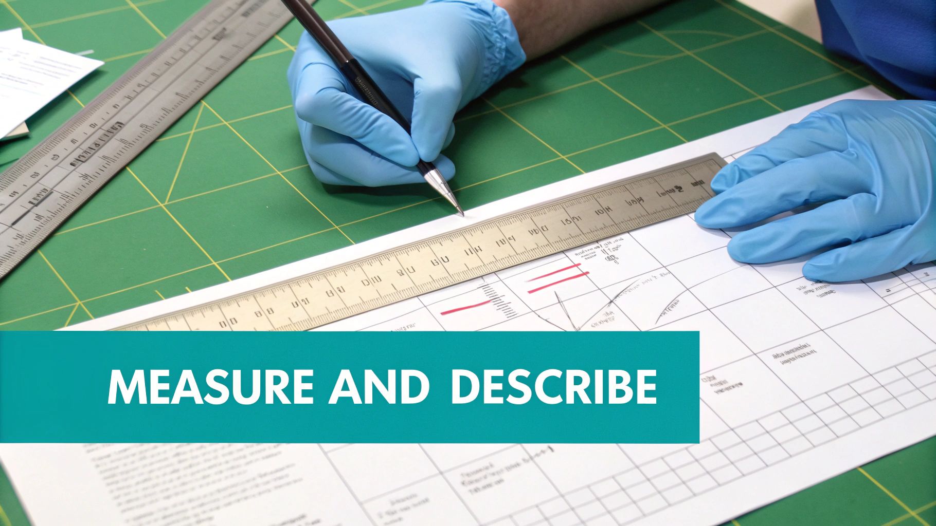
Measuring and Describing Wound Dimensions
Precise, consistent measurements are the only objective way to know if a wound is closing. Always measure in centimeters:
- Length: Measure the longest “head-to-toe” distance.
- Width: Find the widest point “side-to-side.”
- Depth: Gently insert a sterile, cotton-tipped applicator into the deepest part of the wound bed and measure.
Also investigate for undermining (tissue loss parallel to the skin) and tunneling (a narrow channel extending into deeper tissue). Use the clock face method to describe them, noting location and depth (e.g., “3 cm of undermining from 2 o’clock to 5 o’clock”).
Assessing the Periwound Skin
The skin around the wound—the periwound—offers critical clues. Be on the lookout for maceration (white, soggy skin from too much moisture), erythema (redness that may indicate infection), and dryness or scaling. Careful documentation of the periwound helps you protect it, which is vital for healing.
Identifying Biofilm and Other Subtle Signs
Not all healing roadblocks are as obvious as slough or eschar. Biofilm, a slimy community of bacteria, can stall healing and often appears as a subtle, shiny gel on the wound surface. Suspecting and documenting it is the first step toward effective treatment. Don’t forget other clues like odor (a new or foul smell can signal infection) and pain.
The need for this level of detail is only growing. The U.S. wound care centers market is projected to reach $64.49 billion by 2034, underscoring just how critical high-quality, standardized assessment has become. You can learn more about the advanced wound care market and its trends.
Connecting Documentation to Technology and Reimbursement
Your meticulous clinical work has to translate into the language of finance and technology. Precise wound bed descriptions are the bridge between your clinical diligence and the realities of modern healthcare. They justify the care you provide, support the codes you bill, and ultimately, ensure you get paid for your expertise.
From Clinical Notes to Clean Claims
The link between what you write and what you’re paid is direct. Specific terms are the keywords that billing systems and auditors are programmed to find.
Consider these two notes for the same wound:
- Vague: “Patient has a wound on their foot. Dressing changed.”
- Specific: “Diabetic foot ulcer on the plantar aspect of the right great toe, measuring 2.4 x 1.8 x 0.5 cm. Wound bed contains 70% firmly adherent yellow slough and 30% pale granulation. Moderate amount of seropurulent drainage noted, with a mild malodor.”
The second note gives the payer everything they need. It provides clear evidence to code for a higher complexity visit and justifies any debridement you performed. For a deeper dive into making sure your coding is as sharp as your assessments, our ICD-10 and CPT Coding Guide for Wound Care Billing in 2025 is an essential resource.
Your documentation is not just a record of care; it is the primary justification for reimbursement. Every specific detail you include strengthens your claim.
The Rise of Intelligent Automation in Wound Care
For too long, the burden has been on clinicians to manually translate what they see into the rigid data fields of an EMR. AI-powered platforms can now analyze a wound image and handle many of these documentation tasks automatically, closing the gap between the clinical reality and the digital record.
Tools like Ekagra Health AI are completely changing this workflow. You simply take a picture of the wound, and the platform’s AI gets to work:
- Automate Measurements: It instantly calculates length, width, and surface area.
- Analyze Tissue Types: It automatically identifies and quantifies the percentages of granulation, slough, and eschar.
- Structure Data for EMRs: It organizes this data into a structured note, ready for the patient’s chart.
This introduces a new level of standardization and objectivity to wound assessment, taking the subjective guesswork out of documentation. The result is a more efficient day, fewer errors, and a rock-solid, defensible record of care that stands up to payer scrutiny.
Common Questions About Wound Bed Descriptions
Even experienced clinicians run into tricky situations. This section tackles some of the most frequently asked questions to help you sharpen your assessments and document with more confidence.
What Is the Difference Between Slough and Granulation Tissue?
This is the most critical distinction in wound care. Granulation tissue is the healthy, beefy red, bumpy tissue that signals healing. Your job is to protect it. Slough is the yellow or tan, stringy, non-viable debris that must be removed. It is a barrier to healing and must be debrided.
How Do I Accurately Estimate Tissue Percentages in a Wound?
Consistency is more important than perfection. Use the “clock face” analogy: mentally divide the wound into four quadrants and estimate the primary tissue type in each. If the top-right quadrant is mostly slough, chart 25% slough and move on. Using the same method every time allows for accurate progress tracking. This is where AI-powered imaging tools are changing the game by delivering objective percentages automatically.
Why Is Documenting the Periwound Skin So Important?
The periwound skin provides massive clues about your treatment plan’s effectiveness. Maceration (soggy, white skin) indicates too much moisture, meaning your dressing isn’t keeping up. Erythema and induration (redness and firmness) can signal a spreading infection that requires immediate action. Healthy periwound skin is a clear sign that your moisture management is working.
How Can a Clinic Standardize Wound Bed Descriptions?
Getting the entire team on the same page is crucial for continuity of care.
- Adopt a Framework: Make a universal model like T.I.M.E. the standard for every wound.
- Create Shared Templates: Use standardized EMR smart phrases that prompt for the same data points.
- Conduct Peer Reviews: Huddle up and review charts as a group to align on terminology and assessment techniques.
The most powerful way to enforce a standard is to remove the guesswork. Technology that uses objective algorithms for measurements and tissue analysis ensures every assessment is documented the same way, every time.
At Ekagra Health AI, we built our platform to solve these exact problems. Our AI-driven tools eliminate documentation inconsistencies by automatically analyzing wound images to give you precise measurements and tissue percentages, all perfectly structured for your EMR. See how you can standardize care and speed up reimbursement by visiting https://ekagrahealth.ai.
