A wound bed description is the specific language we use in the clinic to paint a picture of a wound's physical state. It’s how we document everything from the types of tissue we see and the amount of drainage to the health of the skin around it. Think of it as creating a detailed map of the healing process. A clear, consistent description is the bedrock of good treatment, solid team communication, and better outcomes for our patients.
Why Mastering Wound Bed Description Matters
Getting the wound bed description right is so much more than a box-ticking exercise in charting. It’s how you tell the wound's complete story. This detailed account is what guides treatment decisions from one clinician to the next, making sure every single person on the care team is on the same page about the wound's status and where it's heading. Without this shared language, care fragments, treatments become inconsistent, and healing stalls.
This skill is what turns a quick look into objective, actionable data. It provides the clinical justification we need for advanced therapies, helps us spot the early warning signs of complications, and creates a solid, defensible record of the care we've provided.
The Foundation of Effective Healing
A precise wound bed description gives you a clinical baseline. It's the starting point that lets you measure progress or spot setbacks with real confidence. A few key elements make up the core of this essential narrative:
- Tissue Types: Being able to identify and quantify the different tissues—like healthy, beefy red granulation versus non-viable slough—is what drives our debridement strategy and treatment goals.
- Exudate Levels: The amount, color, and consistency of wound drainage give us critical clues about the wound's moisture balance and whether an infection might be brewing.
- Wound Edges: Looking closely at the wound's perimeter for things like rolled edges (epibole) can tell you exactly why it isn't closing.
- Periwound Skin: The condition of the skin surrounding the wound offers a ton of insight into how well moisture is being managed and the overall health of the tissue.
When you take a systematic approach to describing the wound bed, the practice goes from being a simple chore to a powerful diagnostic tool. It ensures nothing gets missed and gives you the evidence you need to adapt the care plan when something isn't working.
The Scale of the Challenge
The need for this skill is massive when you consider just how many patients are affected. In the United States alone, chronic wounds impact roughly 6.5 million people. Diabetic foot ulcers make up 1.5 million of those cases every single year.
A precise wound bed description, often guided by a framework like TIME, helps clinicians put a number on the barriers to healing. For example, a wound bed with less than 50% granulation tissue is a huge red flag for stalled healing; we know it correlates with failure rates as high as 40% if we don't intervene. You can learn more about the scale of the wound care market and its continued growth.
By mastering this foundational skill, we can directly improve patient outcomes, make our team communication seamless, and ensure we’re delivering the kind of high-quality, evidence-based care that's essential in a field where precision truly matters.
How to Identify the Four Core Tissue Types
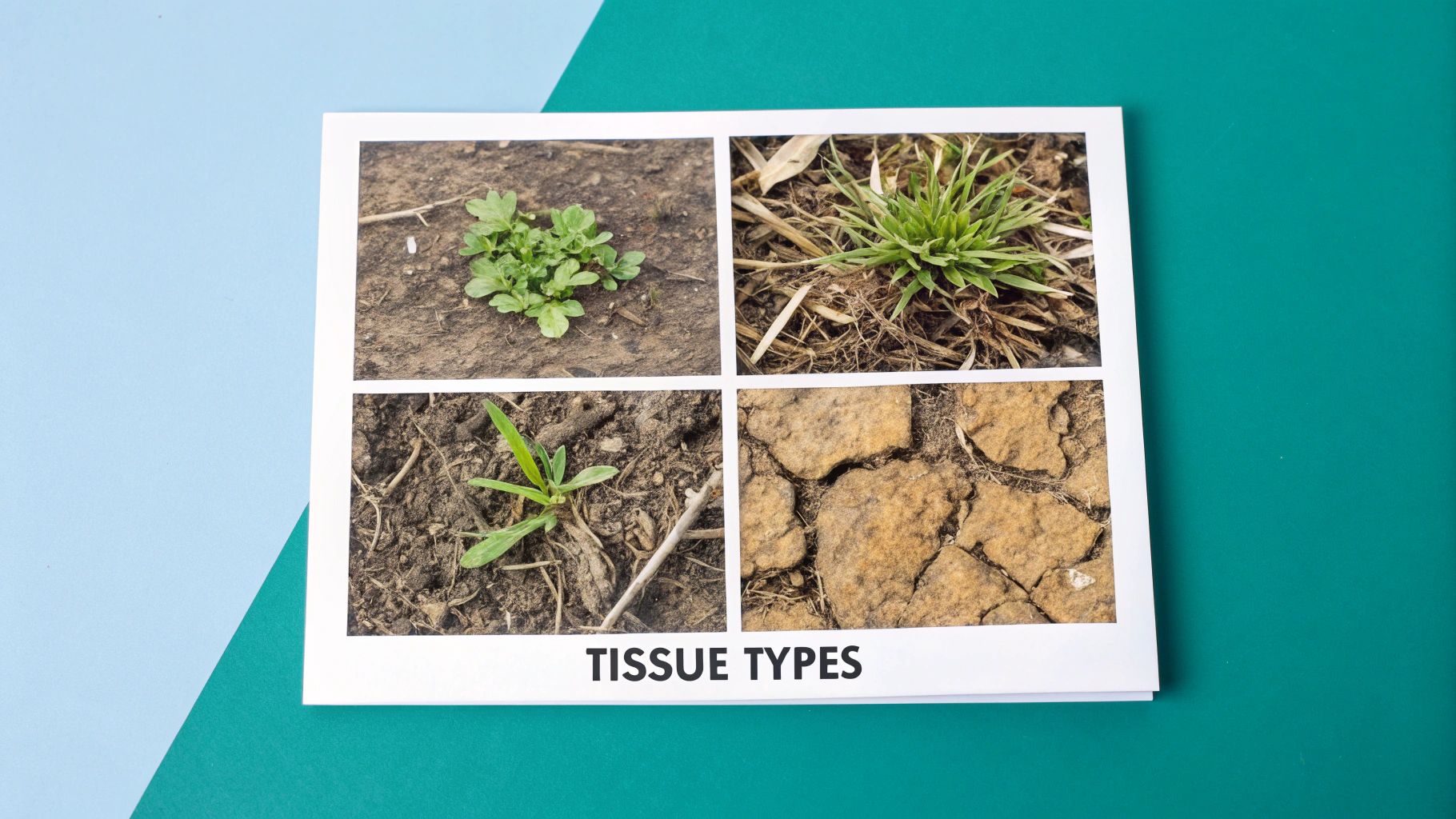
The first step in any good wound bed description is knowing exactly what you're looking at. Think of it like a gardener assessing a patch of soil—what's growing, what isn't, and what needs to be cleared away tells you everything you need to know to bring it back to life.
Learning to spot the four primary tissue types is a core clinical skill. Each one has a unique look and feel that directly guides your next move, whether that's selecting a dressing or deciding it's time to debride. This is the visual language of wound care.
Granulation Tissue: The Healthy New Soil
Seeing granulation tissue is always a good sign. It's the proof that a wound is actively rebuilding from the ground up. In our garden analogy, this is the rich, healthy topsoil that’s full of life and ready to support new growth.
Healthy granulation tissue is typically a vibrant, beefy red or dark pink. Its surface isn't smooth but rather bumpy and granular, almost like cobblestones. This texture comes from the network of new capillaries forming just beneath the surface. It should feel moist and firm, and it won't bleed at the slightest touch. When you see granulation filling the wound bed, you know healing is on the right track.
Slough: The Unwanted Weeds
If granulation is the goal, slough is the obstacle. This non-viable tissue is a major barrier to healing, like stubborn weeds choking out the healthy plants in a garden. To get the wound healing again, the slough has to go.
Slough usually appears as yellow, tan, or grayish gunk. Its texture can vary—sometimes it's stringy, other times it forms a soft, mushy sheet. While it's often loosely attached, it can be surprisingly adherent. Its presence is problematic because it can become a breeding ground for bacteria and physically blocks new skin cells from covering the wound.
A key part of your wound documentation is estimating the percentage of slough. This number is crucial for justifying the need for debridement and for tracking how well your treatments are working to clear the wound bed.
Eschar: The Hard, Dry Crust
Where slough is like wet weeds, eschar is like a thick, dry crust of earth that’s so hard nothing can grow through it. Eschar is a form of dead, necrotic tissue that completely stalls the healing process.
You'll recognize eschar by its dark black or brown color and its thick, leathery texture. It can be hard or sometimes soft, but it's always firmly stuck to the wound bed. Because eschar covers the wound entirely, it’s impossible to know what’s going on underneath—you can't assess the wound’s true depth or condition until that barrier is gone.
Epithelial Tissue: The Delicate New Growth
Finally, we have epithelial tissue, which signals the very last phase of wound closure. Back in our garden, this is the fragile, new growth—the tiny sprouts and blades of grass that show the soil has been fully restored.
This tissue looks like a thin, translucent, or pale pink layer advancing from the wound edges inward over the healthy granulation bed. It often has a smooth, slightly shiny appearance. Seeing this delicate new skin is a clear sign the wound is successfully contracting and resurfacing. The main job at this stage is to protect this fragile new tissue at all costs.
To help you quickly differentiate these tissues during an assessment, here’s a quick-reference guide.
Wound Bed Tissue Identification Guide
This table breaks down the key visual and clinical characteristics of the four primary wound bed tissue types. Use it as a handy cheat sheet to build confidence in your assessments.
| Tissue Type | Visual Appearance | Texture | Clinical Significance |
|---|---|---|---|
| Granulation | Beefy red or dark pink, bumpy | Moist, firm, granular | Indicates active, healthy healing and new blood vessel growth. |
| Slough | Yellow, tan, gray, or white | Stringy, soft, sheet-like | Non-viable tissue that hinders healing and requires debridement. |
| Eschar | Black or brown | Dry, thick, leathery, hard | Necrotic tissue that acts as a barrier and must be removed. |
| Epithelial | Translucent, pale pink, shiny | Smooth, thin, fragile | Signifies the final stage of wound closure is underway. |
Mastering the identification of these four tissues is fundamental. It transforms your wound bed description from a simple observation into a powerful clinical tool that directly informs and justifies your treatment plan.
Using the TIME Framework for a Systematic Assessment

To really understand what a wound is doing, you need to move beyond a quick glance. A simple observation can easily miss crucial details, but a structured framework ensures every key element is assessed, documented, and tracked over time. The most widely adopted and trusted method for this is the TIME framework.
Think of TIME as your clinical checklist. It’s an acronym that stands for Tissue, Infection/Inflammation, Moisture, and Edge, guiding you through the four pillars of a solid wound assessment. When you use this framework consistently, you take the guesswork out of the equation. It helps standardize communication across the entire care team and builds a clear, logical story of the wound's healing journey.
This systematic process transforms your notes from a simple collection of observations into a powerful tool for planning treatments and actually seeing if they're working.
T is for Tissue
The first thing you do in the TIME framework is get a good look at the tissue inside the wound bed. This means identifying the types of tissue present—like granulation or slough—and, just as importantly, estimating the amount of each. A vague description just won't cut it; you need to quantify them as percentages.
For example, a strong wound bed description would sound something like this: "Wound bed is 80% healthy granulation tissue with 20% adherent yellow slough at the superior margin." This simple act of quantification gives you a clear baseline, making it incredibly easy to see if your debridement efforts are paying off at the next visit.
I is for Infection and Inflammation
Next up, you’re on the lookout for signs of infection or excessive inflammation. A little bit of inflammation is a normal, even necessary, part of the healing process. The trouble starts when it sticks around too long or seems out of proportion, as that can bring healing to a dead stop. You have to look beyond the obvious things, like pus.
Often, the first warning signs are much more subtle. Keep an eye out for these clues:
- Friable Granulation: Does the tissue look healthy but bleed at the slightest touch?
- Increased Pain: Is the patient suddenly reporting more pain at the wound site?
- Stalled Healing: Has a wound that was making good progress suddenly hit a plateau?
- Foul Odor: Is there a new or worsening smell, even after the wound has been properly cleaned?
Documenting these subtle but significant findings helps justify why you might need to switch to an antimicrobial dressing or order further diagnostic tests.
M is for Moisture Balance
Wounds are a bit like Goldilocks—they need an environment that's just right. Not too wet, not too dry. The "M" in TIME prompts you to assess the wound's moisture level by carefully evaluating its exudate, or drainage. A complete assessment here means describing both its amount and its type.
The goal is to achieve moisture balance. Documenting exudate levels helps you choose the right dressing—one that can absorb excess moisture without drying out the wound bed, or one that can donate moisture to a dry wound.
Here’s a quick breakdown of how to describe it:
- Amount: Is the drainage scant (the dressing is barely moist), moderate (the dressing is wet but not soaked), or copious (the dressing is saturated and might be leaking)?
- Type: What does the drainage look like? Common types include serous (clear and watery), sanguineous (bloody), serosanguineous (a pinkish mix of the two), or purulent (thick, opaque, and often a tell-tale sign of infection).
E is for Edge of the Wound
The final piece of the TIME puzzle is a close examination of the wound's edge and the surrounding (periwound) skin. The edge is where all the action happens for the final stage of healing, called epithelialization. If the edges aren't healthy and advancing, that wound simply isn't going to close.
During your assessment, you’re looking for critical signs that can halt healing in its tracks. Do you see epibole, where the edges have rolled under on themselves, creating a lip that prevents new skin from migrating across? Are there hidden channels of tissue loss, known as tunneling or undermining, that extend from the wound underneath the intact skin? Describing these features is non-negotiable. To get a deeper look at this, check out our guide on how to provide a complete wound edge description.
By methodically working your way through Tissue, Infection/Inflammation, Moisture, and Edge, your wound bed description becomes truly comprehensive and, most importantly, actionable. This structured approach ensures no detail is overlooked, leading to more precise treatment plans and better outcomes for your patients.
Turning Your Assessment into Bulletproof Documentation
A sharp clinical assessment is one thing, but its real power is unlocked when you translate it into clear, concise, and defensible documentation. This is the moment your careful observations become a permanent part of the patient's story. Great charting weaves all those data points—measurements, tissue types, exudate, and the state of the surrounding skin—into a narrative that justifies your treatment plan and proves medical necessity.
Think of your note as a clinical snapshot in time. It needs to be so clear that any other provider, whether on the next shift or reviewing the chart weeks later, can instantly understand the wound's exact status. This is the bedrock of good continuity of care.
What Great Wound Documentation Looks Like
The best wound bed description tells a complete story in just a few sentences. It’s all about being objective and quantifiable, leaving zero room for interpretation. Let's look at a couple of real-world examples to see how this plays out.
Example 1: Pressure Injury
Pressure injury on sacrum, 3.1 x 2.8 x 0.5 cm. Wound bed is 75% granulation with 25% yellow slough along the superior edge. Scant serous exudate. Periwound skin is intact without erythema. Edges are attached, no undermining noted.
- Why it works: This is a perfect example of precision. It gives exact measurements, quantifies the tissue types, describes the amount and type of drainage, and confirms the status of the surrounding skin and edges. Each detail is a piece of evidence.
Example 2: Venous Leg Ulcer
Venous leg ulcer on the medial malleolus, 4.5 x 3.2 cm. Depth cannot be assessed due to 100% adherent slough. Moderate serosanguineous exudate present. Periwound skin shows 2+ pitting edema and hemosiderin staining. Edges are indistinct and macerated.
- Why it works: This description immediately tells you why debridement is necessary (100% slough) and why you'd reach for a highly absorbent dressing (moderate exudate). The periwound details paint a clear picture of underlying venous insufficiency, connecting the wound directly to its cause.
A Simple Framework for Your Notes
Building consistent, high-quality notes is much easier when you have a go-to structure. Using a template ensures you hit every critical point, every single time. It not only elevates your charting but also saves you precious minutes on a busy day. If you're looking for a solid starting point, check out our comprehensive wound care documentation template.
Here’s a simple, effective structure you can use for any wound:
- Location and Measurements: Start with the basics. Where is the wound, and what are its dimensions (Length x Width x Depth)? Be sure to note any tunneling or undermining here.
- Wound Bed Composition: This is the heart of your wound bed description. Break down the tissue types by percentage (e.g., 60% granulation, 40% slough).
- Exudate Assessment: Describe both the amount (scant, small, moderate, large/copious) and the type (serous, sanguineous, purulent).
- Periwound and Edge Condition: What does the surrounding skin look like (intact, macerated, erythematous)? And the edges (attached, rolled, epibole)?
- Pain and Odor: Don't forget to note any patient-reported pain or the presence of an odor, as these can be critical signs of infection or inflammation.
Sticking to a structure like this transforms your documentation from a chore into a powerful clinical tool. It gives you a clear baseline to track progress, provides solid justification for your treatment choices, and creates a legally sound record of the excellent care you’re providing.
The Financial Case for Precise Wound Documentation
Beyond the exam room, your wound bed description is one of the most powerful financial tools your clinic has. Think of it this way: your chart note is a legal and financial document that must prove why a specific treatment was necessary. Vague descriptions leave money on the table and open the door to claim denials and audits.
On the other hand, a clear, detailed note builds a rock-solid case that justifies your care. When you translate what you see at the bedside into the language of reimbursement, you connect the dots for payers. This isn't just about getting paid; it’s about securing the financial health of your practice so you can keep providing excellent care.
Connecting Clinical Observations to Reimbursement
Every word you write in a wound care note has a potential dollar value attached to it. Certain phrases are absolutely essential for getting prior authorizations approved and justifying the use of advanced therapies. Simply writing that a wound is "not healing" just won't cut it.
You have to provide the evidence.
- Justifying Debridement: Phrases like "70% adherent black eschar" or "40% yellow slough" create an undeniable medical necessity for debridement procedures (CPT codes 97597, 97598).
- Supporting Advanced Therapies: Documenting "epibole at wound edges" or "persistent high bioburden despite standard care" builds the case for using advanced modalities like cellular and tissue-based products (CTPs).
- Demonstrating Need for Antimicrobials: Noting "increased seropurulent drainage with a foul odor" validates the use of antimicrobial dressings and justifies more frequent visits.
By linking this specific clinical language directly to CPT and ICD-10 codes, you create an airtight argument for reimbursement. It's a meticulous process, but it leads to faster healing, less wasted supplies, and a much healthier bottom line for your organization.
The Economic Impact of Accurate Data
The financial pressure for detailed wound documentation is immense. The global wound care market is projected to hit $38.39 billion by 2034, and in the U.S. alone, chronic wounds cost the healthcare system over $25 billion every year. For your revenue cycle team, coding the wound bed status—for instance, "partial thickness with 60% slough"—is what unlocks reimbursement for the products you prescribe.
Modern platforms with ONC certification can help automate this, connecting the dots from patient intake to the final claim. This standardization can slash documentation time by up to 70% and dramatically reduce denials. You can read more about the growing wound care market size on BioSpace.com.
In wound care, if it wasn't documented, it wasn't done—and it won't be paid for. A detailed wound bed description is your best defense against claim denials and the foundation of a healthy revenue cycle.
At the end of the day, speaking the financial language of wound care is just as critical as the clinical assessment itself. It ensures the incredible work you do is recognized, justified, and properly compensated, securing the resources you need to keep healing patients.
How AI Is Modernizing Wound Assessment

The future of wound care documentation is here, and it's moving past manual measurements and subjective estimates. Artificial intelligence is stepping in to take much of the guesswork and administrative burden off clinicians, making the entire process faster, more accurate, and far more consistent.
This isn’t about replacing a clinician's expert judgment. Think of it as giving that expertise a serious upgrade. AI-powered tools handle the tedious, repetitive tasks, which frees you up to focus on what matters: patient care and treatment strategy. The result is a more objective and data-rich wound bed description with a fraction of the effort.
Superhuman Accuracy with AI Image Analysis
One of the most immediate ways AI is changing the game is through wound imaging. All it takes is a simple photo from a smartphone or tablet for an AI platform to instantly perform tasks that are normally time-consuming and ripe for human error. The level of precision is something you just can't achieve by hand.
These systems can automatically:
- Calculate Dimensions: AI algorithms measure length, width, and surface area with incredible accuracy, eliminating the inconsistencies that happen when different clinicians use a ruler.
- Quantify Tissue Types: The software analyzes the colors and textures in the wound bed to calculate the precise percentage of granulation, slough, and eschar, giving you objective data for your note.
- Track Subtle Changes: By comparing images over time, AI can spot tiny changes in wound size or tissue composition that might be missed by the human eye. This offers early clues about whether the wound is on the right track.
This kind of consistent, objective data is a game-changer. It nearly eliminates inter-observer variability—the classic problem where two clinicians describe the same wound differently—and creates a reliable, evidence-based record of healing.
From Spoken Words to Structured Notes
Beyond just images, AI is also reshaping the documentation workflow itself. With ambient clinical voice technology, you can simply describe the wound out loud during a patient visit. The AI listens, understands the clinical context, and turns your words into a perfectly structured and coded chart note in real time.
For example, you could say, "The sacral pressure injury measures about three by two centimeters, with roughly seventy percent granulation and thirty percent slough at the top. There's scant serous drainage, and the periwound skin is intact."
The AI takes that and instantly populates the EHR with a complete, well-formed entry, often mapping your findings to the correct billing codes. This one capability alone can slash documentation time by up to 70%—a huge relief for any busy clinician. There are a variety of modern wound assessment tools for nurses that now include these features.
At the end of the day, AI is becoming an essential partner in wound care. It boosts the accuracy of the wound bed description, simplifies workflows, and gives clinicians better data to make better decisions. This means less time staring at a screen and more time doing what you do best—caring for patients.
Common Questions About Wound Bed Description
Even with a good handle on the basics, a few practical questions always pop up in the clinic. Getting these details right is what separates a good wound assessment from a great one, giving you the confidence that your documentation is spot-on.
Let's walk through some of the most common questions I hear from fellow clinicians.
How Often Should I Document a Wound Bed Description?
The short answer? Every single time you assess the wound. That means a full wound bed description should be part of every dressing change or clinical visit where the wound is evaluated. This consistency is crucial—it creates the clear, chronological story of the wound’s progress (or lack thereof) and gives you the evidence to back up your treatment decisions.
In practice, this looks like a detailed assessment at least weekly for a chronic wound in an outpatient clinic. If you're in an acute care setting, that frequency might jump to daily or even once per shift, especially for unstable wounds or those under an intensive care plan.
What Is the Difference Between Slough and Pus?
This is a classic point of confusion, but the distinction is absolutely critical for diagnosis and treatment.
Think of slough as non-viable tissue. It's solid, stringy, or sheet-like material that's often stuck to the wound bed. It can be yellow, tan, or gray. Essentially, it's dead tissue that the body is trying to get rid of.
Purulent drainage (pus), however, is a liquid byproduct of infection. It's fluid, not solid tissue. You can often wipe or irrigate it away, and it almost always comes with a foul odor. While both can be yellowish, the key takeaway is: slough is tissue, pus is drainage.
What Is the Easiest Way to Estimate Tissue Percentages?
When you're staring at a complex wound bed, estimating percentages can feel like guesswork. The clock face technique is a simple, reliable way to make it more objective.
Imagine the wound is a clock. First, mentally divide it into four quadrants: 12 to 3, 3 to 6, 6 to 9, and 9 to 12. Each of these quadrants represents 25% of the total wound surface.
Then, just estimate the tissue types within each smaller, more manageable section and add them up.
For example:
- The 12-3 quadrant is 100% granulation.
- The 3-6 quadrant is also 100% granulation.
- The 6-9 quadrant looks like half granulation, half slough (50% / 50%).
- The 9-12 quadrant is the same (50% granulation / 50% slough).
Adding it all together, you get a final estimate of 75% granulation tissue and 25% slough. This method breaks the task down and makes your final percentage much easier to calculate and defend. It's worth noting that newer AI tools can now do this automatically from a photo, giving you a precise measurement in seconds.
Ready to turn documentation from a burden into a tool that works for you? Ekagra Health AI uses voice and AI-powered image analysis to create accurate, coded, and bill-ready chart notes in a fraction of the time. Discover how you can cut documentation time by up to 70% and speed up your revenue cycle by visiting https://ekagrahealth.ai.
