
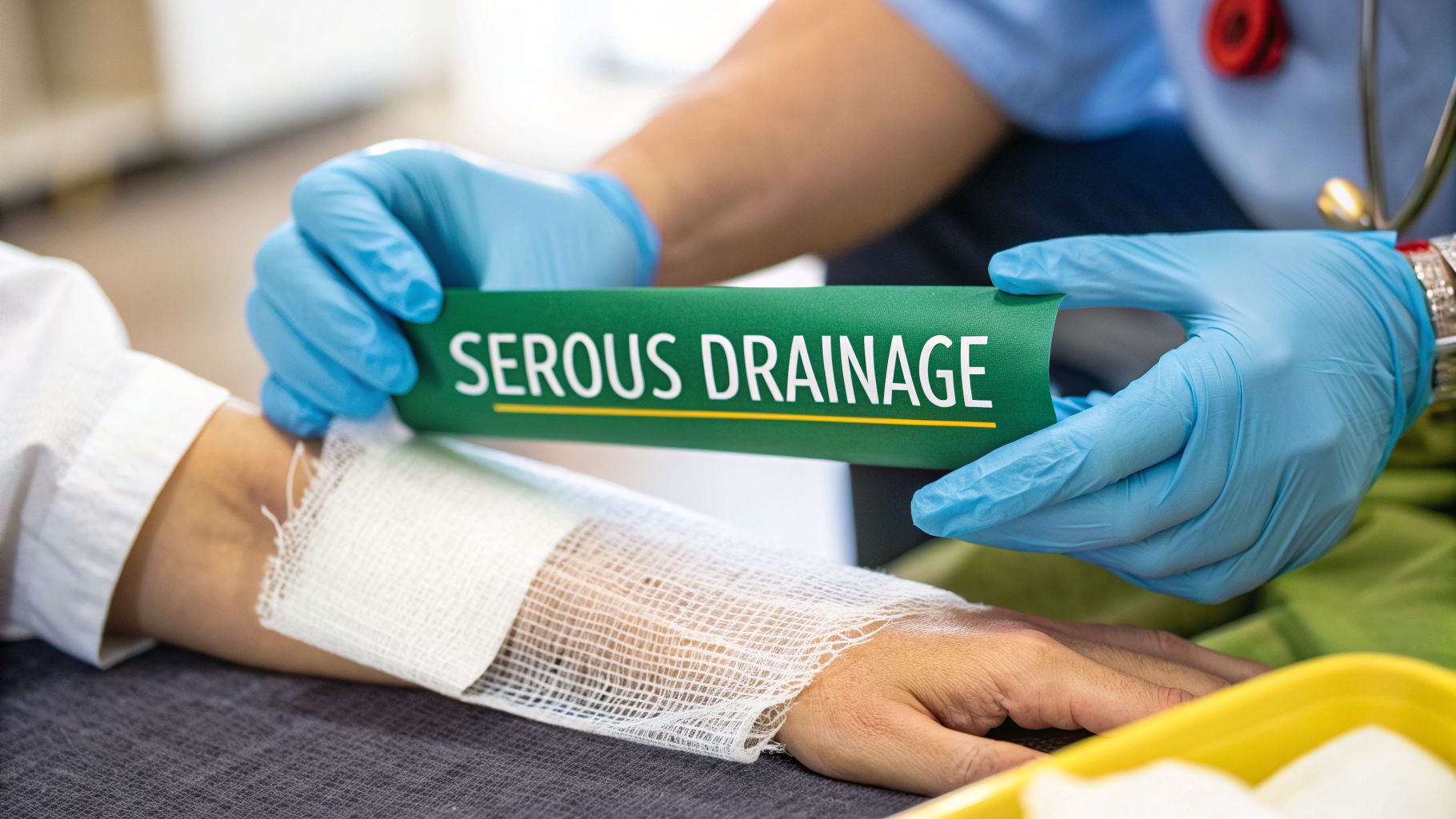
When a wound starts to heal, one of the first things you'll notice is the fluid it produces. Serous drainage is that clear, thin, watery fluid you see in the early stages. Far from being a cause for alarm, it’s a great sign—it means the body's natural repair crew is on the job, working to protect and mend the injured tissue.
Understanding Serous Drainage in Wound Healing

When you see that clear fluid on a dressing, you're essentially looking at the body's first responders in action. This fluid, mostly made of water, electrolytes, and a bit of protein, is all about creating the perfect environment for healing to begin.
Think of it as a natural irrigation system. Its main purpose is to gently cleanse the wound bed, washing away dead cells and any debris that could cause trouble. This process is a critical first step, helping to prevent infection and get the tissue ready for rebuilding. This initial cleansing function is vital because it establishes a clean slate for the more complex stages of tissue repair that follow. A wound cluttered with necrotic tissue or foreign matter is a breeding ground for bacteria and will heal much more slowly, if at all.
The Role of Serous Fluid
This clear or pale-yellow fluid isn't just a passive byproduct of an injury; it’s an active player in the healing game. By keeping the wound moist, serous drainage makes it easier for new cells to travel across the wound surface—a fundamental principle of modern wound care. The moisture creates a slick, friction-free highway for migrating epithelial cells, which are the building blocks of new skin.
It's also a delivery service, bringing crucial supplies right where they're needed:
- Nutrients: It's packed with the sugars, amino acids, and proteins that cells need to fuel their repair and replication processes.
- White Blood Cells: It carries the body's infection-fighting soldiers, primarily neutrophils and macrophages, to guard against invading bacteria and clear away cellular debris.
- Growth Factors: It contains special proteins like platelet-derived growth factor (PDGF) and transforming growth factor-beta (TGF-β) that act as signals, telling cells to start dividing, moving, and rebuilding the tissue matrix.
Serous drainage is a hallmark of the initial inflammatory phase of healing. Its presence tells you the body has successfully kicked off its immune response to clean the wound and prepare for tissue regeneration.
A Foundation for Assessment
Knowing what serous drainage looks like is the starting point for any good wound assessment. It gives you a baseline for what's normal and healthy. This knowledge is absolutely vital because any change in the drainage—whether it's the color, thickness, or amount—is often the first clue that healing might be heading off track. A skilled clinician learns to read the language of wound exudate, and serous fluid is the first word in that vocabulary.
By accurately identifying serous drainage, clinicians can confidently document that a wound is progressing as expected. This skill is also the foundation for spotting other types of drainage, like serosanguinous (pink-tinged) or purulent (thick and cloudy), which signal different stages or potential complications. Getting this right isn't just about good charting; it's essential for providing the right care and maintaining a clear clinical story for every patient. This clear story is crucial for care continuity, especially when multiple providers are involved in the patient's treatment.
How to Identify Serous Drainage by Color and Consistency

Being able to accurately identify wound drainage is a fundamental skill in patient care, and recognizing serous fluid is usually the first step. Think of it as the body’s natural, built-in rinsing agent. Its appearance gives you immediate clues about the wound’s status, and getting that initial assessment right is crucial for good documentation and creating an effective care plan.
Healthy serous drainage has a very distinct look. It should be clear or have a pale, straw-like yellow tint—almost like weak chamomile tea. The consistency is always thin and watery, never thick, sticky, or viscous. Perhaps most importantly, it should have a complete absence of any odor. When you see these signs on a dressing, you're looking at a normal, healthy healing process in action.
Visual and Sensory Cues
To confidently identify serous drainage, just focus on three basic characteristics. These simple observations are the bedrock of a solid clinical assessment and make it easy to tell healthy fluid from other types that might signal a problem.
- Color: The fluid should be mostly transparent. A pale yellow tint is perfectly normal and comes from the plasma proteins, such as albumin, floating within it. If held up to the light, it should be easy to see through.
- Consistency: It should feel just like water. If you were to touch it (with gloves, of course), it wouldn't feel gooey or sticky at all. It will soak readily into a gauze dressing without leaving a thick residue.
- Odor: Healthy serous fluid is completely odorless. Any foul, pungent, or even slightly "off" smell is a major red flag that demands immediate investigation, as it often indicates bacterial colonization.
Serous drainage is essentially the clear, watery part of blood plasma that seeps out of capillaries into a wound during the early inflammatory phase of healing. It’s a sign that the body's immune system is on the job but without any infection present. In a clinical setting, you’ll see this pale yellow or clear fluid in small to moderate amounts, keeping the wound bed moist—an environment that’s perfect for cellular repair. For a deeper dive into its composition, you can find more information from sources like Medical News Today.
Comparing Different Types of Wound Drainage
Spotting serous fluid becomes second nature once you can compare it to the other common types of wound exudate. Each one tells a different story about what's happening inside the wound. An experienced clinician can glance at a soiled dressing and instantly know if things are on track or if trouble is brewing.
A wound's drainage is its way of communicating. Serous fluid says, "I'm cleaning and healing." Other types might be screaming, "I'm bleeding," or "I'm fighting an infection." Learning to interpret this language is fundamental to effective wound care.
To help sharpen your assessment skills, we've put together a simple reference guide. This chart breaks down the key characteristics of different drainage types, making it easier to make fast, accurate calls right at the bedside.
Wound Drainage Identification Guide
This quick-reference chart compares the key characteristics of different types of wound drainage to aid in accurate clinical assessment.
| Drainage Type | Color | Consistency | Odor | Clinical Significance |
|---|---|---|---|---|
| Serous | Clear, pale yellow | Thin, watery | None | Normal, part of the inflammatory phase of healing. |
| Sanguineous | Bright red | Thin, watery | None | Indicates fresh bleeding, common in deep or new wounds. |
| Serosanguinous | Pale pink to reddish | Thin, watery | None | A mix of serous fluid and blood, normal in early healing stages. |
| Purulent | Yellow, green, tan, or brown | Thick, opaque, creamy | Foul, pungent, or musty | Sign of infection, contains bacteria and white blood cells. |
Using a simple guide like this can take the guesswork out of assessing wound exudate. The more you practice, the faster you'll be able to connect the visual cues to the underlying clinical picture. Consistent practice builds the clinical intuition necessary for rapid and reliable wound assessment, which directly impacts patient outcomes.
The Science of Serous Drainage in the Healing Process

To really get a handle on serous drainage, we need to see it for what it is: a crucial part of the body's natural, well-timed healing cascade. This isn't just some random fluid leaking from a wound. It's a purpose-built substance that shows up at a specific time to do a specific job.
Healing isn't a single event; it's a process that unfolds in clear, overlapping stages. The appearance of serous drainage is a direct signpost of where a wound is on that timeline. For clinicians, understanding when and why it appears transforms a simple observation into a powerful assessment tool and helps us explain the healing journey to our patients.
A Key Player in the Inflammatory Stage
The body’s healing process generally follows four main stages. While you might see serous drainage in more than one phase, it really has its moment to shine in the second one.
The four stages are:
- Hemostasis: The immediate reaction. Blood vessels clamp down, and a clot quickly forms to stop the bleeding. Think of it as the emergency first response.
- Inflammation: Once the bleeding is under control, the body launches its defense and clean-up operation. This is where serous drainage is most active.
- Proliferation: The focus shifts to rebuilding. New tissue—that healthy, red granulation—and new blood vessels start filling in the gap.
- Maturation: Finally, the new tissue gets stronger and reorganizes itself. This remodeling can take months, sometimes even years, to complete.
It’s during that inflammatory stage, which usually lasts from day one to about day four after the injury, that serous drainage really ramps up. The body intentionally makes the local blood vessels more permeable, allowing this plasma-like fluid to seep into the wound bed. This fluid, which is over 90% water, acts as a natural irrigation system. It flushes away debris and dead cells while bringing in vital white blood cells to fight off bacteria.
Want to dive deeper into the whole timeline? You can explore the full journey in our detailed guide on the wound healing process.
Think of serous drainage as the wound's own built-in support crew. It keeps the area moist, delivers the defenders, and clears the site so the "construction team" of the proliferation stage can get to work.
The Science of Moist Wound Healing
The incredible importance of this fluid was really brought to light by the work of Dr. George Winter back in the 1960s. For years, the standard practice was to let a wound "air out" and form a dry scab, thinking that was best. Winter’s research turned that entire concept on its head.
He proved, quite simply, that wounds kept in a moist environment healed much, much faster.
This landmark discovery confirmed that the body's own serous fluid creates the perfect setting for healing. In fact, his research showed that keeping a wound moist can actually double the speed of epithelialization—that’s the process where new skin cells migrate across the wound to close it up. Why? A moist environment lets those cells glide easily across the wound bed. A dry, hard scab, on the other hand, is like a roadblock they have to burrow underneath. Modern wound care dressings are all designed around this principle, aiming to manage excess exudate while maintaining a moist interface.
You can read more about how moist environments accelerate healing on woundzoom.com.
When we understand the science, we stop seeing serous drainage as just a passive leakage. We recognize it as an active, therapeutic part of healing. Its presence is a fantastic sign that the body is doing exactly what it's supposed to do, giving us confidence that the wound is on the right track. This understanding also empowers clinicians to educate patients, helping them see the "weeping" as a positive sign rather than a source of worry.
Recognizing When Serous Drainage Signals a Problem

While clear, watery drainage is usually a good sign, it's not always benign. The mark of an experienced wound care clinician is knowing how to spot the subtle shifts that turn this normal healing fluid into a red flag. Catching these changes early can stop a minor issue from spiraling into a serious complication.
Think of serous fluid as a live report from the wound bed. As long as the report is consistent—a small to moderate amount of clear, odorless fluid—you know the healing process is on track. But if that report suddenly changes, it’s a clear signal that something’s off, and it's time to dig deeper.
Key Warning Signs to Watch For
Often, the very first clue that something is wrong is a change in the drainage's volume, color, or smell. Any deviation from the baseline you've been observing should put you on high alert.
Here are the critical changes that demand immediate attention:
- Sudden Increase in Volume: If a wound suddenly starts weeping significantly more fluid, it could be forming a seroma (a pocket of fluid trapped under the skin) or reacting to an underlying inflammatory problem like a foreign body or high bioburden.
- Change in Color or Clarity: If the drainage goes from clear to cloudy, milky, green, tan, or a thick yellow, it's no longer just serous fluid. This is a classic sign of infection, as the exudate is now likely loaded with bacteria and white blood cells, forming pus.
- Development of a Foul Odor: Healthy serous fluid doesn't smell. Any foul, pungent, or unusually sweet scent is a strong indicator of bacterial activity and a brewing infection. Certain bacteria even have characteristic smells (e.g., Pseudomonas has a sickly sweet, grape-like odor).
- Increased Pain and Redness: When drainage changes are paired with worsening pain, swelling, warmth, or redness spreading around the wound (erythema), you're likely looking at an active infection or severe inflammation that needs to be addressed now.
A sudden change in wound drainage is a clinical alarm bell. It's the body's way of telling you its healing plan has been derailed, often by bacteria or another complication.
Actionable Steps for Clinical Teams
When you spot these warning signs, acting quickly and confidently is key to protecting the patient. Ignoring these signals can lead to delayed healing or even systemic infection. That thick, colored, or foul-smelling fluid is a clear signpost of trouble, and you can get more details on how to assess it in our guide on if pus is a sign of infection.
Here are the essential steps to take when you suspect a problem:
- Thoroughly Document the Changes: Get specific. Note the new color, estimate the volume increase (e.g., "dressing 75% saturated in 8 hours vs. 25% previously"), describe the odor, and measure the area of any surrounding redness. A standardized note helps everyone on the team see the trend.
- Cleanse the Wound and Obtain a Culture: If you suspect infection, gently clean the wound with a non-cytotoxic solution, then get a wound culture before applying any new antimicrobial dressings. This is crucial for identifying the specific bug you're fighting and guiding antibiotic therapy.
- Notify the Primary Care Provider or Physician: Escalate your findings. Give the responsible physician or NP a clear, concise report of what you've observed and what you've done so far. Use a structured communication tool like SBAR (Situation, Background, Assessment, Recommendation).
- Adjust the Care Plan: Following the provider's orders, the treatment plan will likely need to change. This might mean switching to an antimicrobial dressing, increasing the frequency of changes, or starting the patient on systemic antibiotics.
Best Practices for Documenting and Managing Wound Drainage
Good documentation is the backbone of great wound care. It’s more than just a legal record; it’s the story of your patient’s healing journey, ensuring everyone on the care team is reading from the same script. If your notes are vague, tracking progress becomes a guessing game, and defending your clinical choices gets a lot harder.
When you're charting any kind of drainage, especially serous fluid, consistency is everything. One of the simplest and most effective ways to achieve this is by using the TACO method. This little acronym is a powerful framework that helps you hit all the key details every single time.
The TACO Method for Drainage Assessment
TACO is a mental checklist that walks you through the four most critical characteristics of wound exudate.
- T (Type): What kind of fluid are you seeing? Is it serous (clear, watery), sanguineous (bloody), serosanguinous (a pinkish mix), or purulent (pus)?
- A (Amount): How much drainage is there? You can quantify this by describing the dressing saturation—think scant, small, moderate, or large/copious. For even more precision, you can weigh the dressing.
- C (Color): Get specific. "Yellow" isn't enough. Is it the "pale, translucent yellow" of serous fluid or the "thick, opaque tan" of purulence? Details matter.
- O (Odor): Does the wound have a smell? Note if the odor is absent, foul, pungent, or musty. Always assess odor after cleaning the wound to ensure you aren't just smelling old drainage on the dressing.
Putting it all together, a solid clinical note sounds something like this: "Moderate amount of serous drainage noted on dressing, roughly 50% saturated. Fluid is clear, pale yellow with no odor." That simple sentence paints a precise, reproducible picture for the next person who sees the patient.
Accurate and consistent documentation isn't just about good record-keeping; it's a direct line to better clinical decisions. It tells a story of healing, or signals the first warning that a wound is in trouble.
Making Documentation Work For You, Not Against You
While the TACO method gives you a solid framework, the reality is that manual charting is a massive time sink. It’s not uncommon for documentation to eat up as much as 50% of a nurse's shift, time that could be spent at the bedside. This is where smarter tools are really starting to change the game.
AI-powered ambient listening technology is a perfect example. Instead of stopping mid-assessment to type everything into the EHR, you can simply talk through your findings as you work.
Imagine a nurse examining a wound and saying, “The wound on the left heel shows a small amount of clear, serous drainage with no odor.” The AI captures that description on the fly, slots it into the correct fields in the patient’s chart, and can even help tee up the right billing codes. If you want to dive deeper into streamlining your workflow, take a look at our guide for building a wound care documentation template.
This kind of technology doesn't just give you back precious time; it drives a higher standard of care. It ensures every assessment is documented with the same level of detail, creating a clear, data-rich history for each patient. That robust timeline is exactly what you need to make sharp clinical decisions and provide the hard evidence required to justify care to payers, which ultimately helps you get paid faster.
Your Questions About Serous Drainage, Answered
When you're on the front lines of wound care, you see it all. But even with experience, questions pop up, especially when you're trying to figure out if what you're seeing is normal or a red flag. Let's tackle some of the most common questions clinicians have about serous drainage.
How Long Is Serous Drainage Normal After Surgery?
For the first 2 to 3 days after surgery, seeing some serous drainage is completely normal—in fact, it's expected. This is part of the body's natural inflammatory response, where the fluid works to clean the site and get it ready for healing.
But this shouldn't last forever. As the wound moves into the next phase of healing (the proliferative stage), you should see that clear, watery fluid start to taper off. If you're still seeing a significant amount of serous drainage after the first week—or if it suddenly increases—it's time to take a closer look. That could be a sign that the wound is stalled, there is an underlying issue like a hematoma, or that a complication like a seroma is brewing.
What's the Difference Between Serous Fluid and a Seroma?
This is a great question because they’re both made of the same stuff. The real difference is all about location and what it means for the patient.
Serous fluid (or drainage) is what you see on the dressing. It’s the liquid actively weeping from a healing wound, which is a normal part of the process. A seroma, on the other hand, is when that same fluid gets trapped under the skin, forming a pocket or collection. This often happens in surgical "dead space" where tissue was removed, leaving a cavity.
You'll usually spot a seroma as a soft, sometimes squishy-feeling swelling near the incision. It’s a problem because that trapped fluid keeps the tissues from knitting back together and creates the perfect, nutrient-rich environment for an infection to take hold. A seroma almost always requires intervention, like being drained with a needle (aspiration) or having a drain placed.
Is It Okay If Serous Drainage Has a Pink Tinge?
Yes, absolutely. A pink or pale red tint is very common, especially in the first 24 to 48 hours after surgery or an injury. We call this serosanguinous drainage.
Think of it as normal serous fluid mixed with just a few red blood cells that have leaked out from tiny, damaged capillaries. It’s a standard part of the healing process. The key is to watch the color over time. In a healthy wound, the drainage should gradually transition from pink (serosanguinous) back to clear or pale yellow (serous). If it becomes bright red, that’s sanguineous drainage, a sign of active bleeding that needs immediate attention.
How Can AI Technology Help with Documenting Wound Drainage?
This is where things get really interesting. AI changes the game by making documentation faster, easier, and far more consistent. Instead of spending time manually typing detailed notes, a clinician can just speak their observations out loud during an assessment.
For instance, you could simply say, "The sacral wound has a moderate amount of serous drainage, which is clear and odorless." A voice-first AI system will instantly understand that, parse it, and place each piece of information into the correct fields in the patient’s chart. This accomplishes a few critical things all at once:
- It cuts documentation time dramatically, giving you more hands-on time with your patients.
- It standardizes the language used, so everyone on the care team is on the same page.
- It creates structured data, which is far more useful for analytics and quality reporting than unstructured text.
- It automatically links those clinical findings to the right billing codes, which is a huge help for the back office.
The end result is a clean, data-rich timeline of the wound's progress. This not only improves clinical decision-making but also helps streamline the billing process and prevent claim denials.
By automating the entire workflow from voice to claim, Ekagra Health AI helps wound care teams reduce documentation time by up to 70%, improve assessment accuracy with AI-powered tools, and accelerate reimbursement. Learn how our end-to-end platform can support your organization at https://ekagrahealth.ai.
