When you're dealing with different types of wounds, the first and most important question to ask is this: is it acute or is it chronic? This isn't just a matter of terminology; it's the fundamental starting point for all effective wound care. Acute wounds are the ones that heal as expected, moving neatly through the body's repair sequence. Think of a clean surgical incision or a paper cut.
Chronic wounds, however, are a different beast entirely. These are the wounds that get stuck. They fail to heal in a predictable, timely manner and often get trapped in one of the healing phases, refusing to progress. This guide will serve as a comprehensive resource, exploring the full spectrum of wounds from a clinical perspective, providing the depth needed for a pillar content piece on this high-competition topic.
The Critical Divide Between Acute and Chronic Wounds

I find it helpful to think of the body's healing cascade as a well-orchestrated construction project. An acute wound is a project that runs like clockwork. The blueprint is clear, the right materials are delivered on time, and the specialized teams—the cells and growth factors—complete their jobs in a precise order. From closing the site to rebuilding the structure, everything proceeds on schedule. The process begins with hemostasis (stopping the bleeding), moves into inflammation (the cleanup crew), then proliferation (rebuilding), and finally maturation (remodeling).
A chronic wound is that same project gone completely haywire. It's stalled, often because of serious underlying problems. Maybe the supply lines are cut off (poor blood flow), the worksite is constantly being compromised (persistent infection or biofilm), or the crew itself is overworked and ineffective (impaired cellular function). These wounds just can't move forward, often lingering in a state of inflammation for weeks, months, or even longer. This stalled inflammatory phase is a hallmark of chronic wounds, preventing the transition to the proliferative, or rebuilding, stage.
For a quick reference in a busy clinical setting, this table breaks down the essential differences.
Acute vs Chronic Wounds At a Glance
| Characteristic | Acute Wounds | Chronic Wounds |
|---|---|---|
| Onset | Sudden, from a specific event (e.g., surgery, trauma). | Can develop slowly over time, often from an underlying condition. |
| Healing Time | Follows a predictable timeline, usually healing within 3-4 weeks. | Fails to progress through the normal healing stages; healing is delayed or stalled. |
| Underlying Cause | The wound itself is the primary problem. | Caused or complicated by underlying issues (e.g., diabetes, poor circulation). |
| Inflammation | A normal, temporary phase of healing. | Prolonged or excessive inflammation that prevents progression. |
| Appearance | Edges are typically clean and well-defined. | Edges may be irregular; may show signs of repeated breakdown. |
| Treatment Goal | Clean, close, and protect to facilitate natural healing. | Identify and manage the underlying cause to get the healing process "unstuck." |
Having this mental checklist helps you quickly size up the situation and start thinking about the right path forward.
Why This Distinction Matters
Making the call between acute and chronic is far more than an academic exercise. This single determination shapes your entire plan of care, from how you assess the wound to how you treat it and even how you code for reimbursement.
- Treatment Strategy: Acute wounds usually respond to straightforward care: cleaning, appropriate closure, and preventing infection. Chronic wounds demand detective work to uncover and manage the root cause that’s holding up the healing process. This may involve vascular assessments, nutritional interventions, and advanced therapies.
- Resource Allocation: Chronic wound management is much more involved. It often requires specialized dressings, advanced therapies like debridement or negative pressure therapy, and much closer clinical monitoring. It's a multidisciplinary effort, often involving specialists in endocrinology, vascular surgery, and infectious disease.
- Patient Expectations: Someone with an acute wound can generally expect a quick, linear recovery. A patient with a chronic wound needs clear communication about a longer, more complicated journey that might have its share of setbacks. Managing these expectations is a critical part of patient-centered care.
The core difference really comes down to progression. An acute wound is on a clear path to closing. A chronic wound is fundamentally stuck. Your main job becomes figuring out why it's stuck.
Recognizing which category a wound falls into is the foundational skill for any clinician in this field. For a deeper dive into the specific biological stages, you can learn more about the complete wound healing process in our dedicated guide. This initial assessment is what guides your clinical judgment, helping you anticipate potential hurdles and create a care plan that addresses the real problem, not just the hole in the patient.
A Deep Dive into Common Chronic Wound Types
When you're dealing with wounds, it's crucial to understand that not all are created equal. An acute wound, like a simple cut, follows a predictable, orderly path to healing. But chronic wounds? They're a different beast entirely. These are the wounds that get stuck, failing to heal in a timely fashion, and they almost always signal a deeper, underlying issue that's sabotaging the body's natural repair process.

The scale of the problem is staggering. Chronic wounds aren't just a footnote in healthcare; they are a dominant force, representing between 59.9% and 65% of the entire global market. As our population ages and conditions like diabetes become more common, the financial toll continues to climb. We're talking about the lion's share of a market valued at over $20 billion, a figure that highlights just how critical effective diagnosis and management are. You can get a closer look at the numbers and global wound care market trends and their financial impact here.
Let's break down the major players you'll see in the clinic every day.
Pressure Injuries
We used to call them pressure ulcers or bedsores, but the term pressure injury is more accurate. It describes localized damage to the skin and tissue, almost always found over a bony spot like the tailbone or heels. The cause is simple physics: intense or prolonged pressure, often combined with shear forces, squashes the tissue and cuts off blood flow.
Think of it like stepping on a garden hose. The pressure completely blocks the supply line, starving the tissue of the oxygen and nutrients it needs to survive. Eventually, it dies. Contributing factors like moisture from incontinence can lead to maceration, further weakening the skin and increasing susceptibility.
- Where You'll See Them: Sacrum, coccyx, heels, hips, and elbows.
- Who's at Risk: Patients with limited mobility, poor sensation, inadequate nutrition, or issues with moisture.
- What They Look Like: The spectrum is wide, from a patch of non-blanchable red skin (Stage 1) to a deep crater that goes all the way down to bone or muscle (Stage 4).
The defining feature of a pressure injury is its cause: pressure. The primary intervention will always be to identify and relieve that pressure. Everything else is secondary.
Diabetic and Neuropathic Ulcers
The term "diabetic foot ulcer" strikes fear in any clinician, and for good reason. These wounds are a devastating complication of diabetes, born from a perfect storm of nerve damage (neuropathy), poor circulation (peripheral artery disease), and uncontrolled blood sugar. Neuropathy is the silent partner in crime here; it robs the foot of the ability to feel pain, pressure, or even a rock in a shoe. High glucose levels also stiffen tissues and impair immune cell function, creating an ideal environment for non-healing wounds.
Because of this, a tiny, unnoticed injury can quickly become a limb-threatening wound. Diabetic foot ulcers are the number one cause of non-traumatic lower-limb amputations. If you need to do a deep dive, our guide on diabetic foot ulcer management is a must-read.
Telltale signs include:
- Location: Almost exclusively on the foot. Look for them on the plantar surface (the bottom), especially over bony prominences like the metatarsal heads.
- Shape: They have a classic "punched-out" appearance, often framed by a thick ring of callus.
- Wound Bed: The base can be pinkish or pale, and surprisingly, there might be very little drainage unless an infection has set in.
The most dangerous feature is the lack of pain. It's not uncommon for a patient to come in with an advanced, infected ulcer they didn't even know was there.
Venous Stasis Ulcers
If pressure injuries are from a blocked supply line, venous stasis ulcers are the result of a faulty drainage system. The root cause is chronic venous insufficiency—the one-way valves in the leg veins have failed, and they can't effectively pump blood back up to the heart.
This causes blood to pool in the lower legs. The constant back-pressure forces fluid to leak out of the vessels and into the surrounding tissue, leading to persistent swelling (edema), skin discoloration (a rusty brown stain called hemosiderin staining), and eventually, the skin just gives up and breaks down. This environment of chronic inflammation and poor nutrient exchange is why the skin becomes so fragile.
- Location: The "gaiter" area is the classic spot—the lower leg, somewhere between the ankle and the knee.
- Shape: These are often big, shallow wounds with messy, irregular borders.
- Appearance: They tend to be very wet, with moderate to heavy drainage. The wound bed itself can be red or covered in yellow slough, and the skin around it will be swollen, firm, and discolored.
Arterial Ulcers
On the opposite end of the circulatory spectrum are arterial ulcers, also known as ischemic ulcers. These are the polar opposite of venous ulcers. Here, the problem isn't getting blood out; it's getting blood in. Severe atherosclerosis (hardening of the arteries) has narrowed the vessels so much that not enough oxygen-rich blood can get to the lower legs and feet.
This is a tissue drought. The area is starved of blood, which causes incredible pain and leads to tissue death. The lack of oxygenated blood flow directly leads to necrosis.
Key identifiers include:
- Location: They show up in the most distal parts of the body—the tips of the toes, the heels, or the bony part of the outer ankle.
- Shape: Very distinct. They have a "punched-out" look with clean, well-defined, and even borders.
- Appearance: The wound base looks pale, gray, or even black, with little or no sign of new tissue. These wounds are dry, with minimal drainage. The surrounding skin is often cool to the touch, shiny, and hairless. A major red flag is the patient reporting severe pain, especially at night or when they elevate their leg.
A Practical Guide to Identifying Common Acute Wounds
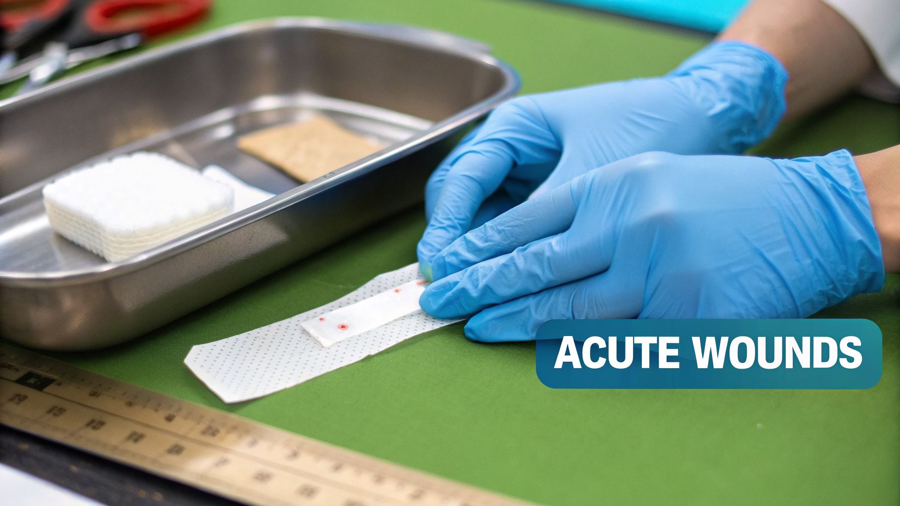
Acute wounds, unlike their chronic cousins, are supposed to heal on a predictable schedule. Think of them as having a clear roadmap to recovery. Your job in the initial assessment is to make sure they stay on that road.
Getting the identification and initial management right is everything. A misstep early on—like a missed sign of infection or a poor closure choice—is the roadblock that can divert a simple acute wound into a complex, chronic problem. Let's break down the most common acute injuries you'll see every day: surgical wounds, traumatic injuries, and burns.
Surgical Wounds and How They're Meant to Heal
Every surgical wound is a story of intention. Created in a sterile, controlled setting, the way a surgeon closes it (or doesn't) is a deliberate choice based on factors like contamination risk and the extent of tissue loss. To properly assess these wounds, you first need to understand the three "healing intentions."
- Primary Intention: This is your best-case scenario. The wound edges are clean and can be brought together neatly with sutures, staples, or adhesives. Think of it like zipping up a jacket—it's a direct closure designed for minimal scarring.
- Secondary Intention: When a wound has significant tissue loss or a high risk of infection, closing it would be like locking bacteria inside. Instead, we leave it open to heal from the base upwards. It’s like filling a hole brick by brick—it's slower and naturally creates more scar tissue.
- Tertiary Intention: This is a hybrid strategy, also called delayed primary closure. The wound is first left open to be cleaned and monitored, just like in secondary intention. Once we’re confident it’s clean and free of infection, we then close it surgically, as in primary intention.
Keep a sharp eye out for Surgical Site Infections (SSIs). The classic red flags—growing redness, localized warmth, swelling, any purulent drainage, or a sudden fever—are alarm bells that demand immediate action.
Traumatic Wounds: Lacerations and Punctures
Where surgical wounds are planned, traumatic wounds are pure chaos. Caused by external force in uncontrolled environments, your first thought should always be contamination. The two main categories you'll face are lacerations and punctures.
A laceration is essentially a tear or cut in the skin. When you're assessing one, you need to look at its depth, how clean it is, and whether the edges are smooth or jagged.
A puncture wound, on the other hand, is a different beast. It’s caused by a sharp, pointed object, and the small entry point can be incredibly deceptive. The real danger lies deep beneath the surface, where bacteria can be plunged into an oxygen-poor environment—a perfect breeding ground for nasty infections.
When a traumatic wound comes in, your priorities are clear:
- First, control the bleeding.
- Next, carefully check for any foreign bodies—glass, dirt, wood splinters, you name it.
- Finally, assess the potential for damage to deeper structures like nerves, tendons, or blood vessels.
The Unique Challenge of Burns
Burns are among the most complex acute wounds you will ever manage. Whether they're caused by heat, chemicals, electricity, or radiation, their severity is all about depth, and getting that assessment right is non-negotiable.
Burn Classifications by Depth:
| Degree | Skin Layers Involved | Appearance | Sensation |
|---|---|---|---|
| First-Degree | Epidermis only | Red, dry, no blisters | Painful |
| Second-Degree | Epidermis & Dermis | Blisters, moist, red/pink | Very painful |
| Third-Degree | Epidermis, Dermis, Subcutaneous | White, leathery, or charred | No sensation |
| Fourth-Degree | All layers plus muscle/bone | Black, charred, with exposed bone | No sensation |
Beyond depth, you have to calculate the Total Body Surface Area (TBSA) involved. The "Rule of Nines" is a quick and effective tool for estimating this in adults, assigning percentages in multiples of nine to different body regions. An accurate TBSA is critical—it dictates fluid resuscitation and helps determine whether the patient needs immediate transfer to a specialized burn center. This is one assessment you have to get right.
Your Toolkit for Wound Assessment and Documentation

Knowing the different types of wounds is where it all begins. But to deliver exceptional care and ensure you get paid for it, you need a rock-solid process for assessment and documentation. This is more than just checking boxes; it’s about crafting a complete, defensible story of the wound from day one.
Great wound care is built on the foundation of a great assessment. Without a clear and detailed picture of what you're treating, your interventions are little more than educated guesses. A methodical assessment process is your best friend for tracking a wound's progress, justifying your treatment choices, and heading off frustrating compliance issues before they start.
The Core Components of Wound Assessment
Think of yourself as a detective at the bedside. You're collecting clues from the wound and the skin around it to build a case for your diagnosis and treatment plan. Every single detail matters.
A thorough, systematic evaluation should always cover these key areas:
- Precise Location: "Left heel" is a start, but it's not enough. "Posterior aspect of the left calcaneus" is what auditors and other clinicians need. Pinpoint the location with correct anatomical terms.
- Accurate Measurements: Consistency is key. Always measure in the same order—length x width x depth—using centimeters. Length is always head-to-toe, and width is side-to-side, no matter how the wound is oriented on the body.
- Wound Bed Tissues: Describe what you see in the wound bed by percentage. Look for healthy, beefy red granulation tissue, stringy yellow slough, or tough, leathery black eschar.
- Exudate (Drainage): How much drainage is there (scant, moderate, copious)? What does it look like (serous, sanguineous, purulent)? This tells you so much about the wound's healing status and potential for infection.
- Periwound Skin: Don’t forget to examine the surrounding real estate. Is the skin healthy, or do you see redness (erythema), swelling (edema), or discoloration?
The story of a wound is written not just in the open area but in the tissues that surround it. Ignoring the periwound skin is like reading only half the page.
Translating Assessment into Airtight Documentation
Clear, specific documentation is your best defense against claim denials and audit headaches. Vague notes like "wound improving" are a huge red flag. Your documentation has to paint a vivid picture that justifies every code you bill and every supply you use.
The financial stakes are incredibly high. For example, diabetic foot ulcers (DFUs) affect 6.3% of diabetes patients worldwide and are a factor in over one million amputations every year. And in the U.S., pressure injuries impact 2.5 million patients annually, with severe cases costing a hospital more than $70,000 each. If your documentation is weak, coding errors can cause 20-30% of your DFU claims to be denied. It’s a clear case for why getting the details right matters.
It's not just about telling acute from chronic wounds apart. You have to master identifying common acute wounds and understand the unique demands of each, like the importance of wound care for foot and ankle injuries.
Pressure Injury Staging: A Critical Skill
When you're dealing with pressure injuries, staging isn't optional—it's a core competency. The classification system from the National Pressure Injury Advisory Panel (NPIAP) gives us a universal language to describe the level of tissue damage.
Common Stages of Pressure Injuries:
| Stage | Description | Key Characteristics |
|---|---|---|
| Stage 1 | Intact skin with non-blanchable redness | A persistent red spot over a bony area that doesn't turn white when pressed. |
| Stage 2 | Partial-thickness skin loss with exposed dermis | A shallow open ulcer with a red-pink wound bed, or an intact serum-filled blister. |
| Stage 3 | Full-thickness skin loss | Fat (adipose) is visible in the ulcer, and granulation tissue is often present. |
| Stage 4 | Full-thickness skin and tissue loss | Exposed or directly palpable fascia, muscle, tendon, ligament, or bone in the ulcer. |
There are two other crucial classifications you need to know:
- Unstageable: The wound bed is covered by slough or eschar, so you can't see how deep it goes. If you can't see the bottom, you can't stage it.
- Deep Tissue Pressure Injury (DTPI): This appears as a persistent, non-blanchable deep red, maroon, or purple area of discoloration on either intact or broken skin.
Every one of these assessments directly guides your care plan, making this a non-negotiable skill. To standardize and improve this process, it's worth exploring modern wound assessment tools for nurses designed to make these evaluations more consistent and efficient.
How AI Can Fix Your Wound Care Workflow
Anyone working in wound care knows the struggle. The constant pressure of paperwork, the fear of a coding mistake leading to a denial, and the sheer volume of documentation can pull you away from the one thing that matters: your patient.
What if you could get that time back? Imagine focusing completely on the person in front of you, confident that the mountain of administrative work is being handled accurately in the background.
That’s not a far-off dream; it’s what’s happening right now with AI in wound care. Platforms like Ekagra Health aren’t just abstract tech—they’re practical tools designed to solve these exact frustrations. They weave intelligent automation into your daily routine, bridging the gap between your clinical skill and the demands of documentation and billing.
The outcome is simple: a faster, more accurate workflow that gets you away from the keyboard and back to the bedside.
From Voice to Claim in Minutes
Think about your last wound assessment. How much of it was spent typing or clicking through an EHR? Now, picture doing that same assessment without ever breaking eye contact with your patient.
This is the reality of a "voice-to-claim" workflow, and it starts with ambient clinical listening. An AI tool securely records the natural conversation during an exam.
You can simply talk through your findings—describing the wound's location, its measurements, the tissue types you observe, and the state of the surrounding skin. The AI listens, understands the clinical context, and organizes it all into a perfect chart note. It’s like having an expert scribe who never misses a detail.
Here’s why this matters in the real world:
- Massive Time Savings: Clinicians using these tools report cutting their documentation time by as much as 70%. That’s hours back in your day to see more patients or dive deeper into complex cases.
- Better Patient Connection: When a screen isn't a barrier between you and your patient, the quality of the interaction skyrockets. You build better rapport and can pick up on subtle cues you might have otherwise missed.
- Richer, More Accurate Notes: The system captures your full clinical narrative, preserving the nuances that often get lost when you’re forced to summarize your thoughts into rigid EHR fields.
Moving from manual data entry to automated voice capture is a game-changer. It puts the human side of medicine first and lets technology handle the tedious administrative tasks that lead to burnout.
AI-Powered Image Analysis for Objective Data
Let’s be honest: manual wound measurement can be all over the place. Two clinicians can measure the same wound and get two different sets of numbers. That inconsistency makes it tough to track healing progress and can be a huge red flag for payers.
AI-powered image analysis puts an end to the guesswork. A simple photo from a smartphone or tablet is all the Ekagra Health platform needs to deliver instant, standardized metrics.
The system automatically:
- Calculates precise measurements for length, width, and surface area.
- Helps identify and quantify different tissue types in the wound bed.
- Tracks changes over time with objective, repeatable data.
This gives you a clear, data-driven timeline of the wound's healing journey. You get a defensible record that proves your treatments are working, backing up your clinical judgment with hard evidence instead of just subjective notes. Of course, when adopting this kind of technology, ensuring data security is non-negotiable, which is why working with HIPAA compliant AI solutions is essential.
Nail Your Coding and Stop Denials
Claim denials are one of the biggest financial headaches in wound care, often stemming from coding errors or documentation that doesn't support the bill. An AI-driven system tackles this problem head-on by linking your clinical notes directly to the billing process.
As the AI structures your narrative, it’s also identifying the correct ICD-10 and CPT codes that match the diagnosis, wound characteristics, and any procedures you performed.
For example, if you describe debriding a certain percentage of non-viable tissue from a full-thickness ulcer, the system automatically suggests the most specific, appropriate code for that action. This ensures every claim is clean, accurate, and fully supported by your documentation, drastically reducing denial rates and accelerating your revenue cycle.
Your Top Wound Care Questions, Answered
Day in and day out, clinicians face a host of practical questions when it comes to wound care. Whether you're on the floor trying to distinguish between two similar-looking ulcers or in the back office trying to figure out why claims are being denied, the challenges are real.
This section tackles some of the most common questions we hear from practitioners. Think of it as a quick reference guide, connecting best practices to the real-world scenarios you encounter every day and showing how the right tools can make all the difference.
How Can I Reliably Differentiate a Venous from an Arterial Ulcer?
This is a classic—and absolutely critical—distinction. Getting it right comes down to looking at two things: location and appearance. I find it helps to think of them as having totally different personalities.
A venous ulcer usually shows up in the lower leg's "gaiter" area, which is the region from mid-calf down to the ankle. These wounds tend to be shallow with messy, irregular borders and often have a lot of drainage. The surrounding skin tells a huge part of the story, too. Look for signs of chronic swelling and that tell-tale rusty brown color from hemosiderin staining, a classic sign of long-term venous disease.
On the other hand, an arterial ulcer is all about poor blood supply. You'll find these on the most distant parts of the body, like the tips of the toes or over bony spots on the foot. They are often deep with a "punched-out" look and smooth, even borders. The wound bed itself is typically pale, and the surrounding skin looks thin, shiny, and hairless—all red flags for severe ischemia.
One of the most telling clues is pain. Patients with arterial ulcers often describe intense pain, especially when they elevate their legs. Venous ulcers, while still uncomfortable, can be surprisingly less painful.
What Is the Biggest Cause of Wound Care Claim Denials?
Hands down, the vast majority of claim denials in wound care trace back to two connected problems: a lack of documented medical necessity and a mismatch between your notes and the codes you bill. Payers need a clear, detailed story that justifies every single thing you do, especially for high-value services like debridement.
Vague documentation is the real killer. A note that just says "debrided wound" is a huge red flag for auditors. It doesn't answer the crucial questions: What instrument did you use? How deep did you go (e.g., to subcutaneous tissue)? What percentage of the wound was covered in non-viable tissue before you started? Without those details, you haven't proven the service was medically necessary or met the standard for the code you're billing.
Common Documentation Failures That Lead to Denials:
- Failing to describe the specific characteristics of the tissue removed.
- Not recording precise, updated wound measurements at each and every visit.
- Lacking a clear care plan that connects your assessment to the treatment you provided.
This is where smart systems can be a lifesaver. Tools that can intelligently connect your clinical narrative directly to the most specific ICD-10 and CPT codes become a powerful defense against these expensive and frustrating denials.
How Can My Team Standardize Wound Measurements?
Inconsistent measurements are a plague on good wound care. When two clinicians measure the same wound and get different numbers, it completely undermines your data. You can't accurately track healing, and you certainly can't prove that a treatment is working.
Relying on old-school rulers is often the source of this problem. The single best way to get everyone on the same page is to adopt digital wound measurement technology.
Platforms like Ekagra Health use sophisticated AI to analyze a simple photo of the wound. With a single tap, the system can instantly and objectively calculate the length, width, and surface area down to the millimeter. This completely removes the guesswork and human error from the equation, ensuring that every measurement is taken the exact same way, every single time.
Suddenly, you have a reliable, objective healing timeline you can actually trust. That's the kind of hard data that holds up under scrutiny from payers and gives your team confidence that you're tracking progress with precision.
What's the Difference Between an Unstageable Injury and a DTI?
This is a frequent point of confusion, but telling the difference between an unstageable pressure injury and a Deep Tissue Injury (DTI) is vital. They have completely different underlying pathologies, and that means they require different management strategies.
An unstageable pressure injury is a full-thickness wound where you can't see the bottom. The base is completely covered by slough or eschar. You know there's significant tissue loss under there, but because it's obscured, you can't determine the true depth—and therefore can't assign a stage (like Stage 3 or 4) until the wound is cleaned out and the bed is visible. The name says it all: it is literally "un-stage-able."
A Deep Tissue Injury (DTPI), however, looks very different. It presents as a patch of intact (or sometimes non-intact) skin with a deep red, maroon, or purple color that doesn't blanch when you press on it. It might also look like a blood-filled blister. The key concept here is that the damage is happening from the inside out. Intense and/or prolonged pressure has injured the underlying soft tissues, but the full extent of that damage hasn't broken through to the surface yet.
Getting these two right is a cornerstone of good clinical practice and a requirement for accurate billing and reimbursement.
Ready to put documentation headaches behind you and bring new clarity to your wound care practice? Ekagra Health AI offers a powerful, voice-first platform that transforms your workflow from assessment all the way to reimbursement. Find out how our AI-powered tools can slash your charting time, ensure coding accuracy, and deliver the objective data you need to drive better patient outcomes. Learn more and request access at Ekagra Health.
