
Normal wound healing isn't a single event; it's a beautifully complex biological process that unfolds in four distinct stages: hemostasis, inflammation, proliferation, and maturation. Think of it less like a simple patch-up and more like a highly coordinated construction project. Each phase has to happen in the right order, building on the work of the last to successfully repair the damaged tissue and restore the skin's protective barrier.
Unpacking the Four Phases of Wound Repair

From the very moment of injury, your body launches a cascade of cellular activity. It's an intricate ballet designed to first control the damage, then clean the site, rebuild what was lost, and finally, strengthen the new tissue.
For any clinician in wound care, truly understanding this biological blueprint is non-negotiable. It's what allows you to make sharp, informed judgments at the bedside, anticipate what should happen next, and, most critically, spot when a wound is starting to veer off course. Getting the stages of normal wound healing down cold is the foundation of everything we do.
The Construction Project Analogy
I’ve always found it helpful to think of the whole process like a four-phase construction project. It really clicks for a lot of people.
- Hemostasis: This is your emergency response. The first crew rushes in to stop the immediate crisis—the bleeding—and secure the site.
- Inflammation: Next, the demolition and cleanup crew arrives. They're tasked with clearing out all the debris and damaged materials, preparing a clean foundation for the rebuild.
- Proliferation: This is where the real building happens. A new framework is put up, new blood vessels (the plumbing) are laid down, and the gap is filled in with fresh tissue.
- Remodeling: Finally, the finishers come in for the renovation. The new structure is strengthened, reorganized, and refined to make it as tough and functional as possible.
Each phase perfectly sets the stage for the next. If you get a delay or a breakdown in one of the early stages, the entire project can grind to a halt, leading to a chronic, non-healing wound.
In this guide, we're going to dive deep into each of these stages. We’ll look at the key cellular players, the expected timelines, and what you should be seeing at the bedside. We'll also connect this back to how modern documentation tools can help you track this journey, turning your visual assessments into structured data that tells a clear, objective story of healing. When you know the patterns of normal repair inside and out, you can intervene with confidence the moment things go wrong, and that leads to better outcomes for your patients.
Hemostasis: The Body's Immediate First Response
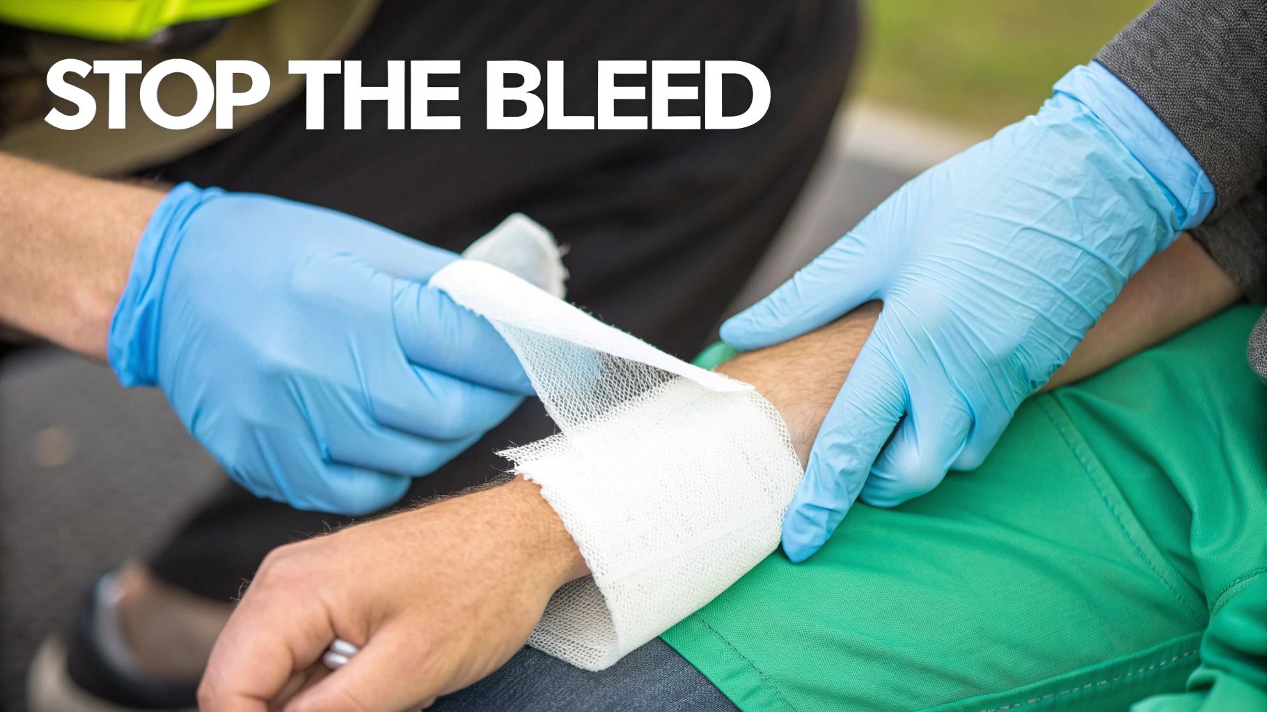
The second the skin is broken, the body's emergency response system kicks into high gear. This first stage, hemostasis, is all about one thing: stopping the bleed. It's the body’s first and most critical priority in the entire wound healing process.
Think of it as the body’s own team of highly efficient first responders. Within seconds of an injury, a cascade of events is triggered. The very first action is vasoconstriction—the blood vessels right around the wound clamp down, immediately narrowing to restrict blood flow to the site. This rapid constriction buys just enough time for the next wave of responders to get to work.
The Platelet Plug and Fibrin Mesh
Platelets, which are tiny cell fragments constantly circulating in our blood, are the next to arrive. Once they sense the damage to a vessel wall, they rush to the scene, transform into a sticky state, and begin clumping together. This initial pile-up forms a temporary platelet plug, a quick but fragile patch to slow the leak.
Of course, this temporary plug isn't strong enough on its own. To build a truly durable seal, the body fires up the clotting cascade, a beautifully complex chain reaction that involves a whole host of clotting factors. The ultimate goal here is to convert a protein called fibrinogen into long, tough, insoluble strands of fibrin.
These fibrin strands then weave themselves through and over the platelet plug, creating a strong, net-like structure. This fibrin mesh is the biological rebar that reinforces the entire structure, trapping more platelets and red blood cells to form a stable, solid clot. This clot doesn't just stop the bleeding; it also provides the essential scaffolding that new cells will use to start rebuilding the damaged tissue.
The hemostasis phase kicks off the wound healing process right after injury, acting like an emergency brake to stop bleeding and set the stage for recovery. This phase typically wraps up in minutes, but it's crucial because it provides the structural foundation for granulation tissue later on. In normal wounds, this rapid response ensures the site stabilizes quickly, with studies showing that effective hemostasis reduces the risk of excessive blood loss by up to 90% in minor injuries. For a deeper dive, you can explore the detailed mechanisms of hemostasis in this in-depth overview from the National Library of Medicine.
Clinical Signs and Implications
From a clinical standpoint, you know hemostasis has been successful when the bleeding stops. This stage is usually over within a few minutes to a couple of hours, though the severity of the wound plays a big role. But it's not always a smooth process; several factors can throw a wrench in the works.
- Anticoagulants: Common medications like warfarin or heparin are designed to interfere with the clotting cascade, which means bleeding will naturally take longer to control.
- Bleeding Disorders: Inherited conditions like hemophilia prevent the body from forming a stable clot, turning even minor injuries into serious events.
- Poor Platelet Function: Some diseases or medications can either lower the platelet count or make the existing platelets less effective, hampering the initial plug formation.
Documenting what you see in these first few moments is absolutely critical. Noting how quickly the bleeding was controlled, any known medical conditions, and the patient's current medications gives you a vital baseline for the entire plan of care. A delay in hemostasis is a major red flag that can signal trouble ahead, making sharp, early observation essential as the wound moves into the next phase: inflammation.
Inflammation: Preparing the Ground for New Growth
Once the bleeding is under control, the body’s focus shifts. The next critical step in healing is the inflammatory phase. Think of this as the body sending in its specialized cleanup and demolition crew to the injury site. This stage is absolutely essential for fighting off infection and meticulously clearing out damaged cells, bacteria, and any other debris. It’s all about creating a clean slate so new tissue can be built.
This whole process kicks off within hours of the injury and usually lasts anywhere from one to six days. As clinicians, we're all familiar with the classic signs: redness, heat, swelling, and pain. These aren't just inconvenient symptoms; they're the visible proof that a powerful and necessary biological response is underway.
The redness and heat are a direct result of increased blood flow as vessels widen to rush immune cells to the area. Swelling happens because those same vessels become more porous, allowing fluid and specialized cells to move into the surrounding tissue. It's a coordinated effort to flood the site with everything needed for the big cleanup.
Key Players in the Cleanup Crew
First on the scene are the neutrophils. These white blood cells are the front-line soldiers, arriving within minutes to hours. They immediately get to work, aggressively consuming bacteria and cellular debris. Their job is intense but brief; after a few days, they’re replaced by the next wave of specialists.
Following the neutrophils are the macrophages. These are the heavy lifters of the cleanup crew. Macrophages are bigger and stick around longer, continuing to clear out debris and any lingering bacteria. But they do more than just clean. They also release powerful growth factors—biochemical messengers that signal the start of the next stage: rebuilding, also known as the proliferative phase.
The inflammatory response is a delicate dance. It’s a vital process for defending against infection and prepping the wound bed. But if this stage drags on for too long or becomes too intense, it can stall the healing process entirely, which is a hallmark of many chronic, non-healing wounds.
Distinguishing Normal Inflammation from Infection
One of the most important clinical skills is telling the difference between the normal signs of inflammation and the warning signs of an infection. While they can look similar at first glance, the key is to watch the context and the trajectory of the symptoms. Normal inflammation is controlled and should slowly decrease as the wound moves into the next phase.
- Normal Signs: Mild to moderate redness right around the wound edges, clear or light pinkish drainage (serosanguinous), localized warmth, and pain that makes sense for the injury and gets better over time.
- Warning Signs: Redness that starts spreading, pain that gets worse instead of better, thick or discolored drainage (pus), a bad smell, and systemic signs like a fever.
Learning to spot the difference is fundamental to good wound care. For a deeper dive into this specific topic, you can learn more about how to tell if pus is a sign of infection in our related article. Careful observation during this phase gives you the critical data needed to track healing and step in at the first sign of trouble.
Proliferation: Rebuilding the Damaged Tissue
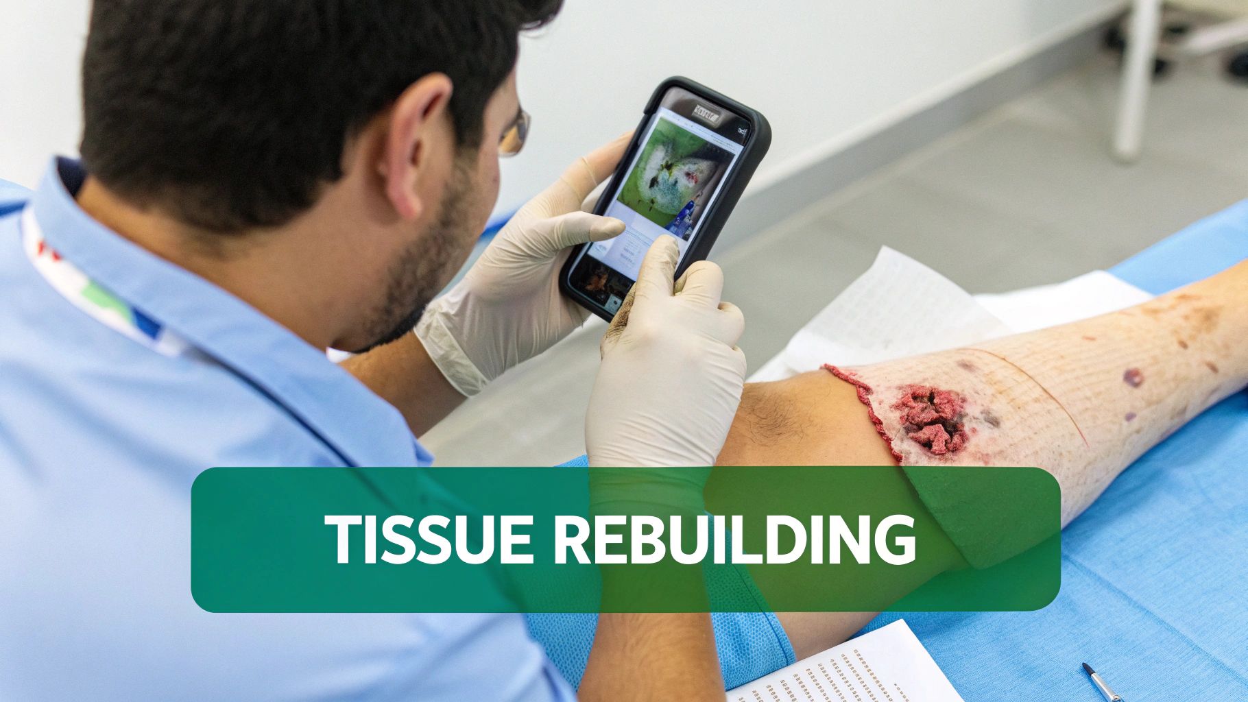
Once the cleanup crew has done its job, the body shifts from defense to construction. This is the proliferative phase, a period of intense rebuilding that kicks off around day four and can stretch for about three weeks. It’s where you can really see the magic happen as new tissue starts to fill the gap left by the injury.
This stage is like a construction site with three critical jobs running at the same time: laying new plumbing, putting up the framework, and finally, adding the roof. Each step is vital for closing the wound successfully. For clinicians, this is one of the most visually rewarding parts of the stages of normal wound healing, offering clear, tangible signs of progress.
Angiogenesis: Laying Down New Pipes
To power this massive rebuilding project, the wound needs a steady supply of oxygen and nutrients. This is where angiogenesis comes in—the formation of brand-new blood vessels. Tiny capillaries start to sprout from the healthy vessels at the wound's edge, branching out into the damaged area like a network of life-giving pipelines. It's these new vessels that give healthy healing tissue that vibrant, beefy red color. A well-vascularized wound bed is non-negotiable for healing.
Fibroplasia and Granulation: Building the Framework
At the same time, specialized cells called fibroblasts swarm into the wound. Their main mission is to produce collagen, the protein that gives our skin its strength and structure. They get to work laying down this new collagen, creating a fresh, though initially disorganized, matrix.
This mix of new blood vessels and fresh collagen creates what we call granulation tissue. As a wound care professional, this is exactly what you want to see. It looks bumpy or granular—almost like cobblestones—and is typically moist and bright red, which is a fantastic sign that the wound is actively healing.
Granulation tissue is the hallmark of a healthy proliferative phase. Its presence confirms a good blood supply and shows the wound is successfully building the scaffold it needs to close. It’s the foundation for everything that comes next.
Epithelialization: Putting on the Roof
The last big job in this phase is epithelialization. Here, new skin cells, or keratinocytes, mobilize from the wound's edges. They begin a slow crawl across the surface of the new granulation tissue, moving from all sides toward the center until they meet up.
This process lays down a new, protective epidermal layer over the wound, basically "putting a roof" on our construction site. This new skin is very fragile at first. Tracking this progress is a huge part of our job, which is why consistent wound measurements and high-quality photo documentation are so important. They help us objectively monitor how quickly granulation and epithelialization are occurring. For more on this, explore these evidence-based wound care practices in our detailed guide.
Maturation and Remodeling: Strengthening the New Tissue
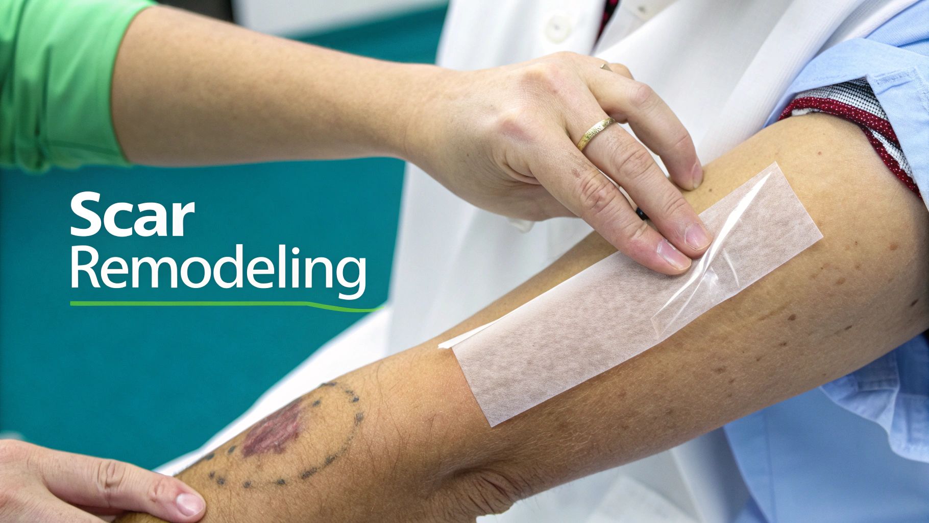
The final chapter in the healing story is maturation and remodeling, a slow and methodical renovation of the newly closed wound. Just because the surface looks sealed doesn't mean the work is done. This is easily the longest of the four stages, kicking off around day 21 and often lasting for a year or even longer.
The body's primary objective now is to strengthen and reorganize the tissue that was just laid down. The collagen put in place during the proliferation phase is a bit like a quick, messy patch job—it’s functional, but it’s not built to last. The body starts a careful, deliberate process of replacing that initial Type III collagen with the much stronger, more organized Type I collagen. This swap is the key to increasing the wound's tensile strength, its ability to withstand stretching and tension without tearing.
From a Quick Fix to a Lasting Repair
Think of this remodeling phase as a constant push-and-pull between collagen production and collagen breakdown. Specialized cells manage this delicate dance, slowly but surely realigning the collagen fibers along the natural lines of stress.
You can actually see the results of this long-term project by observing the scar over many months. At first, the new scar is typically raised, red or pink, and can be quite itchy. This is a sign of intense cellular activity. Over time, as remodeling progresses, the body reabsorbs many of those extra blood vessels. The scar begins to fade to a paler, less conspicuous color. It also flattens out and becomes softer to the touch.
Here’s a crucial takeaway for patient education: even after a year or more of remodeling, healed tissue is never quite the same. A fully matured scar will only ever achieve about 80% of the tensile strength of the original, uninjured skin. This is vital information for preventing re-injury.
The final look and feel of a scar depend on many factors, including age, genetics, and the quality of care received. This is why consistent, long-term documentation is so important for tracking the scar's evolution. Watching how a scar matures gives you incredible insight into the quality of the body's repair job and helps identify issues like hypertrophic or keloid scarring.
How to Document Each Stage of Wound Healing
Good documentation is more than just jotting down what you see. It's about strategically telling the wound's story, stage by stage.
Hemostasis and Inflammation
Right after an injury, your hemostasis notes set the stage. Record the injury time, what was done to stop the bleeding, and how long it took. It's also critical to note patient-specific factors, like if they're on anticoagulants.
As the body moves into the inflammatory phase, your focus should pivot to the wound bed and the surrounding skin. This is where describing exudate—the fluid draining from the wound—becomes incredibly important. Document its amount, type (serous, sanguineous, purulent), and any odor. Pay close attention to the periwound skin, too. Describe any erythema (redness), edema (swelling), and warmth. Using precise language here is what separates normal, expected inflammation from the first warning signs of an infection.
Proliferation and Maturation
The proliferative phase is all about rebuilding, so your documentation needs to capture that progress with numbers. Your notes need objective measurements: length, width, and depth. Even more importantly, quantify the types of tissue you see in the wound bed.
For instance, a powerful note might say: "Wound bed shows 75% pink, granular granulation tissue with 25% yellow slough along the top edge. New epithelial tissue is migrating from the wound edges, covering about 2mm."
This kind of detail gives anyone reading the chart an instant, clear picture of how things are progressing. Our guide on wound assessment tools for nurses is a great resource for this.
Finally, during the maturation phase, your documentation shifts to tracking how the scar is evolving. Your notes should follow its color, texture, and pliability over time, capturing details that can help guide any future scar management treatments.
Unpacking Common Questions in Wound Care
Even with a solid grasp of the four stages of wound healing, real-world scenarios always bring up new questions. Let's tackle some of the most common ones that pop up in clinical practice.
How Can I Tell if a Wound Is Stuck in the Inflammatory Stage?
The tell-tale sign is when those classic signs of inflammation just won't quit, and you're not seeing any real progress. If you're still observing significant redness, dealing with a lot of drainage, and not seeing that healthy, bumpy, bright red granulation tissue pop up after a week or two, you've likely got a stalled wound.
What's the Real Difference Between an Acute and a Chronic Wound?
An acute wound moves right along through the healing cascade as expected. A chronic wound, on the other hand, is the one that gets stuck, almost always trapped in that prolonged inflammatory phase.
As a rule of thumb, if a wound hasn't shown any meaningful signs of healing after 30 days, it’s time to reclassify it as chronic. This is a sign that something bigger is getting in the way, such as an underlying infection, poor circulation (ischemia), systemic issues like poorly controlled diabetes, or constant pressure on the wound bed.
Think of a stalled wound as a warning light. Your job is to play detective, find the root cause, and fix it to get things moving again.
Where Does Technology Fit into All This?
The biggest advantage of modern technology is that it takes the subjectivity out of wound assessment. It provides consistent, objective measurements, which eliminates the "eyeballing it" variability that happens when assessments are done manually. AI-powered image analysis, for example, can precisely calculate the percentage of different tissue types—like granulation versus slough—giving you hard data on whether the wound is truly improving. This helps you build a solid, evidence-based story of the wound's progress.
Streamline your entire workflow from voice to claim with Ekagra Health AI. Our platform uses AI to automate documentation, coding, and billing, reducing administrative burdens by up to 70% so you can focus on patient care. Learn how Ekagra Health AI can transform your practice.
