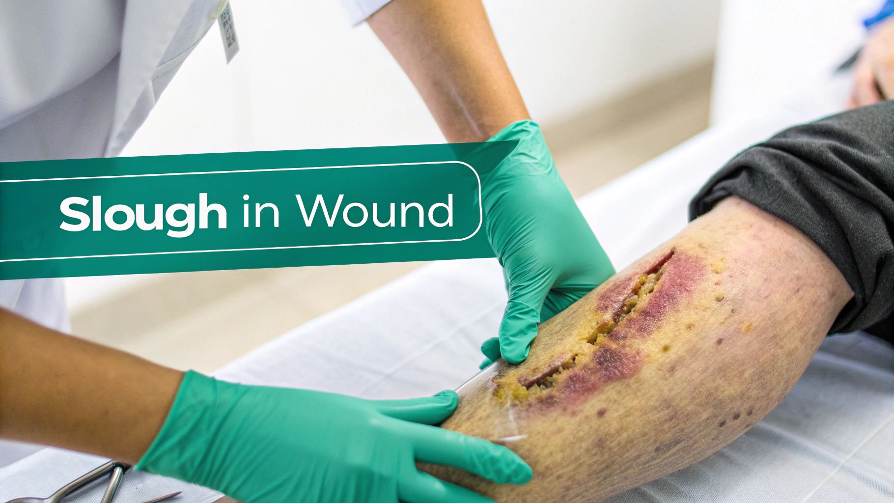
In the world of wound care, slough is a word you'll hear a lot. It's that stringy, yellowish, or tan gunk you often see covering a wound bed. More than just an unsightly layer, slough is a mix of dead tissue, cellular junk, and a protein called fibrin that builds up and stalls the healing process. Getting a handle on what slough means from a medical standpoint is the first step toward effective treatment. Without a clear understanding, clinicians risk misinterpreting wound status, choosing the wrong treatment, and ultimately prolonging patient suffering. This guide will provide a deep dive into identifying, understanding, and managing slough to empower healthcare professionals in their practice.
What Is Slough and Why Does It Stall Healing?
When the body gets injured, it kicks off an amazing, highly coordinated repair sequence. A key part of this is the inflammatory phase, where the body's cleanup crew rushes in to clear out damaged cells and get the site ready for rebuilding. Slough is essentially the leftover debris from this initial cleanup. If the body's natural processes are overwhelmed or compromised, this debris accumulates instead of being cleared away.
Think of it like getting a construction site ready. Before you can pour a new foundation (which is like healthy granulation tissue) or put up walls (new skin), you have to clear away all the rubble and old materials. Slough is that rubble. If it’s left sitting there, it physically obstructs the path for new cells, and the whole project comes to a standstill.
This buildup of non-viable tissue is a major roadblock. It physically prevents new, healthy cells—specifically keratinocytes and fibroblasts—from traveling across the wound to close it. This phenomenon, known as stalled epithelialization, is one of the main reasons so many chronic wounds get stuck and just won't heal. The presence of slough effectively traps the wound in a prolonged inflammatory state, preventing it from advancing to the proliferative phase of healing.
The Composition of Wound Slough
Slough isn't just one thing—it's a complex, sticky mixture of biological materials. Knowing what it's made of helps us understand why it's such a problem from both a biological and clinical perspective. Its composition explains its appearance, texture, and its detrimental effect on the wound environment.
Here’s a look at what’s usually in the mix:
- Fibrin: This is a protein that helps with clotting, but in a non-healing wound, it can form a tough, mesh-like covering over the wound bed, trapping other debris.
- Leukocytes: These are the dead white blood cells, primarily neutrophils, that were initially sent to fight off infection and clean up the area. Their accumulation contributes to the yellowish, purulent appearance of slough.
- Cellular Debris: This is a collection of dead skin, fat, and muscle cells that need to be cleared out. This necrotic material serves no purpose and must be removed for healing to progress.
- Bacteria and Biofilm: Slough creates a perfect home for bacteria. It’s moist and full of nutrients, allowing bacteria to multiply and often form a protective, slimy shield known as a biofilm, which is highly resistant to antibiotics and immune responses.
All these components together create a damp, nutrient-rich sludge that unfortunately is far better at growing bacteria than it is at helping new tissue grow. The presence of this material significantly increases the bioburden of the wound.
Key Takeaway: Identifying and dealing with slough is far more than just cleaning a wound—it's a crucial step that directly impacts how fast a wound heals, the risk of infection, and the patient's final outcome.
The presence of slough is a serious clinical hurdle. A UK study highlighted this, showing that chronic wounds with a suspected or confirmed infection (often linked to slough) had only a 45% healing rate. That's a big drop from the 59% healing rate for uninfected chronic wounds, really driving home how much slough can interfere with recovery. You can learn more about these wound healing findings in the full study.
Because slough can look so different—varying in color, texture, and how stubbornly it sticks to the wound—the first step for any clinician is learning to spot it accurately. This initial assessment is what drives every decision that follows, from picking the right dressing to deciding if debridement is necessary. Accurate identification is a cornerstone of effective wound management.
Slough at a Glance Key Characteristics
To help with that crucial first step, here is a quick reference table. It breaks down the essential attributes of slough to help clinicians identify it quickly and confidently during a wound assessment. This systematic approach promotes consistent documentation and communication among the care team.
| Characteristic | Description | Clinical Implication |
|---|---|---|
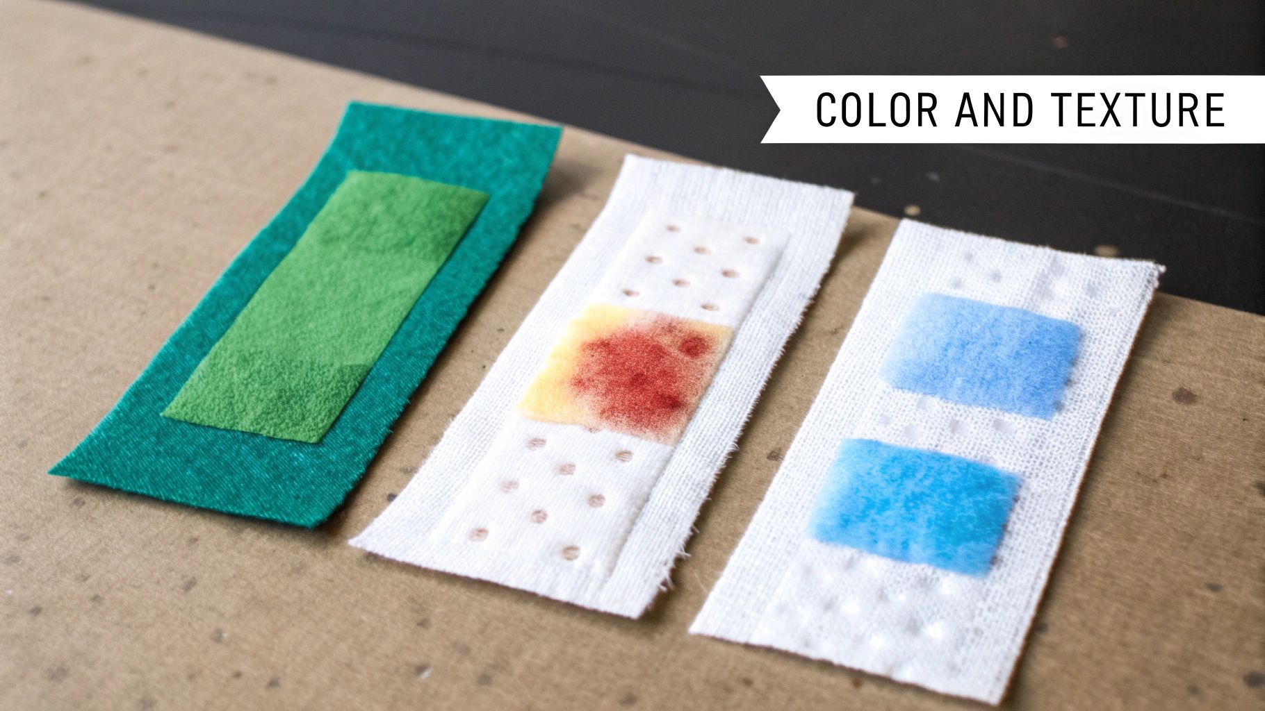
| Color | Typically yellow, tan, beige, or sometimes grayish or green. | A green hue can indicate Pseudomonas infection; gray may suggest poor perfusion. |
| Texture | Can be stringy, fibrinous, gelatinous, or paste-like. | Texture often dictates the best debridement method (e.g., loose slough may respond to irrigation). |
| Adherence | May be loosely attached and easy to remove or firmly adhered to the wound bed. | Tenacious slough may require more aggressive debridement, such as sharp or enzymatic methods. |
| Moisture | Is almost always moist or wet, contributing to its soft, non-viable nature. | The high moisture content supports bacterial growth and can lead to maceration of surrounding skin. |
| Amount | Documented as a percentage of the wound bed it covers (e.g., 25% slough, 75% granulation tissue). | Quantifying the amount allows for objective tracking of treatment effectiveness over time. |
| Location | Found within the wound bed, often mixed with other tissue types like granulation or eschar. | Its location can provide clues about the etiology of the wound and areas of greatest pressure or ischemia. |
This table serves as a practical field guide. By keeping these characteristics in mind, you can build the confidence needed to distinguish slough from other tissues and make sound clinical judgments for your patients.
Learning to Read Wound Slough: Colors and Textures
Knowing the basic medical definition of slough is one thing, but the real clinical skill comes from being able to interpret what you see. Think of yourself as a detective at the bedside. Just like a mechanic can diagnose an engine by its sounds, an experienced clinician learns to "read" the wound bed. This interpretive skill transforms assessment from a simple observation into a diagnostic process.
Slough is never just one-size-fits-all. Its appearance tells a story, offering valuable clues about the wound’s hydration status, bioburden, and overall healing environment. It’s a visual progress report, and learning to read it correctly helps you make smarter treatment decisions that are tailored to the wound's specific needs.
This skill is also your first line of defense against misidentification. For instance, a thin, yellowish film of fibrin over healthy, budding granulation tissue can easily be mistaken for slough. Making that mistake could lead to inappropriately aggressive debridement, removing healthy tissue and causing a major setback for the patient. Conversely, mistaking pus for slough could delay the diagnosis of a serious infection.
Decoding the Color Spectrum of Slough
The color of slough is your first and most immediate clue. While it’s typically some shade of yellow or tan, different colors can signal specific issues, like bacterial colonization or changes in moisture. Paying attention to these nuances is critical for accurate diagnosis.
- Yellow or Tan: This is the classic look. You're seeing a combination of fibrin, dead cells, and white blood cells. It’s a clear sign of non-viable tissue that needs to be managed and is the most common presentation of slough.
- Off-White or Grayish: A duller, gray-toned slough often points to a compromised blood supply (ischemia). It can also mean the tissue has been there for a while and is more devitalized. This finding should prompt a vascular assessment.
- Greenish Hue: A green tint should always get your attention. It's a major red flag, often associated with bacteria like Pseudomonas aeruginosa, which produces a tell-tale green pigment called pyocyanin and has a distinct, sickly sweet odor. If you see green, it’s time to look closely for other signs of infection and consider a wound culture.
Remember, context is everything. A small patch of yellow slough in an otherwise clean, healing wound is a totally different scenario than a wound filled with foul-smelling, greenish slough surrounded by angry red skin (erythema) and warmth. The overall clinical picture must guide your interpretation.
Understanding Slough Textures and Consistency
Beyond color, the texture and consistency of slough are just as telling. These characteristics give you insight into the wound’s moisture balance and will often guide your choice of debridement method and dressing.
Clinical Insight: The texture of the slough often dictates how difficult it will be to remove. Loosely adherent, soupy slough might come away with gentle irrigation, whereas thick, stringy slough will likely require a more hands-on approach like sharp debridement.
Here are a few common textures you'll encounter:
- Stringy or Fibrinous: This type looks like tangled threads or damp cotton fibers clinging tightly to the wound bed. This texture is often due to high concentrations of fibrin and can be tough to remove without accidentally damaging the healthy tissue underneath.
- Soft or Gelatinous: Think of something like melted cheese or a wet paste. This texture is usually less adherent and points to a very moist, high-exudate environment. It's often easier to remove but signals a critical need to manage moisture to prevent maceration of the surrounding skin.
- Mucinous or Paste-like: This thicker, more uniform gunk can sometimes be mistaken for pus. The key difference is that slough is more organized and attached to the wound bed, while pus (purulent drainage) is a sign of active infection and is typically free-flowing, thinner, and often accompanied by a foul odor.
Getting these descriptions right is crucial for communicating with the rest of the care team. Mastering the language of color and texture, along with other wound bed descriptions, turns a simple observation into a powerful diagnostic tool that ensures everyone is on the same page and providing consistent care.
Slough vs Eschar: Differentiating Necrotic Tissue Types
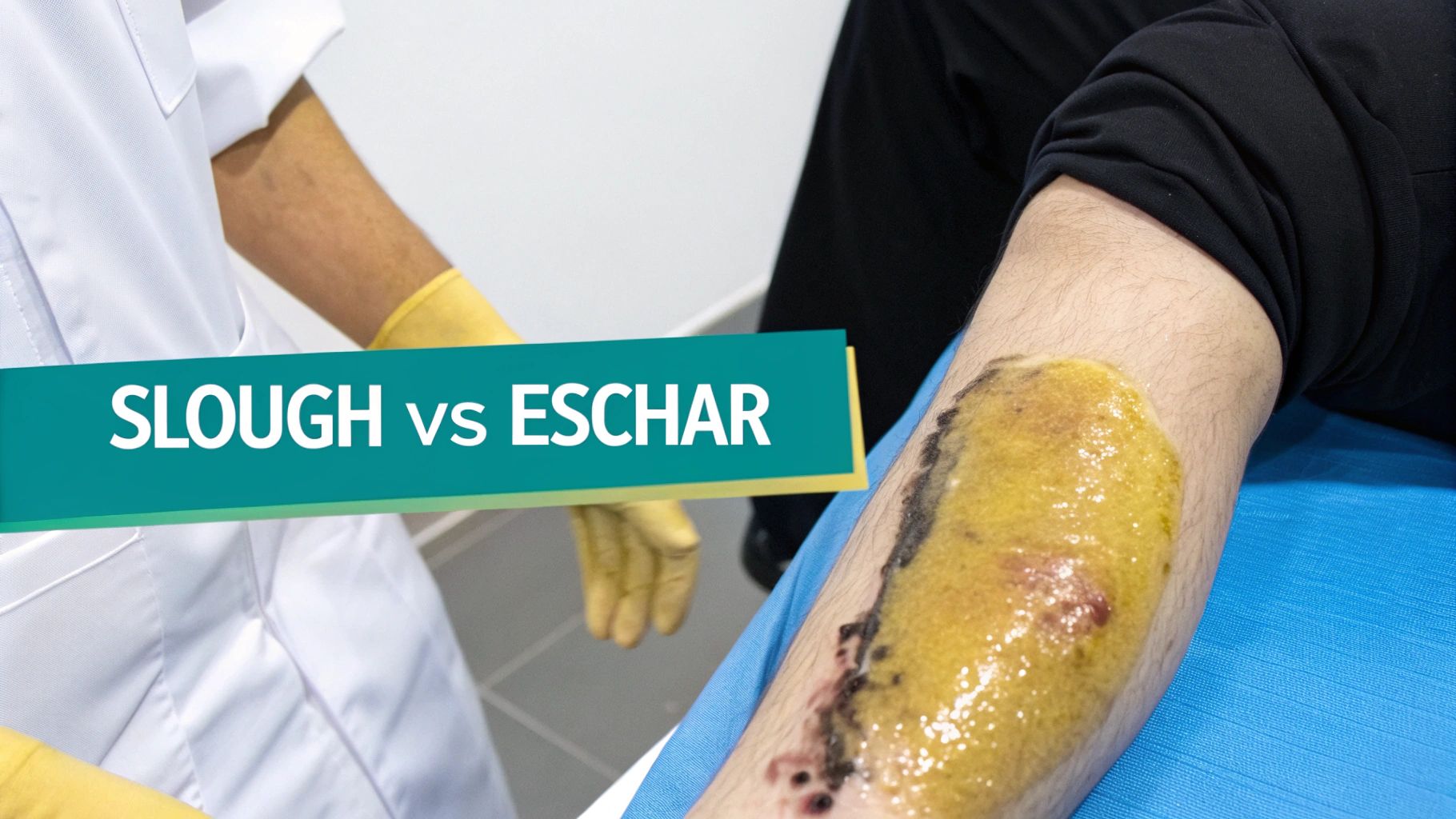
When you look at a wound bed, it's crucial to know that not all non-viable tissue is the same. While you'll frequently see slough, it’s often found alongside another type of necrotic tissue: eschar. Confusing the two is an easy trap to fall into for novice clinicians, but it's a mistake that can derail your entire treatment plan and potentially harm the patient.
Getting this distinction right is a foundational skill in wound care. It’s what helps you decide between reaching for a moisture-donating dressing or a surgical scalpel. Simply put, slough and eschar tell two completely different stories about what the wound has been through, its level of hydration, and what it needs right now.
Think of it like this: slough is a wet, disorganized blanket of gunk thrown over the wound. It’s messy, it harbors bacteria, and it needs to be cleared away for healing to happen. Eschar, in contrast, is more like a hard, dry shield protecting the injury underneath. Sometimes that shield needs to be removed, but other times, particularly in cases of poor arterial supply, it’s best left alone as a natural biological cover.
The Defining Characteristics of Slough
As we've covered, slough is that moist, devitalized tissue that shows up as part of the inflammatory process. It's a mix of fibrin, dead cells, and wound fluid that creates a perfect breeding ground for bacteria and physically obstructs healing.
The biggest giveaways are its moisture and texture.
- Color: Look for shades of yellow, tan, beige, or even grayish-white.
- Texture: It can be stringy, fibrous, gelatinous, or have a paste-like consistency.
- Adherence: You'll find it either loosely attached or stubbornly stuck to the wound bed.
- Moisture: It's almost always moist or wet, reflecting a hydrated state.
Slough is a red flag that the wound is stuck in a prolonged inflammatory phase. Its physical presence is a roadblock, preventing healthy granulation tissue from forming and stopping new skin cells from migrating across the wound to close it. Its management almost always involves debridement and moisture management.
The Defining Characteristics of Eschar
Eschar, on the other hand, points to a more advanced stage of tissue death, usually involving full-thickness damage where the tissue has become desiccated. It’s essentially dried-out—necrotic tissue and is a hallmark of deeper injuries like Stage 3 or 4 pressure injuries or arterial ulcers.
Dryness and firmness are its defining features.
- Color: It’s typically dark brown, black, or a leathery tan.
- Texture: The tissue feels hard, leathery, and dry to the touch, sometimes described as feeling like shoe leather.
- Adherence: Eschar is almost always firmly attached to the wound edges and underlying structures.
- Moisture: It is characteristically dry and dehydrated.
The presence of eschar often signals a more serious injury, like a deep pressure ulcer or an arterial wound where blood flow is severely restricted. Unlike slough, a patch of stable, dry eschar on a heel or an ischemic limb is sometimes left in place to act as a natural, biological cover. Removing it in such cases could expose the underlying structures to infection without adequate blood flow to support healing.
Key Distinction: The fundamental difference comes down to one thing: hydration. Slough is a hydrated form of necrotic tissue, while eschar is a dehydrated form. This single factor drives their appearance, texture, and the clinical approach we take.
Slough vs Eschar: A Clinical Comparison
To make identification easier at the bedside, this side-by-side table breaks down the critical differences between slough and eschar. Mastering these distinctions is vital for accurate assessment and choosing the right debridement strategy.
| Feature | Slough | Eschar |
|---|---|---|
| Primary Color | Yellow, tan, beige, gray | Black, dark brown, leathery tan |
| Texture | Moist, stringy, soft, gelatinous | Hard, dry, leathery, firm |
| Moisture Level | High (hydrated necrotic tissue) | Low (dehydrated necrotic tissue) |
| Adherence | Loosely or firmly attached | Very firmly attached to the wound edges |
| Clinical Story | Prolonged inflammation, bioburden | Deeper tissue injury, ischemia |
| Common Location | Venous ulcers, infected wounds | Pressure injuries, arterial ulcers |
Ultimately, learning to tell these two apart is much more than an academic exercise—it directly shapes your plan of care. Mistaking hard eschar for adherent slough could lead to a harmful debridement attempt on an ischemic limb. Conversely, leaving slough in the wound bed because you think it's a stable covering just lets the wound fester and delays healing indefinitely. Accurate identification is always the first and most critical step toward healing.
How Slough Actively Sabotages the Healing Process
Think of slough as more than just a passive layer of gunk sitting in a wound; it's an active saboteur, constantly working against the body's attempts to heal. Its presence completely derails the natural repair process, turning what should be a straightforward sequence of events into a stalled, frustrating battle. Understanding exactly why slough is so harmful gives us the clinical rationale we need to be aggressive about removing it.
Imagine a road crew trying to pave a street that's still covered in mud, rocks, and garbage. It doesn't matter how skilled the workers are or how good their asphalt is—they simply can't make progress. Slough is that physical barrier, literally blocking new skin cells (the "pavers") from traveling across the wound bed to close it up. It also obscures the clinician's view of the underlying wound bed, making it impossible to assess for signs of new tissue growth or infection.
Even worse, this dead tissue isn't just sitting there. It's the perfect breeding ground for bacteria, providing a warm, moist, and nutrient-rich buffet where microbes can multiply. This is how biofilms get their start—those slimy, stubborn colonies of bacteria that are notoriously difficult to get rid of and are a major cause of chronic wound infections.
The Vicious Cycle of Inflammation and Biofilm
When slough is left in a wound, it traps the injury in a state of chronic inflammation. Your body keeps sending in inflammatory cells (neutrophils and macrophages) to clean up the mess, but they can't keep up and become overwhelmed. This constant inflammatory state doesn't just stall progress; it actively damages the healthy tissue around the wound by releasing destructive enzymes.
This kicks off a destructive, self-perpetuating cycle:
- Slough gives bacteria the perfect surface to latch onto and form a biofilm.
- The biofilm acts like a shield, protecting the bacteria from both antibiotics and the body's own immune system.
- This persistent colony of bacteria triggers a non-stop inflammatory response.
- The inflammation causes more tissue to break down, which in turn creates even more slough.
A wound trapped in this cycle cannot heal. It is stuck, often leading to increased pain for the patient, a higher risk of systemic infection (sepsis), and significantly longer, more costly treatment plans.
This is precisely why understanding the medical meaning of slough is so critical. When you see it as an active enemy, not just a byproduct, you can intervene with the right strategies to break the cycle. To get a better sense of how things should work, you can learn more about the complete wound healing process and its distinct stages. Removing the slough is the key to breaking this vicious cycle.
The Clinical and Economic Consequences
Failing to manage slough has a ripple effect that goes far beyond the wound itself. Stalled wounds mean poor patient outcomes—prolonged pain, a lower quality of life, and the potential for severe complications like osteomyelitis (bone infection) or limb loss.
The financial cost is just as staggering. Chronic wounds place a massive burden on healthcare systems. The global prevalence of chronic wounds is estimated at 1.67 per 1000 population, and the wound care market as a whole was valued at $23.66 billion in 2023. These numbers underscore the pressure to get healing right, and effective slough management is a huge part of that effort. You can discover more insights about the growing wound care market on BioSpace and what it means for clinical practice.
By reframing slough not as something simply in the wound but as something actively harming it, we can make a stronger case for timely and thorough debridement. Getting that barrier out of the way is the first and most important step to restart the healing clock and get the patient back on the road to recovery.
Choosing the Right Debridement Method for Slough
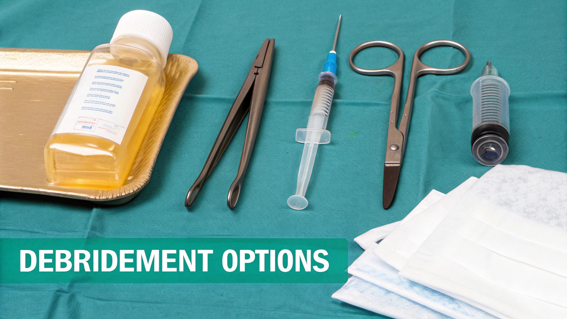
Once you've spotted slough in a wound and understand how it’s actively stalling the healing process, the next step is clear: you have to get it out. This removal process is called debridement, and it’s a non-negotiable part of good wound bed preparation. The entire goal is to clear out all that non-viable junk to give healthy, new cells a clean surface to start rebuilding.
But picking a debridement method isn't a one-size-fits-all deal. The best approach hinges on a handful of factors—the wound itself (amount and type of necrotic tissue), the patient's overall health and pain tolerance, the clinical setting, and of course, your own skill level. Making the right call means matching the right tool to the job, ensuring the method is not just effective but also appropriate for that specific patient's situation.
Autolytic Debridement: Letting the Body Do the Work
Autolytic debridement is the gentlest and most selective option we have. It basically harnesses the body’s own cleanup crew—its natural enzymes (like matrix metalloproteinases) and moisture—to liquefy and separate the slough from the healthy tissue underneath. Think of it like creating a perfect little greenhouse environment where the body can do its own housekeeping.
To make this happen, you apply a moisture-retentive dressing, like a hydrocolloid, a hydrogel, or a transparent film. This dressing traps the wound fluid, which is loaded with the very enzymes needed to break down dead tissue. It’s a fantastic choice for patients who can't handle more aggressive methods or for wounds with just a small to moderate amount of slough.
The trade-off? It's slow, often taking days or weeks. And you absolutely cannot use it on an infected wound, because that warm, moist environment is a perfect breeding ground for bacteria. Autolytic debridement is best reserved for stable wounds in patients whose immune systems are up to the task.
Enzymatic Debridement: A Little Targeted Help
Sometimes the body needs a nudge. That's where enzymatic debridement comes in, offering a more focused approach. This method involves applying a prescribed topical agent—usually a collagenase-based ointment—directly onto the slough. These external enzymes get to work selectively, dissolving the collagen fibers that anchor the slough to the wound bed without messing with the healthy granulation tissue.
It’s definitely faster than letting the body do it alone and works particularly well for softening up and removing that stubborn, stuck-on slough, often in conjunction with other methods.
Clinical Tip: Enzymatic debridement is a great middle-ground strategy. It's perfect when sharp debridement isn't an option (due to the patient's condition or the care setting), but you need faster results than you'd get with autolysis.
Before you go this route, consider a few things:
- Patient Condition: It's a solid choice for patients on anticoagulants where sharp debridement would pose too high a bleeding risk.
- Wound Status: Don't use it on wounds with exposed tendons or bone. Also, be aware that some enzymatic agents are deactivated by certain metals, like the silver found in many antimicrobial dressings, so dressing compatibility is key.
- Cost: These prescription ointments can be significantly more expensive than simple moisture-retentive dressings.
Mechanical Debridement: The Physical Approach
Mechanical debridement is exactly what it sounds like: physically removing slough from the wound bed. It's one of the oldest wound cleaning techniques out there. While it can be effective, its major drawback is that it’s generally non-selective, meaning it can easily take healthy tissue right along with the bad stuff. Because of this potential for collateral damage, we use it much more carefully these days.
Common methods include:
- Wet-to-Dry Dressings: This classic technique involves placing moist gauze in the wound, letting it dry, and then pulling it out, which rips away adherent tissue. It’s often painful and has largely fallen out of favor due to its non-selective nature and potential to damage healthy granulation tissue.
- Wound Irrigation: A much more common and gentle method. It uses pressurized fluid, like normal saline, to flush loose slough and debris out of the wound. This is a standard part of most dressing changes.
- Monofilament Pads: These are specially designed pads with fine fibers that you can use to gently scrub the wound bed, dislodging slough without causing major trauma.
This approach is really only suited for wounds with a lot of loose, easily removable slough. You'd want to steer clear of using it on a clean, granulating wound, where it would just tear up all that fragile new tissue.
Sharp and Surgical Debridement: The Gold Standard for Fast Results
When you’re up against extensive, thick, or firmly attached slough—or if infection is a concern—it's time to bring in the big guns. Sharp and surgical debridement are the fastest, most efficient ways to get a wound clean.
Sharp debridement is a more conservative procedure, often performed right at the bedside by a trained clinician (like a WOC nurse or podiatrist). Using sterile instruments like a scalpel, scissors, or forceps, the clinician can precisely remove the non-viable tissue. It requires a steady hand and a high level of skill to avoid damaging nerves, tendons, and other important underlying structures.
Surgical debridement, on the other hand, happens in an operating room. It's a much more aggressive procedure for removing large amounts of tissue and is typically reserved for severe cases involving extensive necrosis, deep infections, or wounds that go all the way down to bone or tendon. It's the undisputed gold standard for quickly getting to a clean, viable wound bed, but it comes with all the inherent risks of surgery and anesthesia.
Choosing between these methods demands a sharp clinical eye. The urgency of the situation is key. For example, signs of advancing cellulitis or sepsis are a red flag that call for immediate surgical intervention. A deep understanding of what slough means for the wound and the patient is what guides these critical, time-sensitive decisions.
Documenting Slough for Better Care and Reimbursement
In wound care, what you write down is just as important as what you do. Your documentation is the thread that connects the entire care team, and it can make or break not only the patient's progress but also your facility's reimbursement. Accurate, detailed documentation is not just an administrative task; it is a critical component of patient safety and quality care.
When it comes to slough, vague notes just don't cut it. Charting "some yellow stuff" or "gooey drainage" leaves far too much to interpretation and can lead to inconsistent care. The goal isn't just to jot down notes; it's to paint a clear, objective picture of the wound bed so that any clinician picking up that chart knows exactly what's going on.
This is about creating a reliable timeline. Precise, quantifiable descriptions help everyone track the wound’s journey, making it obvious whether your current plan is working or if it's time to pivot. It provides the data needed to justify treatment changes and demonstrate medical necessity.
Moving Beyond Vague Notes
The first step is getting your team on the same page with a consistent, descriptive vocabulary. You need to document the key characteristics of the slough: its color, texture, adherence, and the amount covering the wound bed. This systematic approach takes the guesswork out of wound assessment and ensures that progress can be tracked objectively.
Let's look at the difference this makes.
- Vague: "Wound has yellow slough."
- Specific: "Wound bed is approximately 60% covered with adherent, fibrinous, tan-colored slough concentrated on the medial aspect. The remaining 40% of the wound bed is pale pink granulation tissue."
That second note gives the next clinician a wealth of information. "Adherent" tells them debridement might be a challenge. "Fibrinous" describes its stringy nature. And quantifying the amount provides a concrete baseline to measure against at the next dressing change.
Great documentation is your best defense. It serves as a legal record, justifies your clinical decisions, and provides the hard evidence needed to support billing and coding for procedures and advanced wound care products.
When everyone from the wound clinic specialist to the home health nurse uses the same language, continuity of care improves dramatically. The patient gets a much more coordinated and effective treatment plan. If you're looking to standardize your team's approach, exploring different wound assessment tools for nurses is a fantastic place to start.
Connecting Documentation to Coding and Reimbursement
Let's talk about the financial side. Precise charting has a direct, tangible impact on your organization's revenue. Payers, whether it's Medicare or a private insurance company, need to see detailed documentation that justifies the services you're billing for—especially for procedures like debridement.
Your notes must tell a story that clearly supports the ICD-10 (diagnosis) and CPT (procedure) codes you submit. For instance, if you're billing for sharp debridement (CPT code 97597), your documentation has to describe the non-viable tissue—the slough or eschar—that made the procedure a medical necessity.
Here’s what a solid, defensible note for debridement might look like:
- Pre-procedure Assessment: "Central wound bed is 80% covered with thick, tenacious yellow slough, preventing assessment of underlying tissue. No signs of granulation tissue are visible. Patient verbalizes understanding of the procedure and associated risks."
- Procedure Details: "Sharp debridement was performed using a sterile scalpel and forceps to remove all loosely adherent and tenacious slough down to viable, bleeding tissue. Hemostasis achieved with light pressure."
- Post-procedure Assessment: "Post-debridement, the wound bed is now 100% granular and viable, with no signs of bleeding. Wound cleansed with normal saline and dressed per orders."
This kind of detailed narrative proves that the service you billed for was both necessary and performed correctly. Without it, you’re opening the door to claim denials, audits, and lost revenue. In the end, what you write—or fail to write—directly hits the bottom line.
A Few Common Questions About Slough
Even with a solid grasp of wound care, a few questions always seem to pop up in day-to-day practice. Let's tackle some of the most common ones you'll encounter at the bedside to clarify these key points.
Can a Wound Actually Heal with Slough in It?
In short, no. While you might see some very minor activity at the wound edges (epithelialization), a wound can't truly heal and close if there's a significant amount of slough present. The body's healing mechanisms are fundamentally inhibited by its presence.
Think of slough as a roadblock. It's a physical barrier that stops healthy new tissue from filling in the gap. Even worse, it's a perfect breeding ground for bacteria, which perpetuates a state of chronic inflammation. This combination stalls the healing process completely, which is why debridement is almost always necessary to get things moving again.
Is Yellow Slough Always a Sign of Infection?
Not necessarily, but it’s a great question that often causes confusion. Yellow is actually a very common color for slough, mostly because it's made up of fibrin and old white blood cells. By itself, yellow tissue doesn't automatically mean the wound is infected.
However, it should get your attention. You need to look for other classic signs of infection to confirm your suspicions. Be on high alert if you also see:
- New or worsening pain
- A foul, pungent odor
- Redness (erythema) and warmth spreading around the wound
- An increase in purulent drainage, or pus
- Fever or other systemic signs of illness
Always look at the whole picture—the patient and the peri-wound skin. The color alone doesn't tell the full story.
Clinical Takeaway: Context is everything. Yellow slough is a problem because it stops healing, but it only points to infection when other red-flag symptoms are present.
What's the Best Way to Document How Much Slough There Is?
The gold standard here is to estimate the percentage of the wound bed covered by each tissue type. This method, sometimes called the "clock method" (imagining the wound as a clock face), takes the guesswork out of it and gives you a clear, objective baseline to track progress (or decline) over time.
For instance, a great charting note would be something like: "Wound bed is 70% covered with loose, yellow slough and 30% with red granulation tissue." This creates a precise snapshot that anyone on the care team can understand. This quantification is crucial for demonstrating the effectiveness of your treatment plan, which is vital for continuity of care and proper documentation for reimbursement.
At Ekagra Health AI, we know that managing complex wounds requires more than just clinical skill—it demands efficient, accurate documentation. Our AI-powered platform turns your voice notes into structured, coded charts in minutes, freeing you from administrative burdens so you can focus on what matters most: your patients. Discover how our "voice to claim" solution can help your team improve outcomes and accelerate reimbursement by visiting Ekagra Health AI.
