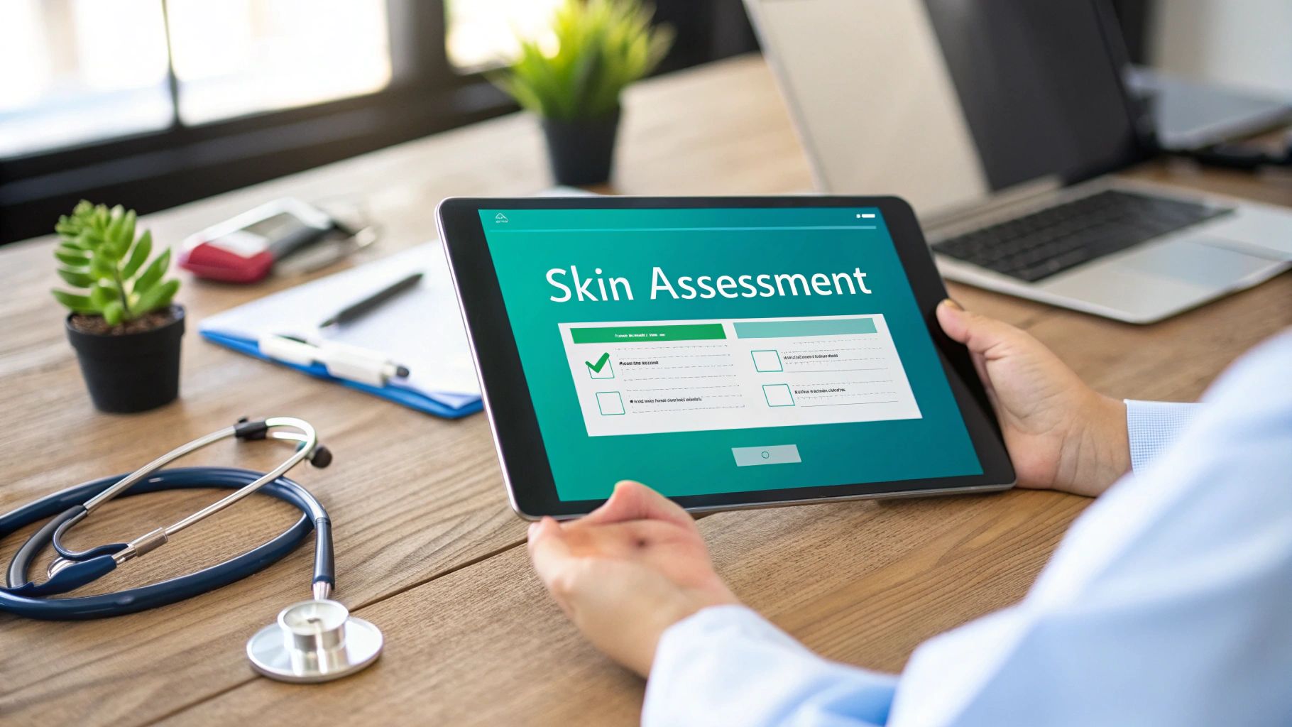
Great wound care isn’t just about the treatment plan; it's built on a foundation of meticulous skin assessment and documentation. We've all seen it: fragmented, inconsistent charting that does more harm than good. This isn't just a paperwork headache—it's a real clinical risk that can stall healing, muddy a diagnosis, and even trigger a painful audit. This guide gets practical, moving past the textbook theories to tackle the challenges we face on the floor every day. We will delve into a new standard for documentation, the nuances of a comprehensive physical assessment, techniques for accurate measurement, and how to craft clinical notes that are both compliant and genuinely useful for the entire care team.
A New Standard in Wound Care Documentation
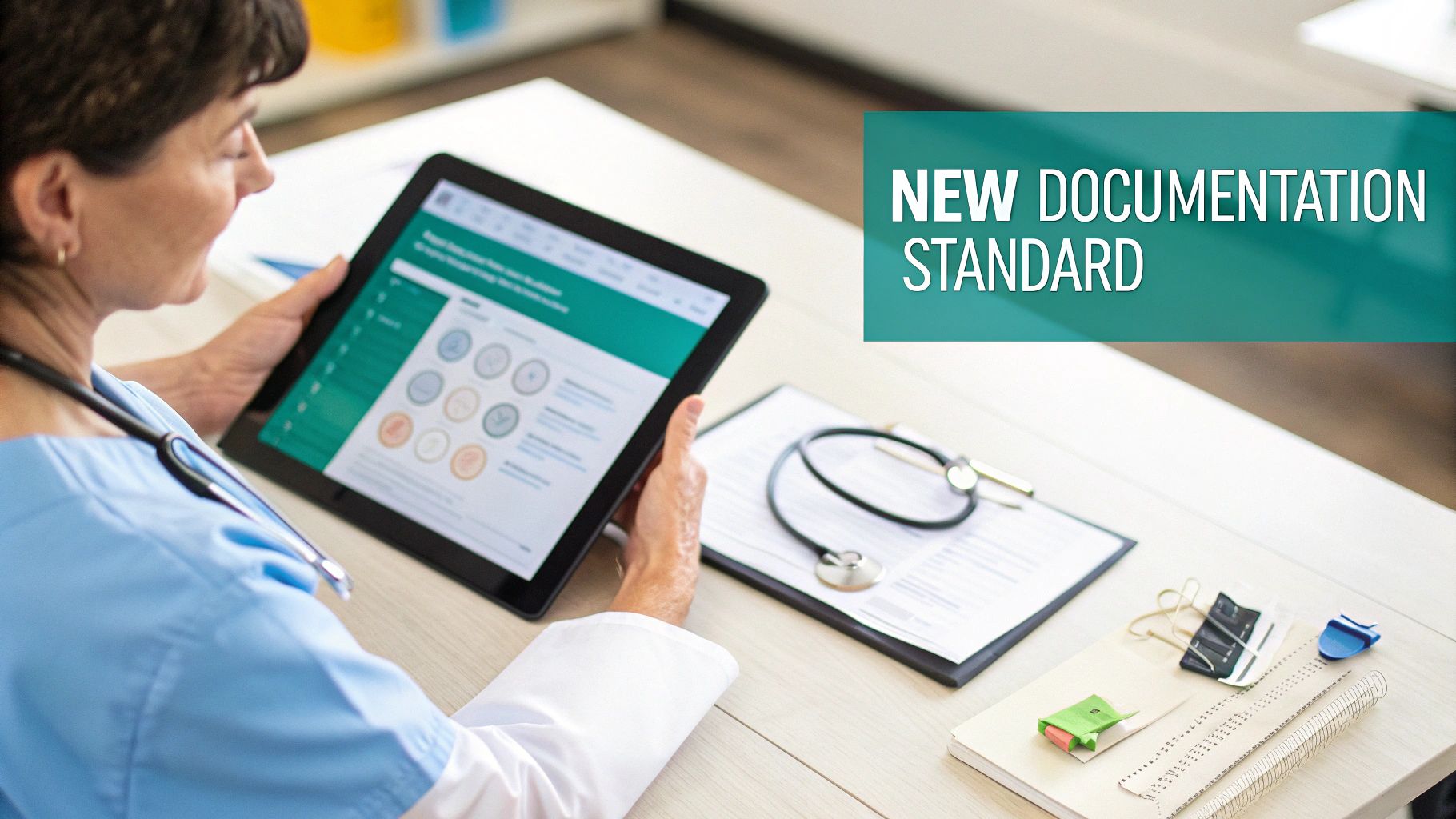
Let's walk through a modern workflow where assessment and documentation are two sides of the same coin, not separate, burdensome tasks. The goal here is simple: turn a time-consuming chore into a sharp, efficient practice that gives you better data. When you get this right, you directly support better patient outcomes while also shoring up your facility's operational and financial health. The evolution from manual, often after-hours charting to real-time, AI-assisted documentation represents a fundamental shift in clinical practice, one that prioritizes data integrity and patient-facing time.
This problem is bigger than just one clinic or hospital. It's a global issue. The landmark GRIDD Study by GlobalSkin collected data from 4,118 people in 96 countries, revealing just how much inconsistent documentation can delay diagnoses and throw off how healthcare resources are allocated. Their findings are a clear call for smarter surveillance and digital tools to get a true picture of skin conditions. The study highlights that systemic data collection is not just a 'nice-to-have' but an essential component of public health strategy, influencing everything from research funding to healthcare policy.
The Problem With Fragmented Charting
In a hectic clinical environment, documentation often gets pushed to the end of the shift, something to be rushed through. This habit creates scattered, incomplete records that don't tell the full story of a patient's healing journey. This "documentation debt" accumulates over time, leading to a cascade of negative consequences that impact patient safety, provider burnout, and organizational liability.
A patient's chart should read like a clear, chronological narrative of their care. When notes are inconsistent or incomplete, crucial details get lost, putting both the patient and the clinician at risk.
This fragmentation has real, immediate consequences for both patient care and your facility’s bottom line:
- Delayed Healing: How can you tell if a treatment is working without a clear, consistent record of the wound's progress? You might stick with a failing strategy simply because the data isn’t there to show you it’s time to pivot. A wound that isn't showing signs of healing within a two-to-four-week period requires a reassessment of the care plan, a process that is impossible without accurate longitudinal data.
- Increased Audit Risk: Incomplete or conflicting documentation is a huge red flag for auditors. It's an open invitation for claim denials, financial penalties, and a whole lot of unwanted scrutiny on your practice. Payers and regulatory bodies require proof of medical necessity, and a fragmented record fails to provide this justification.
- Communication Breakdowns: Vague notes force the next clinician to start their assessment from square one. This wastes precious time and introduces opportunities for errors that can disrupt the continuity of care. It breaks the chain of communication, leading to redundant assessments and potentially contradictory interventions.
The Shift To Integrated Workflows
The solution is to stop treating assessment and documentation as two separate steps. They need to become a single, fluid process. Modern tools are finally making this a reality, letting clinicians capture high-quality data right at the bedside without taking their focus off the patient. This integration means that the act of assessing is simultaneously the act of documenting, creating a seamless flow of information from the patient to the electronic health record (EHR).
A quick look at the old way versus the new way makes the difference pretty clear.
Manual Vs AI-Powered Documentation Workflow
| Metric | Traditional Manual Process | AI-Powered Process (e.g., Ekagra Health) |
|---|---|---|
| Time Spent | 20-30 minutes per patient visit, often after hours. | 2-5 minutes per patient visit, completed in real-time. |
| Accuracy | Prone to human error, typos, and forgotten details. | High accuracy with structured data captured from conversation. |
| Compliance | Dependent on individual clinician's knowledge of coding/regulations. | Built-in compliance checks and standardized templates reduce risk. |
| Data Quality | Often lacks structured, longitudinal data for analysis. | Creates clean, structured data perfect for tracking outcomes. |
The takeaway is straightforward. Tools like Ekagra Health AI are designed to absorb the documentation burden by capturing ambient clinical conversations and instantly generating structured, compliant notes. This isn’t just about saving time; it’s about fundamentally improving the quality and consistency of your clinical records, which is the key to providing truly exceptional care. This technological leap allows for the capture of rich, nuanced patient encounters without the administrative drag, freeing clinicians to practice at the top of their license.
Performing a Comprehensive Skin and Wound Assessment
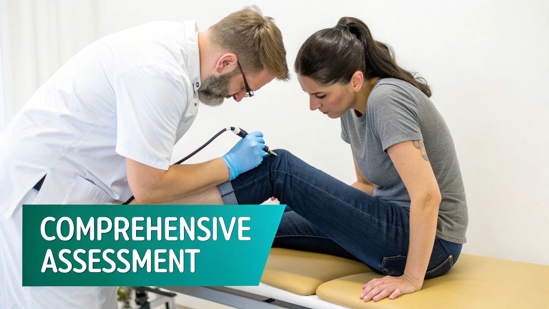
A thorough physical assessment is the bedrock of good wound care. It's where we gather the raw data that informs every single treatment decision we make. This isn't just about glancing at a wound; it’s a systematic, head-to-toe investigation to understand the full story the patient's skin is telling us. It requires a holistic view, considering not just the wound itself but also the patient's overall health, comorbidities, and psychosocial factors.
You have to set the stage for success. That means getting the environment right. Proper lighting is non-negotiable, and so is positioning the patient correctly so you can clearly see every inch of their skin. Pay special attention to those hard-to-see areas like the heels, sacrum, and between skin folds—that's often where the first signs of breakdown are missed. Ensure patient privacy and comfort throughout the examination, explaining each step to build trust and encourage cooperation.
Adopting a Systematic Inspection Process
In skin assessment, consistency is your best friend. A systematic head-to-toe approach is the only way to make sure nothing gets overlooked. It also ensures that every clinician on the team is capturing the same information, every single time, which is crucial for building a reliable dataset to track healing. This methodical approach minimizes the risk of confirmation bias, where a clinician might focus only on the primary wound and miss secondary lesions or early-stage pressure injuries elsewhere.
As you conduct your inspection, you're hunting for subtle but critical clues:
- Changes in Color: Is there erythema (redness)? Is it blanchable or non-blanchable? This is a fundamental distinction for staging pressure injuries. Also look for cyanosis (bluish discoloration indicating poor circulation) or pallor (paleness). In patients with darker skin tones, color changes may be less apparent; look for changes in skin temperature or texture instead.
- Variations in Temperature: Use the back of your hand to feel the skin. Warmth can signal inflammation or infection, while cool areas might point to poor circulation or arterial insufficiency. Comparing the temperature of the affected limb to the contralateral limb is a simple yet effective diagnostic technique.
- Differences in Texture: Palpate for induration (hardness), bogginess (a spongy feel), or edema (swelling). These signs can reveal underlying tissue damage long before the skin ever breaks. Note any dryness, scaling, or maceration, as these can compromise the skin's barrier function.
The whole point of a systematic inspection is to make it reproducible. Whether it’s you or a colleague doing the next assessment, the method should be identical. Only then can you truly compare the data over time and know if you're making progress.
This structured approach is especially vital in facilities with multiple caregivers. Without it, one clinician might zero in on the wound bed while another focuses on the periwound skin, resulting in fragmented and incomplete skin assessment and documentation. Standardization ensures that data collected by different team members across different shifts can be aggregated into a coherent and reliable patient narrative.
Assessing Key Wound Characteristics
Once you’ve found a wound, it's time to get specific. Vague descriptions like "looks better" have no place in a clinical chart. Your documentation needs to be built on objective, measurable data. A detailed assessment is not just for documentation; it directly influences the choice of dressings, debridement methods, and adjunctive therapies.
Think of it like you're dissecting the wound into its core components. Each piece of information tells part of the story and guides what you do next.
Essential Wound Details to Document
- Etiology: First things first, what caused the wound? Is it a pressure injury, a diabetic foot ulcer, a venous stasis ulcer, or a surgical incision? The cause drives the entire treatment plan. A misidentified etiology can lead to ineffective or even harmful interventions.
- Tissue Type: Quantify, in percentages, the different types of tissue in the wound bed. Be specific about healthy granulation tissue (red, bumpy, indicative of healing), slough (yellow, stringy non-viable tissue), or eschar (dry, black necrotic tissue). This quantification is vital for tracking debridement effectiveness.
- Exudate: Describe the amount (scant, moderate, copious) and type (serous, sanguineous, purulent) of drainage. If you note an odor, make sure you've cleansed the wound first so you're not just smelling surface bacteria. The characteristics of exudate can indicate infection or the need for a more absorbent dressing.
- Periwound Condition: Don't forget the surrounding skin—it's just as important. Document any maceration (softening from moisture), erythema, induration, or signs of dermatitis. You can't heal a wound without a healthy periwound.
For example, assessing a diabetic foot ulcer on the bottom of the foot is a world away from evaluating a sacral pressure injury on a bed-bound patient. With the DFU, you’re looking for neuropathic signs and checking their offloading strategy. With the pressure injury, your focus is on moisture management and pressure redistribution. Your assessment has to reflect these clinical nuances.
Using Standardized Tools for Consistency
To nail down that consistency, you should be using standardized assessment tools. The Braden Scale is the gold standard for predicting pressure injury risk, and the Wagner Classification system is a go-to for diabetic foot ulcers. When everyone uses the same tools, you're all speaking the same clinical language. These instruments provide a common framework for communication and decision-making among interdisciplinary team members.
Unfortunately, documentation gaps are still a huge problem. High pressure ulcer prevalence—hitting 28% in long-term care and 29% in home care—often comes back to poor documentation. The reliability of tools like the Minimum Data Set (MDS) for pressure ulcers is shockingly low at just 0.62. Narrative notes still make up 48.5% of documentation, which makes objective tracking nearly impossible compared to what photos (52.9%) can provide. You can find more details on these documentation challenges and their impact at https://ekagrahealth.ai/wound-assessment-tools-for-nurses/.
Using established wound assessment tools for nurses and other clinicians helps standardize the data you collect. This makes it far easier to track outcomes accurately and defend your clinical decisions, which is essential for both improving patient care and meeting today's regulatory demands.
Capturing Accurate Measurements and Quality Images
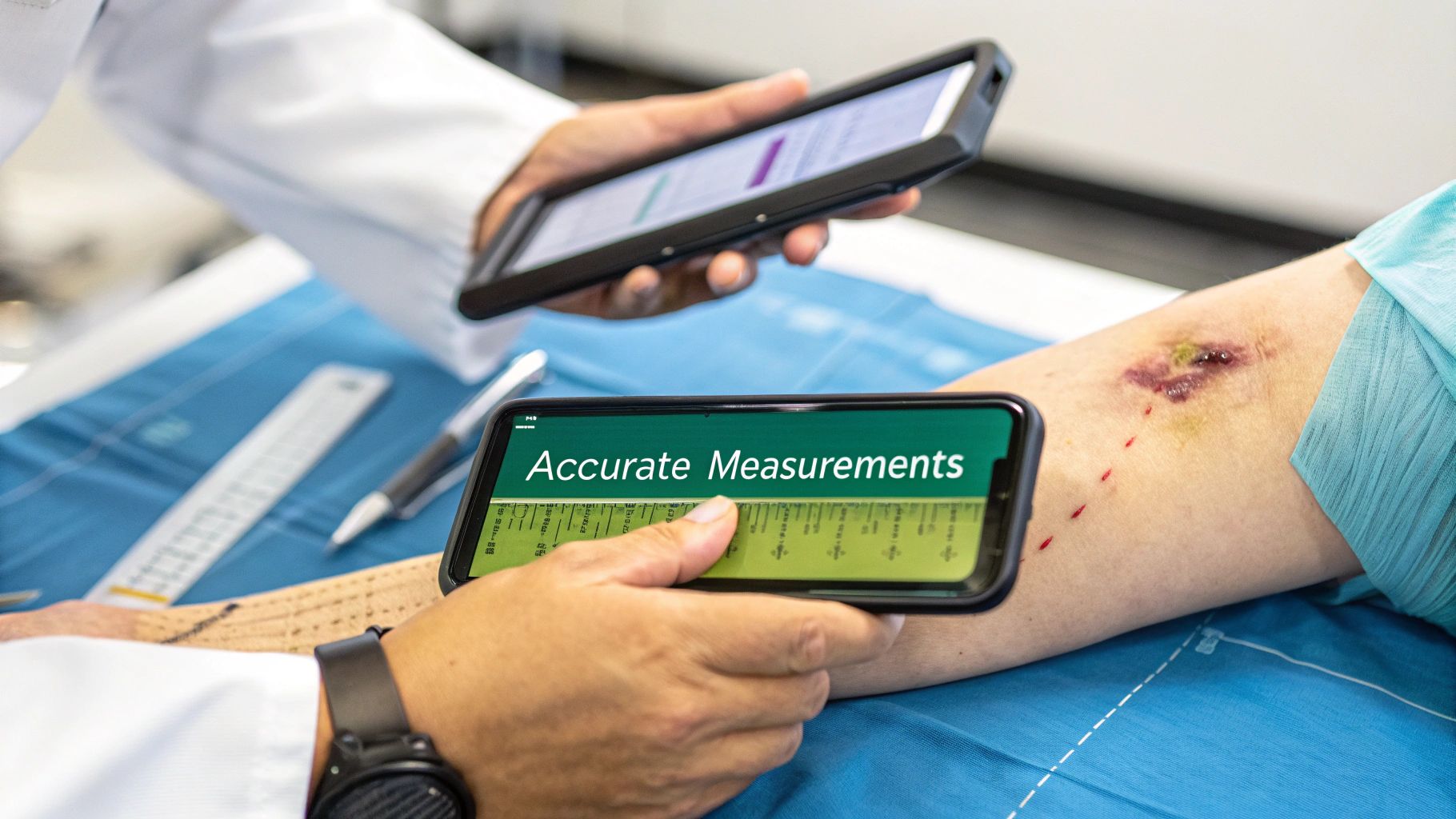
Once you’ve finished the hands-on inspection, it's time to capture the hard data. This is where we turn our observations into objective evidence that tracks healing—or lack thereof—over time. Let’s be clear: precise measurements and high-quality, consistent images are non-negotiable for solid skin assessment and documentation. They tell a story that words alone never could, providing an undeniable visual baseline against which all future assessments are compared.
Think of a wound's dimensions as its vital signs. A slight change in size can be the first clue that a wound is getting better or worse. But you’ll only catch these subtle shifts with accurate, repeatable measurements. These metrics are not just for the chart; they are crucial for justifying continued treatment to payers and demonstrating the efficacy of your care plan.
Mastering Linear Measurement Techniques
The go-to method for most clinicians is linear measurement—essentially finding the greatest length and width. It sounds simple, but consistency is everything. To nail it every time, the clock method should become second nature. This standardized approach eliminates ambiguity and ensures that measurements taken by different clinicians are comparable.
Imagine a clock face laid over the wound. 12 o'clock always points toward the patient's head, and 6 o'clock points toward their feet. This simple orientation keeps everyone on the same page.
- Length: This is your head-to-toe measurement, from 12 to 6 o'clock.
- Width: Measure the widest part from side-to-side, or 3 to 9 o'clock, making sure it's perpendicular to your length measurement.
- Depth: Gently probe the deepest part of the wound with a sterile, soft-tipped applicator. Mark the depth and then measure the applicator against a ruler. Be careful not to apply excessive pressure, which could damage delicate tissue.
When you find any undermining or tunneling, use the same clock orientation to describe its location. For example, documenting "2.5 cm of tunneling at the 7 o'clock position" leaves no room for interpretation. This level of detail is critical for understanding the wound's true volume and complexity.
A Practical Checklist for Clinical Photography
A picture is worth a thousand words, but a bad one can be dangerously misleading. A blurry, poorly lit photo can make a healthy wound look necrotic or hide early signs of infection. To make sure your images are clinically useful, you need a consistent protocol. Photographic documentation should be treated with the same rigor as any other clinical procedure.
Tips for Taking Quality Wound Images:
- Get Consent, Every Time: Always get the patient’s explicit consent before you even think about taking a photo. Just as important, document that consent in their chart. It’s a legal and ethical must. Explain why the photo is being taken and how it will be used and stored securely.
- Light It Right: Find a consistent light source that doesn’t cast harsh shadows. A ring light is great, but a well-lit room often does the trick. Stay away from the camera’s built-in flash—it causes glare and washes out important colors, obscuring tissue detail.
- Keep Your Distance (and Angle): To make photos comparable over time, you have to shoot from the same distance and angle every single time. A 90-degree angle directly over the wound is the standard.
- Add a Ruler for Scale: Always place a disposable, single-use ruler next to the wound. This immediately gives a sense of scale and backs up the measurements you’ve documented.
- Focus, Focus, Focus: Make sure the image is sharp, with the wound bed and periwound skin in clear view. A blurry photo is worthless. Use the tap-to-focus feature on your device to ensure the most critical areas are sharp.
Your goal with every photograph is to create a true, unbiased visual record. Consistency is the only way to achieve this. An image taken today must be directly comparable to one taken next week to have any real clinical value.
This systematic approach is more important than ever. The Global Burden of Disease Study revealed fungal skin diseases are the most common globally, affecting a staggering 578.1 million people. Reporting biases often lead to underestimation and delayed care, a gap that structured data capture, as seen in platforms like Ekagra Health AI, can help close.
Beyond Manual Methods: The Rise of Digital Tools
Let’s face it, manual measurements have their limits. Two clinicians can measure the same wound and come up with slightly different numbers—a problem known as inter-observer variability. This is where technology is really changing the game, introducing a level of precision and objectivity that was previously unattainable.
Digital planimetry and AI-powered image analysis are taking the guesswork out of wound care. With platforms like Ekagra Health AI, you can take a photo with a smartphone, and the software automatically calculates the wound's surface area with incredible accuracy. This technology can also analyze color and texture to help quantify tissue types, providing a level of granular data that is impossible to achieve manually.
You can see for yourself by looking at these wound documentation examples how much richer the data becomes. This tech doesn't just save time; it creates a perfectly consistent, reliable record of healing for every single patient.
Crafting Clinical Notes That Are Both Compliant and Actionable

You’ve finished the physical exam and collected all your data. Now comes the critical part: translating those findings into a clinical note that is clear, compliant, and actually useful. This document is far more than just a record—it’s a communication tool for the entire care team, a legal document, and the very foundation of your treatment plan. It is the official narrative of the patient's journey and your clinical decision-making process.
Your goal should be to create a narrative that tells the complete story of the patient’s wound at that specific point in time.
Every detail, from the patient’s history to your planned interventions, has to be documented with objective, concise language. This isn't just about CYA; it protects you legally and ensures that any other clinician can pick up the chart and immediately grasp the wound’s status and the “why” behind your care decisions. In my experience, effective skin assessment and documentation are two sides of the same coin—one is useless without the other.
Structuring the Perfect Wound Assessment Note
A structured note is an effective note. Period. When you organize your findings logically, you make the information easier for others to digest and, just as importantly, you create a safety net for yourself so critical details aren't missed. Using a consistent template for every patient is a game-changer for team communication and tracking progress over time. This structured format facilitates quick information retrieval and reduces the cognitive load on clinicians reviewing the chart.
Think of your note as building a case for your clinical decisions, supported by all the evidence you just gathered.
Essential Components of a Wound Assessment Note
Every wound note needs to contain specific, standardized information to be clinically useful and compliant. I've found that using a mental checklist or a structured template is the best way to ensure nothing falls through the cracks. Here’s a breakdown of the must-have elements.
| Documentation Component | Key Details to Include | Example |
|---|---|---|
| Wound Location & Etiology | Use precise anatomical terms and state the determined cause. | "Stage 3 pressure injury on the left ischial tuberosity." |
| Measurements & Shape | Document length, width, depth, and any undermining/tunneling using the clock method. | "5.2 cm L x 4.8 cm W x 1.5 cm D. Undermining of 2.1 cm from 1 o'clock to 3 o'clock." |
| Wound Bed Description | Quantify tissue types in percentages. This is key for tracking progress. | "Wound bed composed of 70% granulation tissue, 30% adherent yellow slough." |
| Drainage (Exudate) | Describe amount, type, and color. Always note odor only after cleansing. | "Moderate amount of serosanguineous drainage, no odor noted after irrigation." |
| Periwound Skin | Detail the condition of the surrounding skin—it tells a huge part of the story. | "Periwound skin is intact with mild erythema extending 1.5 cm around the wound margin." |
| Pain Assessment | Use a validated scale and describe the pain character, especially related to care. | "Patient reports 3/10 sharp pain during dressing change, 0/10 at rest." |
| Current Treatment & Response | List the interventions performed during this visit. | "Wound cleansed with NS, silver alginate dressing applied, covered with foam." |
| Treatment Plan | Outline the next steps, including dressing change frequency and goals. Be specific. | "Continue current regimen, change dressing every 3 days. Re-evaluate in 1 week." |
Adopting a structured approach like this transforms a simple note into a powerful clinical asset that truly serves the patient and the care team. For more in-depth examples, you can find a comprehensive wound care documentation template that can help you refine your charting process.
The Power of Objective Language
The quality of your note lives and dies by your word choice. Stick to objective, factual descriptions, not subjective opinions. Vague terms like "looks better" or "less drainage" create ambiguity and can be dangerously misinterpreted by other providers, auditors, or in a legal review. Subjectivity introduces variability and undermines the scientific basis of your practice.
Your documentation should paint a clear picture with data, not feelings.
What Not to Write: “Wound on back looks a lot better. Less drainage. Changed the dressing.”
What to Write Instead: “Sacral pressure injury now measures 3.1 cm x 2.5 cm x 0.8 cm (previously 3.5 cm x 2.9 cm). Wound bed is 90% granular with 10% slough at the 12 o’clock edge. Scant serous drainage noted. Periwound skin intact. Patient denies pain. Cleansed with NS, applied hydrogel and foam dressing. Will continue M-W-F dressing changes.”
The difference is night and day. The second note provides measurable data, specific locations, and a clear plan that anyone can follow. It demonstrates professional diligence and leaves zero room for guesswork. This level of detail creates a defensible legal record and supports high-quality, continuous care.
Connecting Your Documentation to Accurate Coding
Let's talk about reimbursement. Excellent documentation is the bedrock of accurate medical coding, which is how you get paid. Every detail you include helps justify the CPT (Current Procedural Terminology) and ICD-10 (International Classification of Diseases, Tenth Revision) codes you select. Your clinical note must contain the specific language that supports the codes billed for the visit.
Take wound debridement, for instance. To correctly choose a CPT code from the 97597-97602 series, your note must specify the depth of tissue removed (e.g., skin, subcutaneous tissue, muscle) and the total surface area. ICD-10 codes demand the same level of precision. Instead of a generic code for a leg ulcer, your notes should support something specific like "L97.211 – Non-pressure chronic ulcer of right calf with fat layer exposed."
This is exactly the level of detail payers are looking for. I can't tell you how many times I've seen claims denied due to inaccurate or insufficient documentation—it’s a direct hit to revenue and a massive administrative headache.
This is where AI-driven tools are really starting to change the game. Platforms like Ekagra Health AI can listen to a patient encounter and automatically generate a structured clinical note. The technology is smart enough to pull out key terms, measurements, and treatment details from a natural conversation and map them to the right CPT and ICD-10 codes. This slashes the risk of errors and significantly cuts down on administrative burnout, letting you focus on what actually matters: your patients.
Avoiding Common Documentation Pitfalls
Even the most meticulous clinicians can fall into documentation traps that put patient care and compliance at risk. Getting skin assessment and documentation right isn’t just about what you include; it's also about what you consistently leave out. These common mistakes often creep in during the rush of a busy day, but they can snowball into serious problems, from delayed healing to denied claims. Awareness of these common errors is the first step toward building a more robust documentation practice.
One of the most frequent slip-ups I see is inconsistent measurement. One clinician on the team measures a wound using the 12-to-6 o'clock orientation. A week later, another simply records the longest diameter, whatever its axis. This creates a completely unreliable dataset, making it impossible to actually know if the wound is healing. All of a sudden, a wound might look like it has grown when it was just measured differently. That can cause a lot of unnecessary alarm and lead to misguided changes in the treatment plan.
Neglecting the Periwound Skin
The skin around a wound is just as important as the wound bed itself, yet it’s so often treated as an afterthought in documentation. Failing to properly assess and describe the periwound condition is a major oversight. This area provides crucial clues about moisture balance, infection, and potential allergic reactions to dressings.
Think about a venous stasis ulcer where the surrounding skin is getting more macerated and red. If this isn't documented with specific measurements of the affected area, the care team might miss the early warning signs of too much moisture or a creeping infection. The note might just say "dressing changed," completely missing the deteriorating context that should have triggered a new moisture management strategy.
This kind of oversight has real consequences:
- Delayed Intervention: Without clear documentation of periwound breakdown, that crucial switch to a more absorbent dressing or the application of a moisture barrier gets put off.
- Wound Enlargement: Maceration can literally cause the wound borders to melt away, making the wound bigger and setting the healing clock back by weeks.
- Missed Diagnosis: Changes in the periwound can also point to bigger problems, like contact dermatitis from a dressing or advancing cellulitis, which demand immediate medical attention.
Using Vague and Non-Standard Terminology
Another major pitfall is leaning on subjective or non-standard language. Phrases like "looks better," "less drainage," or "healing well" are clinically useless without objective data to back them up. This kind of fuzzy terminology just creates confusion and makes it impossible for the next clinician to grasp the wound's true status. It introduces a high degree of interpretation, which is the enemy of standardized care.
Picture this: A home health nurse documents that a patient's sacral pressure injury "seems to be improving." The next nurse who visits sees what they believe is a stalled wound with subtle signs of decline. Because the previous note lacked specifics—like updated measurements or tissue percentages—the second nurse has no objective baseline to compare against, delaying a necessary tweak to the care plan.
This is where standardized language becomes your best defense. Ditch the subjective phrases and use concrete, measurable descriptions that leave no room for guesswork.
Quick Guide to Objective Terminology
| Instead of This (Vague) | Use This (Specific) | Why It Matters |
|---|---|---|
| "Less drainage" | "Drainage is now scant serous, previously moderate serosanguineous." | Quantifies the change in both amount and type. |
| "Looks cleaner" | "Wound bed is now 90% granulation, previously 70% granulation and 30% slough." | Provides hard data on tissue type progression. |
| "Edges look good" | "Wound edges are well-defined with no epibole or undermining noted." | Uses precise clinical terms to describe the wound margin. |
Misclassifying Wound Etiology
Finally, getting the wound's origin wrong because of an incomplete patient history is a critical error. For example, documenting a diabetic foot ulcer as a pressure injury just because it’s on the heel leads you down a completely wrong treatment path. A pressure injury needs offloading, sure, but a DFU treatment plan also has to address glycemic control, neuropathy, and potential arterial insufficiency. The entire approach is different. The initial diagnosis dictates the entire care trajectory, and an error here can have profound, long-lasting consequences for the patient.
This is where technology can be a great safety net. AI-powered platforms like Ekagra Health AI, for instance, use structured templates that force you to fill in every critical field—including periwound condition and wound etiology. By scanning clinical notes for completeness and consistency, these tools can flag missing information or non-standard terms, helping ensure every record is solid and accurate before you even sign off on it.
Frequently Asked Questions
Even with the best workflow, questions always pop up in the middle of a busy clinic day. Dealing with these real-world challenges is what separates good documentation from great documentation. Let's tackle some of the most common questions I hear from clinicians in the field.
Think of this as a way to fill in the gaps that standard protocols don't always cover. These scenarios often require clinical judgment that goes beyond textbook examples, highlighting the importance of experience and continuous learning.
How Often Should a Chronic Wound Be Assessed?
There's no single magic number for reassessment frequency—it really hinges on the wound itself, its stability, and how it's responding to treatment. That said, for most chronic wounds you're seeing in an outpatient clinic, long-term care, or home health setting, a weekly reassessment is the accepted standard of care.
This regular check-in gives you a consistent rhythm to spot trends and make informed decisions. Of course, if you're dealing with an acute wound, a post-op site, or any wound that's starting to look worse—maybe there's more pain or new drainage—you’ll need to step that up to a daily assessment. The key is to tailor the frequency to the patient's specific clinical situation and the goals of care.
The most important principle here is consistency. You must document every single time you assess the wound. No exceptions. This creates an unbroken clinical narrative and legal record, which is absolutely critical for justifying your treatment choices and getting paid for the care you provide.
If you don't have that regular, documented touchpoint, you're flying blind. It becomes nearly impossible to prove that your care plan is evidence-based and responsive to the patient's condition.
What Is the Best Way to Document Wound Pain?
Simply writing "patient has pain" in a note is a huge red flag. It’s not enough. Good pain documentation tells a story, combining objective data with the patient's subjective experience to create a full clinical picture. Your note should give the next clinician actionable information. It should also demonstrate that pain management is an integral part of your wound care strategy.
First, you need to quantify it. Use a validated tool like the 0-10 Numeric Rating Scale. For patients who struggle with that, the Wong-Baker FACES scale is a great alternative. This gives you a clear, simple metric to track from visit to visit.
But the number is just the beginning. The real substance comes from your narrative description. Be sure to include:
- Character of the Pain: What does it actually feel like? Use the patient's own words if you can. Is it "sharp," "dull," "throbbing," or "burning"? This can provide clues to the underlying cause (e.g., neuropathic vs. nociceptive pain).
- Timing and Triggers: When does it hurt? Is the pain constant, or does it flare up during a dressing change or with movement? Understanding the context of the pain helps in planning interventions like pre-medication.
- Interventions: What did you do about it? Document any actions you took, like giving pain medication before debridement or helping the patient find a more comfortable position. This shows you are actively managing the patient's symptoms.
When you document pain this thoroughly, you not only improve your patient's comfort but also build a rock-solid defense against any potential claims of neglect. It proves you're listening and responding.
Can AI Tools Disrupt Patient Interaction?
That’s a fair question, and one I hear a lot. The fear is that tech will create a barrier between you and the patient. But the new generation of AI tools is designed to do the exact opposite—they're built to be "ambient," fading into the background so you can be more present.
The whole point is to get your head out of the computer and back to focusing on the person in front of you. The goal of this technology is not to replace the clinician but to augment their abilities, removing administrative friction so they can focus on human connection and complex decision-making.
Imagine a voice-first system. It simply listens to the natural conversation you're already having during the exam. The AI intelligently pulls out the important clinical details—wound measurements, tissue types, exudate amount, what you plan to do next—and structures it all for you.
This information gets automatically dropped into a compliant, well-organized note without you having to type a single word. By letting the tech handle the tedious administrative work, you're free to maintain eye contact, build rapport, and give your patient the undivided attention they deserve. You end up with better documentation and a stronger patient connection.
Ready to put an end to documentation headaches and spend more time with your patients? Ekagra Health AI has a voice-to-claim platform that automates your clinical notes, coding, and billing in just minutes. See how you can slash your documentation time by up to 70%.
