
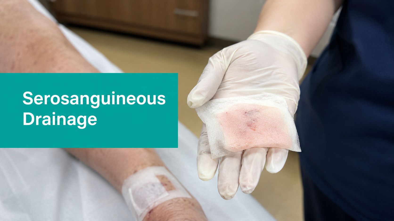
A fresh dressing can fool you if you're not looking at the whole picture. One visit the incision is damp with a pale pink stain, the next visit the gauze is more soaked, the skin edge looks angry, and someone has already written “infected” in the chart without documenting much else. That's where serosanguineous drainage deserves proper attention, because the fluid itself is not the diagnosis, it's the clue.
What Serosanguineous Drainage Actually Looks Like
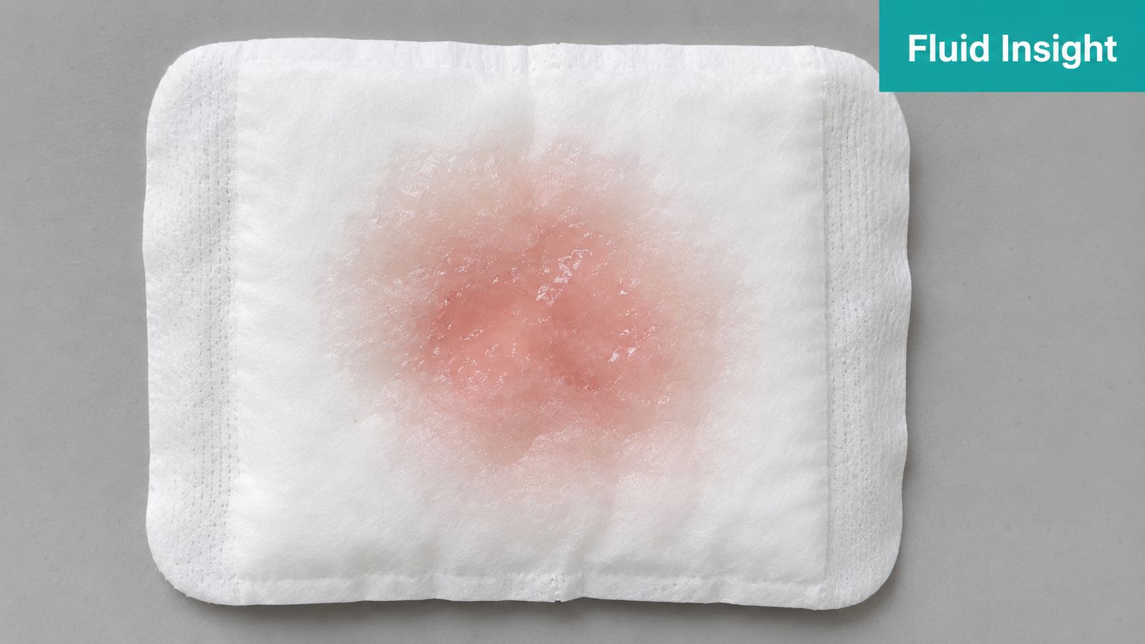
At the bedside, the first clue is usually a dressing, not a label in the chart. A postoperative incision may leave a light pink ring on the gauze, or a dehisced diabetic foot ulcer may show a thin, watery stain after cleansing and packing. The point is straightforward, serosanguineous drainage is a descriptor, not a diagnosis, and it means the wound fluid contains serum plus a small amount of blood. MedlinePlus describes it as yellowish fluid with small amounts of blood, which is why the drainage may look pink, light red, or pale yellow-red instead of frankly bloody or cloudy (MedlinePlus).
Read the dressing, not just the color
A note that says “pink drainage” does not give anyone much to work with. Better documentation separates amount, appearance, and trend, because those details drive interpretation at handoff and in the claim. For a practical visual reference, wound bed descriptions can help tie what you see on the dressing to the tissue environment underneath.
In wound charting, minimal drainage wets less than 25% of the dressing, moderate drainage saturates 25% to 75%, and copious drainage exceeds 75% saturation. Use those terms carefully, because the same pink stain can mean very different things depending on the wound and the surrounding tissue. A small amount after debridement is often expected. A broader wet area that expands from one shift to the next deserves reassessment and a cleaner note.
If the team needs a clinician to review a wound in Australia, they can find a wound care nurse in Australia for assessment and follow-up.
Practical rule: chart the fluid as serosanguineous only when it is truly mixed. If the blood is just a superficial smear from strike-through, call it what it is and do not inflate the wound.
Serosanguineous vs Serous, Sanguineous, and Purulent Drainage
The confusion usually comes from people using color words loosely. Serous drainage is clear or straw-colored. Sanguineous drainage is frankly bloody. Purulent drainage is thick, opaque, and often associated with odor. Serosanguineous sits between them, a thin watery fluid with a pink or light red tint from a small amount of blood.
| Type | Color and Consistency | Typical Clinical Meaning |
|---|---|---|
| Serous | Clear, pale yellow, watery | Common in healing wounds, especially when drainage is light |
| Serosanguineous | Pink, pale red, yellowish with light blood tinge, thin | Mixed serum and blood, often seen during normal healing |
| Sanguineous | Bright red, bloody | Active bleeding or fresh vascular disruption |
| Purulent | Thick, opaque, creamy, often malodorous | Concerning for infection and needs prompt review |
Don't confuse mixed drainage with strike-through
Notes can get sloppy. A dressing that picked up blood from friction, probe contact, or a wipe can look blood-tinged without the wound producing mixed exudate. That's not the same thing as true serosanguineous drainage, which is coming from the wound bed and should be documented as such.
The distinction matters for severity, billing, and handoff. If you write “serosanguineous” when the fluid is just a superficial smear, the note suggests a wound process you didn't really observe. That becomes a problem when a reviewer compares your description to the dressing change, the wound size, and the treatment plan.
I also like to pair drainage type with odor and peri-wound appearance in the same sentence. It keeps the note honest. A wound can have serosanguineous drainage and still look clean, or it can have that same drainage plus new odor and increasing erythema, which is a different clinical picture entirely.
For a quick internal cross-check on how exudate categories are separated in practice, keep an eye on types of exudates. That kind of shorthand helps your whole team use the same language.
Common Wound Types and Situations That Produce Serosanguineous Output
You'll see this fluid in a predictable set of wounds. The first is the early postoperative incision, especially in the first several days when hemostasis is still settling and the tissue is remodeling. A small amount of mixed drainage is often expected there, provided it's tapering and not shifting toward bright red bleeding or cloudy output.
Where it shows up most often
- After sharp debridement: A Wagner 2 or Wagner 3 diabetic foot ulcer can ooze serosanguineous fluid for a period after debridement. That alone doesn't prove infection.
- In venous leg ulcers after compression begins: As edema starts to move, drainage can increase briefly before it settles.
- At donor sites and graft dressings: These areas often produce thin mixed drainage while the surface closes.
- In postoperative wounds with fragile tissue edges: Motion, tension, or delayed hemostasis can keep the dressing lightly pink.
A Wagner 2 or Wagner 3 ulcer with persistent serosanguineous output is not automatically infected. What changes the reading is the pattern around it, the odor, the peri-wound erythema, and the pain story. A wound that's granulating cleanly behaves differently from a wound that's reopening or building a hematoma.
The wound doesn't need to be dramatic to need attention. A subtle increase in output after debridement can be the first clue that the tissue is unhappy.
Watch the clinical context
In a dehisced incision, persistent mixed drainage can reflect tension, incomplete hemostasis, or deeper space issues. In a venous ulcer, it may reflect edema-driven leakage rather than infection. In a graft or donor site, the drainage pattern often tracks with how well the surface is sealing. The source is less important than the trajectory.
That's why I teach staff to ask one question before they label anything: is the wound following the expected healing path, or has the drainage pattern changed? If you can answer that clearly, the rest of the plan usually makes more sense.
Bedside Assessment and Measurement Techniques That Hold Up in a Note
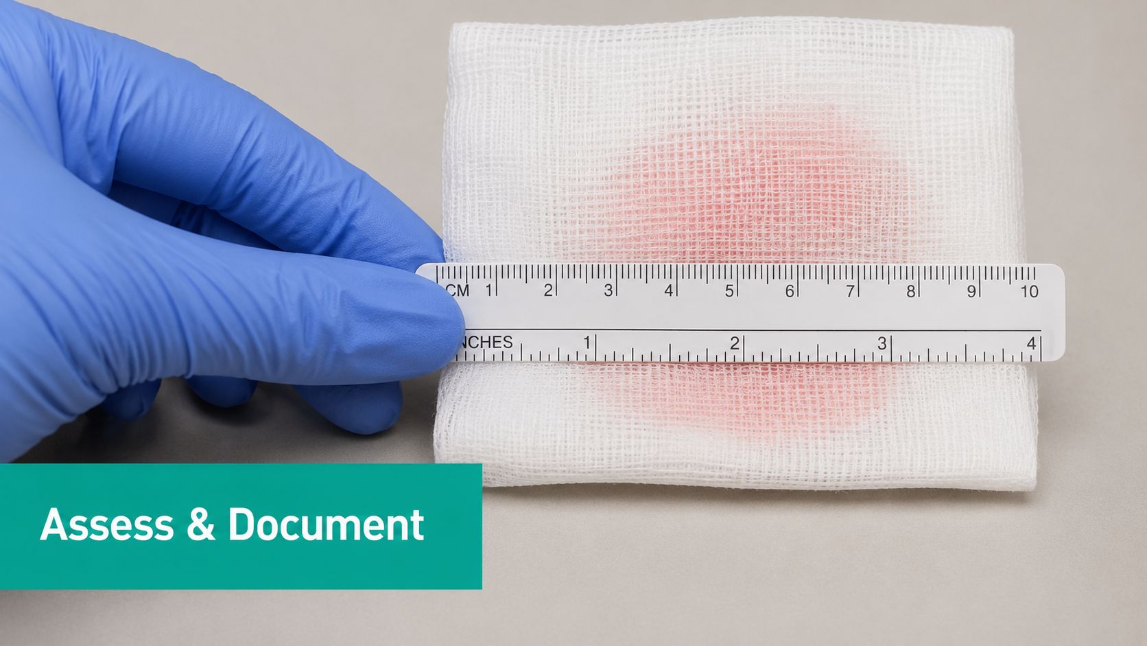
The note has to stand on what you saw, not on a decorative adjective. Start with the dressing saturation, then move to color intensity, then to surrounding tissue. If you do it in the same order every time, your documentation gets tighter and your handoffs get faster.
What to chart at the bedside
- Amount: Minimal, moderate, or copious, tied to dressing saturation.
- Color intensity: Pink, pale red, light red, or brighter red, not just “mixed.”
- Peri-wound skin: Maceration, erythema, warmth, or intact skin.
- Odor: Present or absent, and describe it without editorializing.
- Change from the prior visit: Better, worse, or unchanged.
Write like someone else will have to defend the note later, because they might. “Moderate serosanguineous drainage noted on old dressing, peri-wound skin intact, no odor, drainage decreased from prior visit” tells a very different story than “drainage present.” The first note is usable. The second one invites a denial.
A clean documentation habit is to compare each visit with the last. That's how you separate a single-shift observation from a meaningful trend. A wound that looks stable today but has doubled its output over the last few visits is not the same as a stable healing wound.
If your team uses structured workflows at the point of care, EkagraHealth AI can support wound documentation that captures drainage type, amount, odor, and infection indicators in the note itself. The value isn't the software label, it's the discipline of forcing the clinician to record what's real.
Charting language that holds up: “Serosanguineous drainage present, moderate amount, dressing 50% saturated, peri-wound intact, no malodor, output decreased from prior assessment.”
Normal Healing Phase or Early Warning Sign
A fresh wound can look messy before it looks clean. With serosanguineous drainage, the key question is whether the output is tapering as granulation develops, or whether the pattern is drifting toward continued bleeding, infection, or a wound that is not closing the way it should.
A useful bedside split
A Wagner 2 diabetic foot ulcer after debridement may show mixed drainage that gradually slows over the next several days. That fits an early healing course. If the drainage becomes brighter red, increases instead of tapering, or saturates dressings faster than expected, the wound needs a closer look.
The older surgical-site literature is useful at the bedside. A PubMed-indexed series on isolated serosanguinous incision drainage after surgery reported that many patients presenting within six weeks of surgery were managed successfully with antibiotics alone, so a pink incision does not automatically mean a return to the operating room (PubMed). A later clinical summary cited 88% conservative-management success for serosanguineous drainage without systemic infection signs, with escalation if drainage persists beyond 5 to 7 days or the patient worsens (PubMed).
When I would escalate
- Drainage persists or increases beyond the expected early window
- Color shifts toward brighter red
- Peri-wound erythema spreads
- Pain ramps up instead of settling
- The patient looks systemically unwell
Chest tube literature adds one more practical point. A PubMed study found it was safe to remove chest tubes once drainage became macroscopically serosanguineous, because that appearance indicated cessation of active bleeding (PubMed). Different wound, same logic. The color matters only when the trend matches the exam.
For home health teams or aides trying to understand why a wound is being watched rather than ignored, a plain-language review of what is paramedical service can help place wound observation within broader patient support. The point stays the same. Do not chase the color alone, chase the pattern.
Documentation and Coding So the Claim Survives the Denial Queue
The claim lives or dies on the note. If the drainage description is vague, the depth is missing, or the procedure note doesn't support medical necessity, the payer has an easy denial. That's especially true after debridement, where the reviewer wants to see the wound characteristics, the tissue level removed, the surface area, and why the service was needed.
Sample language that works
“Post-debridement wound with moderate serosanguineous drainage, no purulence, peri-wound skin mildly erythematous without spreading cellulitis, wound bed with viable tissue, continued sharp debridement medically necessary to remove devitalized tissue and promote healing.”
That type of language supports the visit because it connects the observation to the intervention. If the debridement reached subcutaneous tissue, the relevant CPT family is 11042 with add-on 11045 for additional surface area. If the work extended to muscle, the family is 11043 with 11046 for additional surface area. If it reached bone, use 11044 with 11047 for additional surface area. Pair those codes with the appropriate ICD-10-CM diagnosis for the underlying wound or ulcer, and make sure the wound depth in the note matches the level billed.
What reviewers and surveyors look for
- Medical necessity: Why was debridement needed today?
- Depth documented: Did the note support subcutaneous, muscle, or bone level?
- Surface area recorded: Was the additional area justified?
- Drainage detail: Was the drainage serosanguineous, and how much was there?
- Change over time: Did the note show a trend, not just a snapshot?
Most denials around drainage notes come from one of three places. The documentation doesn't support the procedure, the wound description is too generic, or the drainage wording is sloppy enough that the reviewer can't tell whether it was mixed fluid, blood strike-through, or something more concerning. That's why medical necessity documentation has to be built into the bedside habit, not patched in after the fact.
If you want the claim to survive scrutiny, write the note as if a coder, a payer, and a surveyor will all read the same line. Because they will.

Management Steps and When to Escalate
Management is driven by volume, trend, and skin risk, not by the word pink. A scant amount of mixed drainage in a clean postoperative wound often just needs protection and observation. Once the output starts to burden the dressing or macerate the peri-wound skin, you need a different dressing strategy.
Match the dressing to the burden
- Foam dressings fit moderate output well because they handle moisture without overpacking the wound.
- Alginate is a better match when drainage is heavier and you need more absorbency.
- Negative pressure wound therapy makes sense when volume, depth, or maceration risk justifies a stronger fluid-management approach.
- Compression is the right response when venous edema is driving leakage rather than infection.
That last point matters. If the leg is swollen and the drainage is pooling, more gauze is not the answer. You need a plan that addresses the edema, protects the peri-wound skin, and keeps the dressing from becoming a wet dressing that sits against the tissue all day.
Escalate for the red flags
Bright red drainage. Rapid saturation. Spreading erythema. Fever. Pain out of proportion. Those are physician-level changes, not just dressing-change issues. If the fluid is becoming briskly bloody, think active bleeding, vessel injury, reopened tissue, or delayed hemostasis, not ordinary healing.
Practical rule: if the dressing is saturating faster than the wound's usual pattern, stop calling it routine drainage and reassess the wound, the hemostasis, and the plan.
When I see a wound that starts with serosanguineous output and then turns more aggressive, I don't argue with it. I look for the reason the tissue changed behavior. That might mean loosening the dressing plan, bringing in the prescriber, checking whether a dehisced edge has widened, or reconsidering whether the current wound environment is helping at all.
Practical Takeaways and Common Questions From the Field
Keep the terminology tight. Serosanguineous means mixed, not just blood-tinged gauze. Culture when the clinical picture points to infection, photograph and document when the wound is changing in a way you need to track, and defend your dressing choice by tying it to the amount and trend of output. The three things I want every visit are amount, color trend, and peri-wound status.
A CTA for EkagraHealth AI.
