
When we talk about sacral wound staging, we're doing more than just identifying a wound. We're classifying the severity of a pressure injury on the tailbone, and that classification is the single most important first step in a patient's healing journey. Accurate staging is non-negotiable—it dictates the care plan, impacts patient outcomes, and ensures our documentation and billing are on point. For clinicians, mastering this skill is not just a professional responsibility; it's a critical component of delivering effective, compassionate care.
Why Accurate Sacral Wound Staging Is Non-Negotiable

Getting the stage right isn't just a box-ticking exercise. It's the foundation of effective patient care. For any clinician on the front lines, from hospital nurses to home health aides, correctly staging a sacral pressure injury from the very first assessment determines the entire path to healing. The precision of this initial assessment directly influences treatment decisions, resource allocation, and ultimately, the patient's recovery trajectory.
If we misidentify the stage, we risk choosing the wrong treatment, which can stall healing and open the door to serious complications like infection, sepsis, or osteomyelitis. The sacrum is a prime target for these injuries—its bony prominence takes a beating when patients are supine or sitting for long stretches, making it a well-known hotspot for pressure-related tissue damage. This anatomical vulnerability demands heightened clinical vigilance and diagnostic accuracy.
A Modern Problem with a Long History
The devastating potential of sacral wounds is hardly a new discovery. For centuries, clinicians have recognized this area as one of the most vulnerable spots for pressure injuries, documenting the severe consequences of tissue breakdown in this region. The historical context underscores the persistent challenge these wounds present.
Back in the 19th century, renowned French neurologist Jean-Martin Charcot even coined the term decubitus ominous for sacral eschar lesions. He observed that patients who developed these dark, necrotic wounds over their buttocks and sacrum faced an almost certain death, highlighting just how grim the prognosis was. This historical perspective serves as a stark reminder of the stakes involved. If you're interested in the background, you can review the full historical research on these challenging injuries.
This long-standing struggle is exactly why mastering modern sacral wound staging is so critical today. It’s not about memorizing definitions; it’s about having a practical roadmap to manage these complex wounds effectively, leveraging contemporary knowledge and tools to overcome a centuries-old problem.
The ultimate goal in treating sacral injuries is to facilitate and expedite a patient’s return to their pre-injury level of function. To achieve this, it is essential to promote healing in proper alignment while optimizing neurologic recovery and accelerating post-injury rehabilitation.
The Clinical and Financial Stakes
Accurate staging sends ripples across both patient care and your organization’s financial health. When you get it right, you set off a cascade of positive events that benefit the patient, the clinician, and the healthcare facility. The implications are far-reaching and interconnected.
It Dictates the Care Plan: A Stage 1 injury requires aggressive pressure redistribution and proactive skin care, while a Stage 4 may necessitate surgical intervention, specialized dressings, and a multidisciplinary team approach. The stage tells you what to do next, ensuring the treatment matches the injury's severity.
It Prevents Wound Progression: Spotting a wound early and accurately lets you intervene before a minor issue deteriorates into a deep, full-thickness crisis. Early and correct identification is the cornerstone of preventative wound care, stopping a potential Stage 1 from becoming an unstageable wound.
It Secures Proper Reimbursement: Correct staging and documentation are crucial for aligning with ICD-10 codes. This helps prevent claim denials, justifies the use of advanced therapies, and helps avoid the steep financial penalties tied to hospital-acquired pressure injuries (HAPIs). Accurate documentation is the language of reimbursement.
It Improves Patient Outcomes: This is the most important part. A precise diagnosis leads to a tailored treatment plan, which means faster healing, reduced pain, lower risk of complications, and a better quality of life for your patient. This is the ultimate goal of all clinical practice.
In the end, mastering sacral wound staging turns a routine assessment into a powerful clinical tool—one that improves care, minimizes risk, and supports the financial stability of your practice. It is the pivotal point where clinical observation becomes effective action.
Applying the NPIAP Staging System to the Sacrum
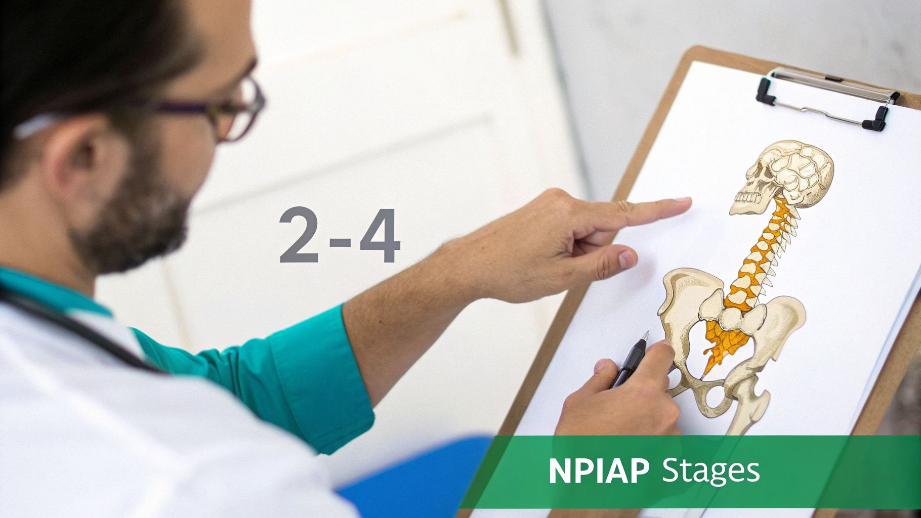
While the National Pressure Injury Advisory Panel (NPIAP) provides the universal framework for staging, applying it to the sacrum is where theory meets reality. This unique anatomical spot, with its bony prominence right next to the gluteal cleft, just doesn't behave like other areas of the body. To get sacral staging right, you have to learn how to translate the standard definitions into the specific visual and tactile cues you’ll encounter at the tailbone. The sacrum presents a unique microenvironment where pressure, shear, and moisture converge.
It's about going past the textbook and understanding the "why" behind what you're seeing. For instance, the NPIAP's 2016 shift from "pressure ulcer" to "pressure injury" was a major step forward, acknowledging damage that occurs long before the skin breaks. This is especially important for the sacrum, which is ground zero for 30-40% of all hospital-acquired pressure injuries. A key 2012 NPUAP position statement also clarified that any sacral wound exposing cartilage is automatically a Stage 4, as cartilage over a bony prominence is functionally equivalent to exposed bone. You can review the specifics in these official CMS training materials. This context is vital for accurate assessment.
Stage 1 Pressure Injury
A Stage 1 pressure injury on the sacrum is your earliest warning. The skin is completely intact, but trouble is brewing in the tissue underneath. The telltale sign is persistent, non-blanchable erythema—that patch of redness that stays red even when you press on it. This is a critical signal of underlying ischemia.
Think of it like pushing your thumb into a piece of clay. When you pull away, the impression remains. With healthy tissue, pressing on a red spot forces the blood out, turning it white (blanching) for a second before it refills. In a Stage 1 injury, the tiny capillaries are damaged, so they can't blanch. The redness is locked in, signaling compromised microcirculation.
Over the sacrum, this redness might come with other subtle changes. The area may feel warmer or cooler than the surrounding skin, or have a different texture—either boggy and soft or firm and indurated. For patients with darkly pigmented skin where redness is hard to spot, these tactile changes are your most reliable clues. Pay close attention to patient complaints of localized pain or discomfort, which can also indicate a developing Stage 1 injury.
Stage 2 Pressure Injury
Once a pressure injury hits Stage 2, we're dealing with partial-thickness skin loss. The damage has broken through the epidermis and now involves the dermis. This represents a breach in the skin's primary protective barrier, increasing the risk of infection.
The best analogy is scraping a fresh tomato. A Stage 2 is like that scrape that peels off just the thin outer skin, revealing the moist, pinkish-red surface below. It can look like a shallow, open ulcer with a red-pink bed, or it might present as an intact, serum-filled blister. The key is that the injury is confined to the upper layers of the skin.
What you won't see in a Stage 2 is slough (yellow, tan, or green dead tissue) or eschar (hard, black necrotic tissue). The wound is superficial, and adipose (fat) tissue is not visible. Be careful here, as Stage 2 injuries on the sacrum are very easily confused with moisture-associated skin damage. A thorough assessment of the wound's shape, location, and cause is essential for an accurate diagnosis.
NPIAP Definition of a Stage 2 Pressure Injury: "Partial-thickness loss of skin with exposed dermis. The wound bed is viable, pink or red, moist, and may also present as an intact or ruptured serum-filled blister."
Stage 3 Pressure Injury
A Stage 3 injury means you have full-thickness skin loss. The damage has punched all the way through the dermis and into the subcutaneous fat. This is a significant injury that requires a more intensive treatment plan.
Think of an iceberg. What you see on the surface of a Stage 3 might just be the tip of a much larger problem hidden underneath. At this stage, you'll see adipose (fat) tissue, and often, healthy red granulation tissue. The presence of fat is the defining characteristic that separates a Stage 2 from a Stage 3.
Because the sacrum has a good amount of fatty tissue, Stage 3 injuries can get surprisingly deep. You should also be on the lookout for undermining (where the wound extends underneath the visible edges) or tunneling (a narrow channel burrowing deeper from the main wound). However, muscle, tendon, ligament, or bone are not yet visible or palpable. It is crucial to probe these wounds gently to assess for hidden pockets of tissue destruction.
Stage 4 Pressure Injury
This is the most destructive stage: full-thickness skin and tissue loss. The damage is so extensive that it exposes or allows you to directly feel underlying structures like fascia, muscle, tendon, ligament, cartilage, or even bone. These wounds represent catastrophic tissue failure.
If Stage 3 is an iceberg, Stage 4 is what's left after the berg has completely broken apart, revealing the deep structures beneath. These wounds are typically deep and cavernous. Slough and eschar are often present, and undermining and tunneling are common and can be extensive, creating complex wound geographies that are difficult to manage.
Given the sacrum's position directly over the coccyx and sacral vertebrae, exposed bone is a common and dangerous finding. This dramatically raises the risk of osteomyelitis, a serious bone infection that can be life-threatening and requires aggressive, long-term antibiotic therapy and often surgical debridement. For a complete overview of all injury classifications, take a look at our comprehensive pressure injury staging guide.
Unstageable And Deep Tissue Injury
Finally, two other classifications are critical for any clinician assessing sacral wounds. Don't misclassify these, as doing so leads to incorrect treatment and poor outcomes.
Unstageable Pressure Injury: You know it’s a full-thickness wound (either a Stage 3 or 4), but you can't see how deep it goes because the base is covered by slough or eschar. Until that necrotic tissue is debrided and the wound bed is visible, you can't definitively call it a Stage 3 or 4. It's simply "unstageable." The priority becomes preparing the wound for accurate staging through debridement.
Deep Tissue Pressure Injury (DTPI): This is a sinister injury that starts deep in the muscle layers next to the bone. On the surface, it looks like a persistent, non-blanchable spot of deep red, maroon, or purple skin. It might even look like a dark, blood-filled blister. Don't be fooled by the intact skin—the damage underneath is severe, and these injuries can deteriorate with shocking speed into a massive Stage 3 or 4 wound, sometimes within hours or days.
Your Step-by-Step Sacral Assessment Checklist

A truly accurate sacral wound assessment isn’t a guessing game—it's a repeatable skill. When you follow a structured process every single time, you build consistency. This ensures your documentation is rock-solid and forms the foundation of a care plan that actually works. This checklist is your clinical guide to a foolproof sacral wound staging.
Think of it like a pilot's pre-flight check. Every step is there for a reason, and skipping one can have serious downstream effects. From digging into the patient's history to documenting the final details, this methodical approach makes sure nothing gets missed and elevates the standard of care.
Step 1: Gather Patient History and Context
Before you even lay eyes on the wound, start by talking to your patient and digging into their chart. Understanding the backstory—the "how" and "why" of the injury—is just as critical as the physical exam itself. A wound does not exist in isolation; it is a manifestation of a patient's overall health status and circumstances.
Put on your detective hat. You're looking for clues that will guide your staging decision. What's their mobility and continence status? How is their nutritional intake? Are there comorbidities like diabetes or peripheral vascular disease? Has there been a recent fall or an unusually long time spent in one position? This context is absolutely vital for accurate sacral wound staging and for developing a holistic care plan.
Step 2: Prepare for the Assessment
Good preparation is half the battle. Taking a minute to set the stage properly ensures the patient is comfortable and, just as importantly, that you can actually see what you need to see. A rushed or poorly prepared assessment is a leading cause of misdiagnosis.
- Ensure Adequate Lighting: Let's be honest, the sacrum is often in a poorly lit spot. Grab a gooseneck lamp or a good penlight to kill any shadows. You need to be able to accurately assess subtle changes in skin color and tissue type. Good lighting is non-negotiable.
- Gather Your Supplies: Have everything you need within arm's reach before you start. This means gloves, measuring tools (a disposable paper ruler), sterile saline, gauze, and a few sterile, single-use cotton-tipped applicators.
- Position the Patient Correctly: You simply can't get a full picture if the patient is flat on their back. The gold standard is a 30-degree side-lying position. Use pillows to support their back and keep their knees separated to fully expose the entire sacrum and gluteal cleft. Explain the process to the patient to ensure their comfort and cooperation.
Step 3: Cleanse and Visually Inspect the Wound
Once your patient is positioned, it’s time to gently cleanse the area. This isn't just about hygiene; it's a crucial step to remove any debris, slough, or thick drainage that might be hiding the true state of the wound bed. A clear view is essential for an accurate assessment.
Use sterile saline and gentle pressure to irrigate. This is not the time for harsh scrubbing, which can easily traumatize fragile, healing tissue. After it's clean, pause and do a full visual inspection, taking in the overall appearance.
A clean wound bed is a non-negotiable for accurate staging. Slough, eschar, or even heavy drainage can completely mask the true depth of the injury, setting you up for a critical misdiagnosis.
Now, it’s time to systematically break down what you see:
- Tissue Type: Identify every type of tissue in the wound bed. Do you see that healthy, beefy red granulation tissue? Pink epithelial cells migrating from the edges? Or is it covered in stringy yellow slough or hard, black eschar? Document the approximate percentage of each.
- Exudate (Drainage): Describe the drainage. Note the amount (scant, moderate, copious), its color, and consistency. Is it serous (thin, clear), serosanguinous (pink-tinged), sanguineous (bloody), or purulent (thick, opaque pus)?
- Odor: After cleansing, note any distinct odor. A foul smell, especially a sickly sweet or pungent one, is a major red flag for infection, particularly with anaerobic bacteria like Pseudomonas.
- Periwound Skin: Don't forget to examine the skin around the wound. Look closely for redness (erythema), maceration (whitish, waterlogged skin from moisture), induration (abnormal firmness), or warmth to the touch. These signs can indicate spreading infection or moisture damage.
Step 4: Measure and Probe the Wound
With a clean view of the wound, you can now capture its dimensions. These numbers are your baseline, allowing you and the rest of the team to track progress—or lack thereof—over time. Consistent measurement is key to evaluating treatment efficacy.
- Length: Measure the longest point from head-to-toe.
- Width: Measure the widest point from side-to-side.
- Depth: Gently insert a sterile, cotton-tipped applicator into the deepest part of the wound. Pinch the applicator at the skin level, pull it out, and measure it against a ruler.
Next, you need to check for any hidden damage. Using a fresh applicator, gently probe around the edges of the wound. You're looking for undermining, where the tissue has separated under the skin's edge, and tunneling, which is a narrow channel that extends from the wound deeper into the surrounding tissue.
A fantastic way to document this is with the clock-face analogy. Imagine the wound is a clock, with 12 o'clock pointing toward the patient's head. You can then describe the location and depth of any hidden cavities with precision (e.g., "3 cm of undermining from 2 o'clock to 5 o'clock"). Walking through these steps methodically will elevate the quality and reliability of your sacral wound staging every time.
Avoiding Common Staging Pitfalls and Mimickers
Let’s be honest—even the most seasoned clinician can get tripped up when assessing the sacrum. It’s a high-stakes area where pressure, shear, and moisture create a perfect storm for skin breakdown. This often leads to conditions that look frustratingly alike, making differential diagnosis a critical skill.
Accurate sacral wound staging is about more than just identifying a pressure injury; it’s about having the confidence to rule out what it isn't. Getting it wrong means the wrong treatment plan, delayed healing, and a patient who suffers unnecessarily. This is where a sharp diagnostic eye becomes your most valuable tool.
Pressure Injury vs. Moisture-Associated Skin Damage
One of the most common mix-ups is telling the difference between a Stage 2 pressure injury and Moisture-Associated Skin Damage (MASD), which you’ll often see documented as incontinence-associated dermatitis (IAD). They can both look like angry, broken skin, but they come from completely different places and require different management strategies.
Think of it this way: a pressure injury is an "inside-out" problem caused by intense, focused pressure cutting off blood flow, usually right over a bone. MASD, on the other hand, is a "top-down" issue. It happens when moisture from urine or stool breaks down the skin’s protective barrier over a wider, more superficial area.
This table breaks down the key visual cues to help you distinguish between these two very different conditions.
Pressure Injury vs. Moisture-Associated Skin Damage (MASD)
| Characteristic | Sacral Pressure Injury | Moisture-Associated Skin Damage (MASD) |
|---|---|---|
| Cause (Etiology) | Intense, prolonged pressure and/or shear. | Exposure to urine, stool, or other moisture. |
| Location | Typically over a bony prominence like the coccyx. | More diffuse; may affect the gluteal cleft, perineum, and thighs. Not necessarily over a bony point. |
| Shape | Distinct, often circular or oval shape with clear, regular borders. | Irregular, splotchy, or "blotchy" with poorly defined, diffuse edges. |
| Wound Bed | Presents as a shallow, partial-thickness ulcer with a pink/red bed. | Presents as inflammation, erosion, or denudement; may be patchy with satellite lesions. |
The key is to look at the whole picture. A pressure injury is like a pothole—a distinct point of failure with clear edges. MASD is more like a flood plain, where the damage is widespread, superficial, and follows the path of the moisture.
Unraveling the Unstageable Wound
The term "unstageable" can feel intimidating, but the concept is actually very simple. An unstageable pressure injury is a full-thickness wound where you simply can't see the bottom. The wound bed is so covered by slough or eschar that judging the true depth is impossible right now. It is a temporary classification pending debridement.
An unstageable wound isn't a mystery; it's just an obstructed view. Until that necrotic tissue is debrided and you can actually see the base of the wound, you can't accurately call it a Stage 3 or 4.
Imagine trying to measure the depth of a muddy pool. You know it’s deep, but you can’t say exactly how deep until the mud is cleared away. It's the same principle here. The presence of non-viable tissue obscures the full extent of the damage. Once the wound is debrided and the base becomes visible, you'll be able to re-stage it as either a Stage 3 or Stage 4 injury.
Distinguishing Other Common Look-Alikes
Beyond MASD, a few other skin issues love to masquerade as pressure injuries on the sacrum. Knowing what to look for is the key to getting the diagnosis right from the start and applying the correct treatment.
- Intertriginous Dermatitis (ITD): This is inflammation that pops up in a skin fold, like the gluteal cleft. It’s caused by trapped moisture and friction where skin rubs against skin. Look for redness and maceration that is linear and stays within the "crease," often presenting as a mirror image on both sides of the fold.
- Skin Tears: You'll see these often in older adults with fragile skin. They are traumatic wounds from friction or shearing forces. The hallmark of a skin tear is often a "flap" of skin (viable or not) and a jagged, irregular shape—very different from the defined borders of a pressure ulcer.
- Medical Device-Related Injury: Always play detective. Don't forget to check for culprits like bedpans, positioning wedges, oxygen tubing, or even the tubing from a catheter. These injuries will often perfectly mirror the shape of the object that caused the pressure. If you see a red, damaged area that looks exactly like the rim of a bedpan, you’ve probably found your cause.
By methodically looking at the cause, location, and shape of sacral skin damage, you can sidestep these common diagnostic traps. This systematic approach doesn't just lead to the right diagnosis—it ensures you're building the right plan of care from day one.
Connecting Clinical Staging To Documentation And Reimbursement
In wound care, we have a saying that’s more of a hard truth: if it wasn't documented, it didn't happen. All the meticulous work you do assessing a sacral wound at the bedside can completely fall apart if it doesn't translate into precise, compliant documentation. This is where your clinical expertise directly intersects with the financial and legal realities of healthcare.
Think of accurate sacral wound staging as the foundation for everything that comes next. It’s what secures reimbursement and justifies the plan of care. When documentation uses vague terms like "sore" or "skin breakdown" or shows inconsistent measurements, it raises immediate red flags for auditors and puts you on the fast track to a claim denial.
From Staging to Coding and Claims
Every detail you chart—from tissue type to measurements—directly feeds into ICD-10 coding. A clearly documented Stage 4 pressure injury tells a completely different story to a payer than a generically described "open sacral wound." One justifies the need for advanced treatments, specialized support surfaces, and higher reimbursement; the other just invites questions and delays.
This level of precision is absolutely critical when it comes to hospital-acquired pressure injuries (HAPIs). A wound that was present on admission but was documented poorly can easily be misidentified as a HAPI later on. When that happens, it can trigger major financial penalties and drag down your facility's quality scores. Your initial assessment is truly your first and best line of defense against these negative outcomes.
Proper documentation isn’t just about getting paid; it’s about building the clinical case for the patient. It validates the complexity of care and proves why someone needs specialized dressings, nutritional support, or surgical debridement, ensuring you have the resources to actually heal the wound.
Clear, accurate records prove medical necessity and serve as your shield against audit risks. When the chart tells a complete and consistent story, it creates an unassailable record of care. To build this kind of solid foundation, a good wound care documentation template can give your team a structured and reliable starting point.
Optimizing the Revenue Cycle
The link between what you see at the bedside and the facility’s financial health is undeniable. Clean, accurate documentation leads to clean claims, which in turn means faster payments and fewer denials. This whole process, the revenue cycle, is the financial engine of any healthcare organization, and wound care is a significant component.
For practices looking to shore up their finances and stop losing money to wound care denials, working with an outsourced revenue cycle management team can make a huge difference. These specialists know how to navigate the maze of medical billing, freeing up clinicians to focus on patients rather than coding nuances.
But here’s the catch: even the world's best billing team can't fix what’s missing from the source documentation. It all begins with the clinician’s hands-on assessment and their ability to translate that assessment into a clear, comprehensive note.
The Role of Technology in Clean Claims
This is where the business side of wound care has to meet clinical reality. Trying to bridge the gap between a detailed bedside assessment and a perfectly compliant insurance claim is a huge administrative headache that eats up far too much of a clinician's time.
Fortunately, modern tools are now being built to handle this translation. Imagine speaking your observations aloud during a sacral wound assessment and having that instantly converted into a structured, coded, and compliant note. AI-powered platforms can automate this process, making sure the nuances of your clinical judgment are captured perfectly for billing.
This technology helps ensure that:
- Staging is consistent: AI-assisted analysis can help standardize how wounds are measured and classified across your entire team, reducing inter-observer variability.
- Codes are correct: The system automatically maps your documented findings to the right ICD-10 and CPT codes, minimizing human error and maximizing coding accuracy.
- Claims are clean: By validating all the necessary information right at the point of care, these tools help generate claims that are right the first time, which gets you reimbursed much faster and reduces the burden of appeals.
By directly linking precise sacral wound staging to the documentation and billing workflow, you not only secure the resources your patients need but also strengthen your organization’s financial stability.
How AI Is Transforming Sacral Wound Management

Artificial intelligence in wound care isn't science fiction anymore. It’s here, and it’s fundamentally changing how we handle sacral wound staging and management. We're moving away from purely manual assessments and toward a more objective, data-backed approach that supports our clinical expertise, rather than replacing it.
Think about it: you point your facility’s device at a sacral wound, and an AI platform gives you an instant analysis. It can measure wound dimensions with incredible precision, help identify different tissue types in the wound bed, and track subtle changes over time, all without the variability of the naked eye. This brings a new level of objectivity to wound assessment.
Boosting Accuracy And Saving Time
In the day-to-day reality of patient care, this technology acts as a reliable second opinion. AI-powered image analysis helps standardize assessments across a care team, significantly reducing the chances of staging errors that can happen between shifts. It provides objective data to back up what you’re seeing, which creates a much more solid care record for legal and reimbursement purposes.
The real win, though, is the time it gives back. For any clinician who feels buried in paperwork, these tools are a lifesaver. Automating measurements and initial tissue analysis can slash documentation time—in some facilities, we're seeing reductions of as much as 70%.
This isn’t just about working faster. It's about reallocating your most valuable asset: your clinical expertise. When you’re not tied to a keyboard, you can spend more quality time on hands-on care, patient education, and the complex problem-solving that no algorithm will ever be able to do.
From Spoken Words To Clean Claims
Where this technology really shines is in the documentation workflow. Many modern systems use a "voice-to-claim" function that lets you dictate your findings during an assessment. The AI then instantly translates your words into the structured, coded data needed for billing and compliance, ensuring nothing is lost in translation.
This creates a seamless and highly accurate record of care.
- Objective Data Capture: AI delivers consistent, unbiased measurements and tissue analysis every single time, creating a reliable data trail of the wound's progress.
- Reduced Administrative Load: Automating documentation frees you up to focus on direct patient interaction and care, which improves both clinician satisfaction and patient outcomes.
- Improved Compliance: These systems help build a perfect, unassailable record of healing for each patient, which is your best defense against audit risks and claim denials.
This kind of AI integration goes beyond the bedside, too. It’s reshaping how we train our teams, which shows how AI is transforming corporate training and continuous skill development in healthcare. From education to application, these advancements are making our work more efficient and our patient care more effective. To learn more about how these platforms work, you can explore the benefits of clinical decision support systems in our detailed article.
Common Questions from the Field: Sacral Wound Staging
Even with the best training, staging sacral wounds in a busy clinical setting can be tricky. We all run into those head-scratching situations that don’t quite fit the textbook definition. Let's tackle some of the most common questions that come up on the floor.
Think of this as a conversation with a seasoned colleague. These are the real-world tips that help you stay confident, accurate, and focused on what matters most—your patient.
How Do I Stage a Sacral Wound If I Can't See It Under a Dressing?
Simple answer: you don’t. If a dressing is stuck tight (e.g., a hydrocolloid that is not scheduled for a change) or a device is covering the wound, you can't see the wound bed. And if you can't see the wound bed, you can't stage it. Period. Staging requires direct visualization and assessment of the wound's base.
For both your clinical notes and proper coding, the right move is to document that the wound base is obscured by the dressing. You’ll classify it as unstageable until that dressing comes off and you can get a clear look. Guessing at what’s underneath is a recipe for errors in the care plan and creates inaccurate documentation.
What's the Deal with "Reverse Staging"? Why Is It a No-Go?
Reverse staging is when someone tries to down-stage a healing wound—for instance, calling a healing Stage 4 a Stage 3 because it has filled with granulation tissue. This is a common mistake, but it's clinically wrong because a deep wound doesn't heal backward by magically regenerating lost tissue.
A Stage 4 pressure injury heals by filling the crater with granulation tissue and, eventually, a scar. It doesn’t regrow the original muscle, fat, and dermal layers. That’s why you should always document it as a "healing Stage 4 pressure injury," not a Stage 3 or 2. The original injury's depth defines its history and its maximum stage for life.
How Can I Be Sure I'm Looking at a Deep Tissue Injury (DTPI)?
A Deep Tissue Pressure Injury (DTPI) on the sacrum is the one you really don’t want to miss, as it can go from bad to worse in a hurry. Think of it like an iceberg—the real damage is hidden beneath the surface, and what you see is only a hint of the underlying tissue necrosis.
Here’s what to look for:
- Color: The skin will have a persistent, non-blanchable deep red, maroon, or even purple hue. This discoloration is a key sign of deep tissue damage.
- Presentation: It might look like intact skin, or you might see a dark, blood-filled blister. The surface may be intact, but the injury is severe.
- Feel: This is a big clue. The area often feels different—painful, firm, mushy, or boggy—when you compare it to the skin around it. These temperature and texture changes are critical diagnostic indicators.
Spotting and documenting a DTPI is critical. It’s a major red flag that signals a high risk of rapid deterioration into a full-blown Stage 3 or 4 wound. Your correct assessment is the trigger for aggressive offloading and intervention to prevent further decline.
At Ekagra Health AI, we’re focused on giving clinicians back their time. Our AI assistant transforms your spoken clinical notes into perfectly structured, coded documentation in minutes. It helps you master the nuances of sacral wound staging without getting bogged down in paperwork. Learn more about how Ekagra Health AI can help.
