
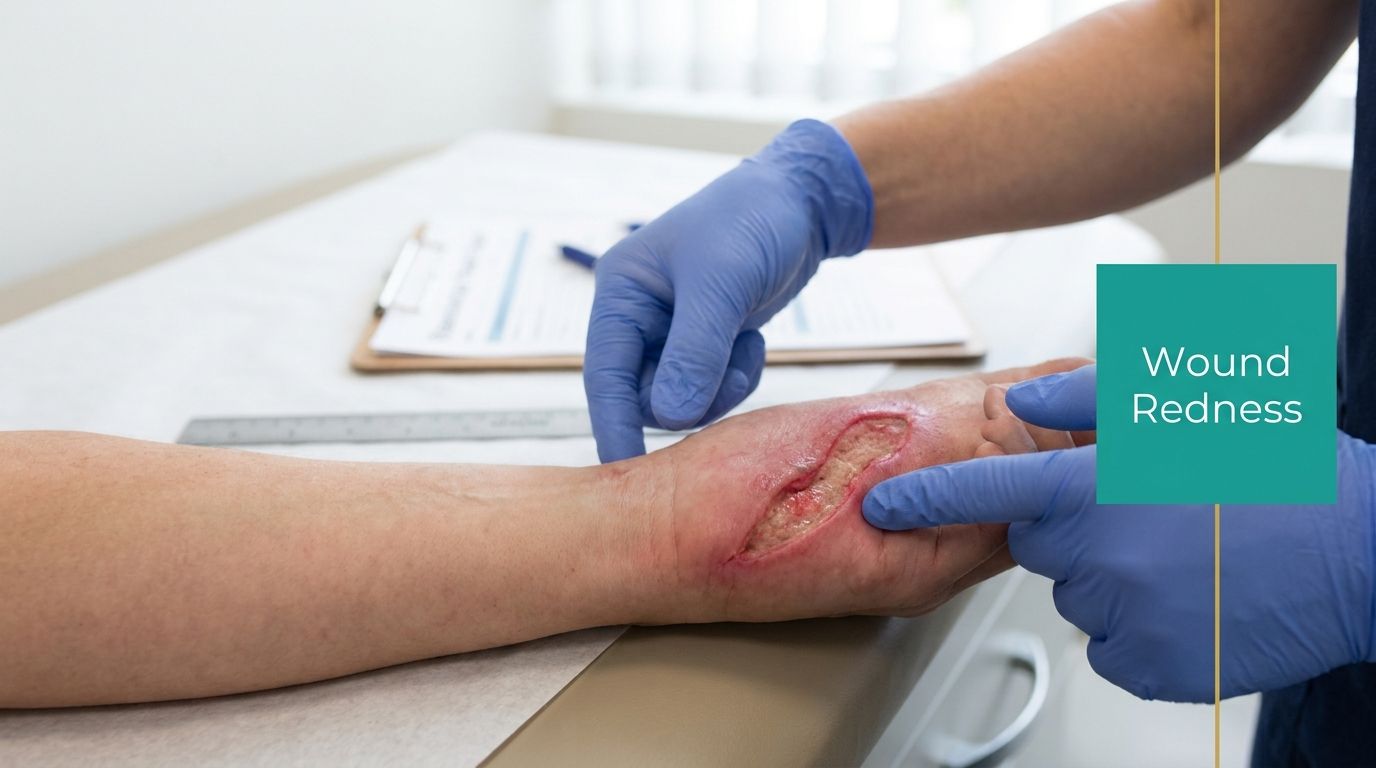
You're standing at the bedside with a chronic wound that looked stable last visit. Today there's a brighter red halo around it. The patient says it feels “a little hotter.” The nurse wrote “redness noted.” That's where people get into trouble.
Some redness around wound edges is expected. Some is the first sign you're about to miss an infection, document it badly, and then fight a denial on the back end. Clinical judgment matters here, but disciplined measurement matters just as much. If you can't tell me how far the erythema extends, whether it's advancing, whether the tissue is indurated, and what changed since the last assessment, you haven't finished the wound exam.
The Clinical Crossroads of Periwound Redness
A patient comes back for follow-up, the wound bed looks roughly the same, and the only new finding is a red rim at the margin. That single detail can push the case in two very different directions. One path is expected inflammatory change. The other is early infection, an antibiotic decision, a different ICD-10 picture, and a chart that either supports payment or falls apart under review.
Periwound redness sits at that decision point because it is easy to minimize and even easier to document badly. Fresh wounds often show a narrow band of erythema near the edge. Chronic wounds can also carry a baseline blush from ongoing inflammation, edema, adhesive trauma, or moisture damage. The job is to decide whether the erythema fits the wound's stage and history, or whether it signals a process that now changes treatment and coding.
That distinction has money attached to it.
If the note says only “mild redness,” the claim reviewer has no reason to accept infection-related treatment, no reason to support higher-acuity supplies, and no reason to believe the wound worsened since the last visit. Surveyors read that kind of charting as incomplete assessment. Payers read it as unsupported medical necessity. Both are usually right.
I tell new clinicians to document erythema the way they want it defended later. Record what you saw, where you saw it, how far it extends, and whether it is new, stable, or advancing compared with the last exam. A standardized wound assessment chart helps keep that consistent across visits and across clinicians, especially when the record later has to justify treatment changes, coding, and recertification. Use a structured format such as these wound assessment charts for clinical documentation.
Diabetic foot ulcers are where sloppy thinking shows up fast. Neuropathy blunts pain. Older adults and immunocompromised patients may not mount a fever. The first useful warning may be periwound erythema that is spreading, warmer, more tender, or paired with edema and induration. Once lymphangitic streaking or rapidly advancing redness appears, you are no longer documenting a minor local finding. You are documenting a change in risk, a change in management, and a chart that has to support why you escalated.
The practical takeaway is simple. Periwound redness is not a decorative wound description. It is a clinical finding with direct consequences for treatment, coding, claim defense, and survey readiness.
Systematic Bedside Assessment of Erythema
Good wound assessment starts with a ruler, not an adjective. If you want a note that another clinician can trust and a payer can follow, assess erythema the same way every time.

Use STONEES like you mean it
In the STONEES mnemonic, E stands for erythema or edema, and erythema extending beyond the wound margin is a marker of localized infection. Normal healing shows localized redness that subsides by days 5 through 7. Infected wounds show spreading redness, increasing warmth, and worsening pain, especially when the erythema radiates more than 2 cm from the wound edge, as described in this STONEES-based wound infection reference.
That means your exam has to answer four things:
Where is the redness located
Measure from the wound edge, not from some imaginary center point. Document the greatest extension in centimeters and note the clock-face position or directional margin.Is it advancing
Compare to the last visit. If there's no prior measurement, mark the border today so the next clinician has something real to compare.What else is traveling with it
Warmth, edema, induration, tenderness, exudate shift, odor, friable granulation tissue. Redness alone is a clue. Redness plus these findings is a pattern.What's the timeline
A little erythema right after injury or debridement is expected. Redness that persists or worsens instead of settling is not reassuring.
The bedside method that actually holds up
Here's the sequence I teach new NPs and staff nurses:
- Expose the whole limb: You can't assess periwound tissue if you only look at the dressing-sized window.
- Clean first, then inspect: Dried drainage and residue distort color.
- Measure the wound before the redness: Get length, width, depth, undermining, and then assess the periwound zone.
- Use a disposable ruler: Record the maximum erythema extension in centimeters from the wound margin.
- Palpate with the back of your fingers: Compare warmth to adjacent tissue and the contralateral side if appropriate.
- Check for induration: Soft pink tissue behaves differently than firm, expanding cellulitic change.
- Map it if you're worried: A skin marker around the border gives you a defensible comparison point.
- Photograph with annotation if your workflow allows it: A plain photo helps less than clinicians think. A measured, annotated image is far more useful.
For practices that want more consistency across staff, using standardized wound assessment charts helps keep the language and measurements from drifting from one visit to the next.
If your note says “erythema present,” you've documented almost nothing. If your note says “erythema extends 2.1 cm beyond lateral margin, warm, mildly indurated, increased from prior assessment,” now you've said something useful.
What not to do
Don't write “looks infected” without supporting physical findings. Don't call it cellulitis if you haven't described spread into the surrounding soft tissue. Don't rely on color alone in a patient with chronic venous disease, hemosiderin staining, or inflammatory dermatoses.
And don't skip reassessment language. A single snapshot is weak. Trend is what makes the exam defensible.
Differentiating Inflammation Infection and Cellulitis
Clinicians often struggle with the distinctions. Normal inflammatory erythema, localized infection, and cellulitis sit on a spectrum, but they are not the same problem and they should not be documented the same way.
Spreading redness appears in 75% of infected chronic wound cases, and 40% of patients with infected wounds were initially misdiagnosed because clinicians attributed the redness to normal inflammation, delaying treatment by an average of 5–7 days. That delay raises the risk of biofilm formation, according to this review on identifying infected wounds.
Redness differential diagnosis
| Clinical Sign | Normal Inflammation | Localized Infection | Cellulitis |
|---|---|---|---|
| Redness pattern | Limited to immediate wound edge, non-advancing | Extends beyond margin, more intense, progressive | Diffuse spreading erythema into surrounding soft tissue |
| Warmth | Mild | Noticeably increased near wound | More extensive heat across involved area |
| Pain or tenderness | Stable or improving | Worsening tenderness, sometimes out of proportion | Deeper tenderness, often more diffuse |
| Edema | Mild, expected early | Increasing periwound swelling | Broader soft tissue swelling |
| Exudate | Serous or scant | May shift toward purulent or cloudy drainage | Variable, depends on wound source |
| Granulation tissue | Healthy appearing | May become friable or bleed easily | Wound bed findings may be overshadowed by surrounding tissue infection |
| Borders | Well localized | More irregular, advancing from wound edge | Less tied to the wound border, spreading outward |
| Systemic features | Absent | Usually absent early | May develop fever, malaise, lymphangitic streaking |
The easiest trap is the chronic leg ulcer with baseline inflammation. The second easiest is the neuropathic diabetic foot ulcer where the patient doesn't report much pain. That's why a structured exam beats intuition.
What usually separates them in practice
Normal inflammation tends to calm down. Infection doesn't. Cellulitis expands.
Localized infection usually brings company with it: more warmth, worsening edema, tenderness, drainage changes, or a wound bed that starts looking unhealthy. If you're also seeing purulence and you need a quick clinical refresher on exudate patterns, this review on whether pus is a sign of infection is worth keeping in your teaching stack.
A wound can be colonized, critically colonized, and then frankly infected without giving you a dramatic early presentation. The redness trend often tells you where on that path the patient is sitting.
Cellulitis is a bigger statement. Use it when the surrounding tissue supports it. If the red area is advancing beyond the periwound zone, becoming more diffuse, warmer, tender, and clinically extending into subcutaneous tissue, document that clearly. If it's not there, don't overcall it.
Bulletproof Documentation for Payers and Surveyors
Claims don't get denied because you cared too little. They get denied because your note was vague. “Wound red” is useless. “Possible infection” is useless. Payers and surveyors want objective findings tied to medical necessity, treatment choice, and code selection.
A 2024 Journal of Wound Care study found that 38% of wound care claims were rejected due to insufficient documentation distinguishing infection from inflammation, including vague or missing erythema measurement. The same discussion emphasizes examples like “erythema 1.2 cm beyond margin with induration” to support coding decisions such as CPT 97597 or ICD-10-CM L03.90 for cellulitis, as summarized in this wound care documentation discussion.
What the note must say
If you want your debridement, antibiotic rationale, and follow-up intensity to make sense on review, include these elements:
- Exact location: “Periwound erythema greatest at lateral and superior margins.”
- Measurement in centimeters: “Extends 1.8 cm beyond superior margin and 0.6 cm laterally.”
- Progression statement: “Increased from prior assessment” or “stable compared with prior tracing.”
- Associated signs: Warmth, induration, tenderness, edema, odor, exudate type, tissue friability.
- Clinical interpretation: Inflammatory change, localized infection, cellulitis concern, or reactive erythema after procedure.
- Action taken: Observation, culture, topical antimicrobial plan, systemic antibiotic start, debridement, referral, escalation.
- Response plan: Follow-up timing and specific triggers for urgent reassessment.
Language that helps and language that hurts
Bad documentation:
- “Redness noted.”
- “Area looks worse.”
- “Possible infection.”
- “Cellulitis?”
Better documentation:
- “Periwound erythema extends 2.3 cm beyond distal margin, increased since last visit, warm and mildly indurated, with worsening tenderness and transition from serous to purulent drainage.”
- “Localized erythema confined to immediate wound border without spread, no induration, no increased warmth, consistent with expected post-debridement inflammatory response.”
- “Diffuse advancing erythema beyond periwound tissues with tenderness and edema, concerning for cellulitis.”
That level of specificity protects both the clinical decision and the code.
Why surveyors care
Surveyors look for evidence that infection surveillance happened at each wound encounter. That means your chart needs trend language, not one-off adjectives. If the wound hasn't improved, the note should show that you recognized it, reassessed it, and changed the plan when appropriate.
I'd also add a separate operational point. If your team is using any automated coding workflow, someone still needs to sanity-check the code logic against the actual note. This primer on detecting medical code hallucination is useful because it addresses a very real problem: generated coding suggestions that sound plausible but aren't fully supported by the documented findings.
Documentation pearl: The phrase that gets claims paid is not “infected wound.” It's the chain of findings that proves why you concluded that.
For clinicians who need examples their team can copy from without turning every note into a free-text mess, these wound documentation examples are the kind of templates that keep everyone on the same page.
And yes, serial photography helps. But only if it's dated, clinically relevant, and paired with written measurements. A picture without a documented assessment won't rescue a weak note.
Clinical Scenarios Treatment Choices and Coding
Friday at 4:30 p.m., a wound suddenly looks "more red," someone reaches for an antibiotic, and the chart gets one vague line about erythema. That is how bad treatment decisions get made, debridement claims get denied, and surveyors decide the team is not reassessing wounds in a disciplined way.

Wagner grade 2 diabetic foot ulcer with advancing erythema
Start with a common high-risk case. Plantar forefoot ulcer. Wagner grade 2. Neuropathy present. Last week, the periwound tissue was callused with only limited reactive pink change. Today, the redness extends beyond the wound edge, the skin is warmer, drainage is purulent after cleansing, and the granulation tissue is friable and bleeds with light contact.
Treat that as a management change, not a cosmetic observation. As noted earlier, infection diagnoses in wound care are often made from the bedside exam rather than a lab result handed to you in advance. In this scenario, spreading erythema plus friable tissue and purulence supports debridement if clinically appropriate, culture when indicated, and antimicrobial treatment based on risk, depth, and overall presentation.
The treatment plan needs to match the findings:
- Debride nonviable tissue when the wound and patient can tolerate it: Sharp debridement may support CPT 11042 if the documented depth, tissue type removed, and medical necessity match the code.
- Culture correctly when the result will change management: Cleanse first. Debride if indicated. Then obtain the specimen. Surface debris is not a useful answer.
- Start antimicrobial therapy based on the exam and patient risk: Diabetes, depth, drainage, tissue quality, and progression matter more than wishful charting.
- Offload aggressively: Continued pressure will keep the bacterial burden up and the wound stalled.
- Document the infection indicators you observed: Erythema extent in centimeters, warmth, drainage character, friable tissue, odor if present, tenderness if present, exposed structure, and whether probing suggests deeper involvement.
One practical coding point. Wagner grade documents ulcer depth and structure involved. It does not code the surrounding infection for you, and it does not justify antibiotics by itself. If you want the infection-related diagnosis and treatment to hold up, the note must separately describe the surrounding tissue findings.
A defensible note sounds like this: "Periwound erythema extends 1.8 cm circumferentially beyond wound margin, increased from prior visit, with warmth, purulent drainage after cleansing, friable granulation, and concern for local infection." That wording supports the clinical plan and gives coding staff something they can use.
Venous leg ulcer with reactive erythema and stasis change
Now the opposite problem. Medial gaiter region ulcer. Chronic edema, hemosiderin staining, irregular borders, shallow depth, serous drainage. The surrounding skin is pink-brown with mild erythema, but the redness is not advancing, there is no meaningful increase in warmth, no induration, and the patient reports no change in tenderness.
That pattern fits venous inflammation or stasis dermatitis far better than infection.
Treatment should follow the physiology:
- Compression therapy when not contraindicated
- Edema control and limb elevation
- Moisture management
- Periwound barrier protection
- Serial reassessment for spread, warmth, induration, or a drainage change that would suggest secondary infection
Inadequate documentation often leads to self-inflicted denial. If the note says only "leg red" and the plan is antibiotics, the payer has every reason to question medical necessity. If the note says "non-advancing periwound erythema with chronic venous skin change, no increased warmth, no induration, no change in pain, no purulent drainage," conservative treatment is easy to defend.
The coding connection
Redness is not billable by itself. Interpretation is.
For debridement codes such as 11042 through 11047, depth, tissue removed, and medical necessity have to line up with the procedure note. For cellulitis or other infection-related coding, the chart must describe surrounding tissue involvement clearly enough that another clinician can see why that diagnosis was chosen. For antibiotics, the record should support suspected infection with actual findings, not a reflex response to color change.
Surveyors look for reassessment. Payers look for medical necessity. Both are reading the same sentence about erythema. Write that sentence like it matters, because it does.
Clinical honesty usually gets the claim through faster than dramatic language.
Escalation Criteria and Final Takeaways
Some findings end the debate. Red streaking tracking proximally, rapidly advancing erythema, diffuse warmth and swelling beyond the periwound area, systemic instability, or a wound that keeps worsening despite initial treatment all need escalation. Don't sit on those cases and write a prettier note.
If the redness around wound margins keeps intensifying, the tissue becomes more tender or indurated, drainage worsens, or the patient fails to respond to early management, increase the level of care. That may mean same-day physician reassessment, culture, imaging, ED referral, or inpatient evaluation depending on the full picture.
Keep the bedside checklist simple:
- Measure it: Every time. In centimeters. From the wound edge.
- Map it: If spread is a concern, mark the border and compare.
- Name the pattern: Normal inflammation, localized infection, or cellulitis. Pick one and support it.
- Document the companions: Warmth, induration, tenderness, edema, exudate, granulation quality.
- Tie findings to action: Debridement, culture, antimicrobial plan, compression, offloading, escalation.
- Write for the next reviewer: Another clinician, a payer, or a surveyor should be able to follow your reasoning without guessing.
Good wound care isn't just about seeing redness. It's about interpreting it correctly, documenting it precisely, and acting before a local problem turns into a hospital problem.
EkagraHealth AI helps wound care teams turn bedside findings into clean, defensible documentation without burying clinicians in charting. If your practice is tired of vague notes, coding misses, and preventable denials, take a look at EkagraHealth AI to see how it supports wound image measurement, SOAP documentation, CPT and ICD-10 alignment, and cleaner claims from the start.
