
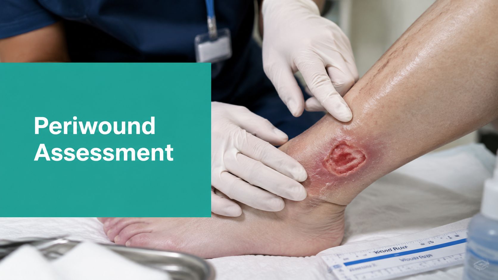
A venous leg ulcer can look obedient on Monday, all pink granulation and no obvious trouble, then the patient walks back in with a white, soggy rim, more pain, and a dressing that's already been fighting strike-through for days. The wound bed got the attention. The skin carrying the load around it didn't. That's how a lot of wounds slow down in real life, and it's why periwound assessment has to happen every time the dressing comes off.
Periwound compromise is part of the wound burden, not a side issue. In a large international survey of 2,018 patients with chronic wounds, 25% reported pain around the wound, which the authors linked to periwound skin damage, and Wounds UK has summarized older international data showing that this kind of compromise is common enough to be routine rather than exceptional (Wounds International reference). If you're only charting the bed, you're missing a major reason a “healing” ulcer returns looking worse.
The Periwound Problem Most Clinicians Miss
A chart note can make a wound sound stable right up until it isn't. I've seen the pattern too many times, a clinician documents healthy granulation tissue in a venous leg ulcer, the dressing plan stays the same, and two weeks later the patient returns with edge breakdown, more tenderness, and a rim that looks irritated from the inside out.
The miss usually isn't the ulcer itself. It's the assumption that the wound bed tells the whole story. In chronic wounds, the periwound is where moisture overload, adhesive trauma, friction, and inflammatory change often show up first, and those changes can stall epithelial migration before the center of the wound looks alarming.
Practical rule: if the wound bed looks acceptable but the margins are white, tender, shiny, or denuded, the wound is not under control yet.
That matters clinically and administratively. A wound that deteriorates at the edge often needs a different dressing strategy, a different cleansing approach, and better documentation than “intact periwound” gives you. It also matters for the record, because surveyors and auditors don't just want to see that a wound was seen, they want to see that the tissue most likely to fail was assessed.
Defining the Periwound Zone
The most useful working definition is simple. The 2025 International Consensus document defines the periwound as the skin and tissue immediately adjacent to the wound bed, extending up to 4 cm from the wound, including tissue under the dressing (International Consensus reference). That zone matters because it's not a decorative border, it's part of the wound environment.
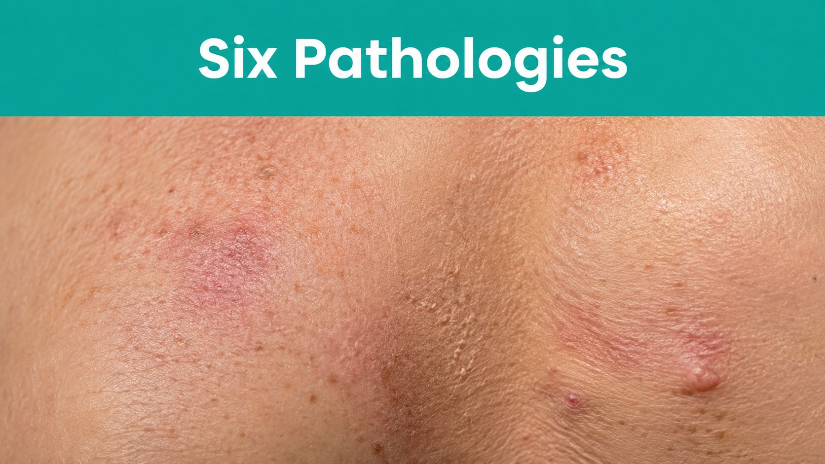
I teach staff to document the periwound the same way they document the bed. Color, temperature, edema, texture, integrity, and edge shape all belong in the note. If the margin is rolled, undermined, wet, or denuded, that's not a minor add-on. It changes what you do next.
The 4 cm boundary is a convention, not a sacred anatomic line, and difficult wounds can extend beyond it. A practical bedside habit is to inspect outward from the wound until the tissue stops behaving like part of the wound environment and starts behaving like surrounding intact skin. In a heel ulcer, that may be a short distance. In a heavily draining leg ulcer, it may be more.
The margin is part of the wound problem until proven otherwise.
That framing keeps you from under-documenting the injury. It also forces better bedside thinking. The question is never just whether the wound is open. The question is whether the surrounding tissue can support closure.
Recognizing Periwound Pathologies
Periwound changes don't all mean the same thing, and that's where charting gets sloppy. Wounds International has pointed out that periwound care is often overlooked and that definitions are still inconsistent, which leaves room for mislabeling venous eczema, adhesive injury, infection, or moisture-associated skin damage as generic “maceration” (Wounds International recommendations). At the bedside, the distinction matters.
The six patterns I watch for
- Maceration: The skin looks white, soft, and waterlogged. It feels boggy rather than firm, and the border may look as if it's been soaked too long. Heavy exudate is the usual driver.
- Callus formation: Thickened, hard tissue builds at the edge, especially in plantar diabetic foot wounds. It can shield the ulcer from a casual glance while concentrating pressure and delaying closure.
- Hyperkeratosis: This is broader, more diffuse thickening and scaling. The skin feels rough and dry, and the edge can look stacked or raised.
- Dermatitis: Red, itchy, inflamed skin around the wound often points to contact irritation, venous eczema, or adhesive sensitivity. The key clue is irritation that extends beyond a simple moisture halo.
- Infection: Look for increasing pain, friable tissue, odor, warmth, or a change in the character of drainage. Periwound infection can show up before the center of the wound looks dramatically different.
- Ischemia: Pallor, coolness, dusky color, or pain out of proportion should make you think perfusion. A wound edge that looks starved will not epithelialize well.
A white rim from moisture and a pale rim from poor blood flow are not the same problem. A tender, shiny border after tape removal is not “just a little redness.” A purple heel margin is not a dressing issue, it may be pressure and shear until proven otherwise.
The practical move is to separate what you see from what you think caused it. That's especially helpful in Wagner 1 and 2 diabetic foot ulcers, dehisced surgical wounds, and venous ulcers with heavy drainage, where several processes can coexist and hide each other.
For a deeper look at one frequent moisture problem, see this focused discussion of skin maceration.
Documenting the Periwound Correctly
Vague documentation is where good clinical judgment gets lost. “Periwound intact” tells a reviewer almost nothing. “Slight erythema” isn't much better. Neither phrase tells the next clinician whether the margin is dry, inflamed, callused, or exposed to repeated adhesive trauma.
Use language that ties the finding to the likely process. If the edge is white and soft, say it. If the patient has itching and scattered redness under the border of the dressing, say dermatitis is suspected. If the rim is hyperkeratotic and raised, document that it's thickened and callused. If the skin is stripped after dressing removal, call it what it is, medical adhesive-related skin injury when that's the clinical picture.
| Vague Documentation | Specific Documentation | Clinical Impression |
|---|---|---|
| Periwound intact | Periwound skin intact, but erythematous and moist at the inferior edge | Moisture imbalance with early maceration |
| Slight redness | Sharply demarcated erythema under adhesive border with pruritus | Contact dermatitis or adhesive injury |
| Wound margins abnormal | Thickened, circumferential hyperkeratosis at plantar edge | Pressure-related callus burden |
| Periwound moist | White, boggy periwound skin extending beyond the wound edge | Exudate exposure with maceration |
| Periwound tender | Warm periwound skin with increased pain and odor | Possible local infection |
SOAP notes work best when they read like a decision tree, not a checkbox. A useful sentence might be, “Periwound skin is macerated at the medial border with mild denudation, dressing strike-through noted on removal, barrier film applied before re-dressing.” That tells the next person what happened, what you saw, and what you did.
For templates that help keep this kind of language consistent, the wound care documentation template can be adapted to your team's workflow without flattening the clinical detail.
Charting tip: write the tissue description first, then the suspected cause, then the intervention. That sequence is easier to defend than a generic impression followed by a vague dressing change.
Coding the Periwound Assessment
Periwound findings don't get coded in isolation, but they absolutely affect coding accuracy. Debridement codes 11042 through 11047 depend on what tissue was removed and how extensive the debridement was, and periwound necrotic or devitalized tissue can influence that selection when it's part of the treated area. If you don't describe the margin correctly, it becomes hard to justify the work you did.
ICD-10-CM selection should follow the clinical picture, not the note template. Pressure injury cases often sit in the L89 series. Excoriation can fall under L87 when the skin findings fit. Bullous disorders may point you toward L51 when that's the actual diagnosis, and E03.9 can matter if hypothyroidism-related myxedema is part of the presentation. The diagnosis has to match the documented finding, not the other way around.
Payer denials often come from the same few weaknesses. The note says debridement happened, but it doesn't specify whether the treated tissue was wound bed, periwound slough, eschar, or nonviable margin. The record mentions redness, but never states whether it was moisture injury, pressure injury, or dermatitis. The surface story is there, the medical necessity story isn't.
Surveyors and auditors usually want three things. They want a clear description of the periwound, a connection between findings and intervention, and enough specificity to show why the chosen code was appropriate. If the edge was denuded, inflamed, or necrotic, your note should make that obvious.
For code alignment basics and diagnosis selection context, use the wound diagnosis code reference as part of your internal education workflow.
Selecting Dressings and Interventions
Periwound preservation starts with moisture control. Wounds UK recommends matching dressing capacity to the exudate burden, using an appropriate moisture vapour transmission rate, and adjusting dressing size and change frequency to the amount of fluid the wound is producing (Wounds UK reference). That principle is essential. You don't force the wound to fit the dressing, you choose the dressing that fits the wound.
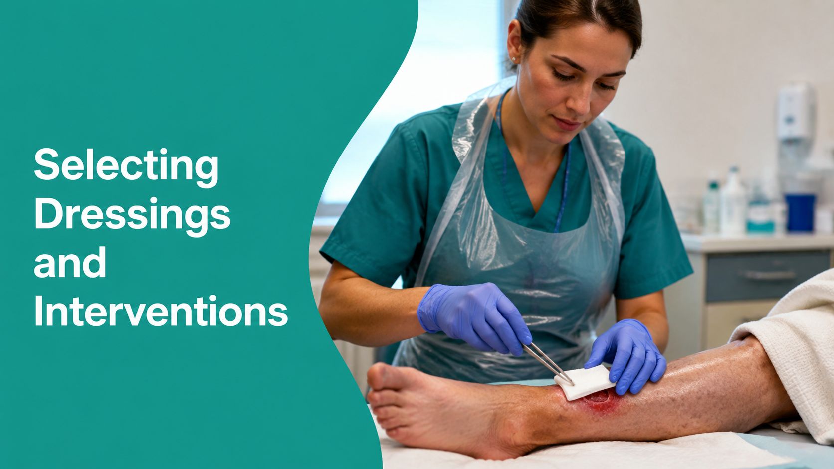
What usually works better
- Heavy exudate: Use higher absorbency, often with a superabsorbent secondary layer when strike-through is starting. If the edge is whitening by the second day, the current setup is underpowered.
- Fragile periwound skin: Favor atraumatic silicone-based interfaces and reduce adhesive trauma. Repeated tape removal can do as much harm as drainage.
- Moisture injury: Cleanse, pat dry, apply a barrier film, then re-dress. That sequence is basic, but people skip it when the clinic is busy.
- Venous leg ulcers: Prioritize drainage control and compression planning when appropriate. If exudate is left to pool against the skin, the margin pays the price.
- Diabetic foot ulcers: Watch callus, pressure points, and plantar edge stress. Offloading matters as much as absorbency.
- Pressure injuries: Look carefully for shear, adhesive injury, and buried margin damage before you choose the next dressing.
Barrier film is useful, but it's not a license to ignore fluid load. Silicone helps, but it won't fix an overloaded dressing. Superabsorbents help once strike-through is appearing, yet they're not a substitute for a better overall plan.
The cleanest practical rule is this. If the periwound is becoming white, shiny, or eroded, stop adding tape and start solving moisture and friction. That usually means a different interface, more absorbency, shorter wear time, or a reassessment of whether the current primary dressing matches the wound type.
Building a Periwound Assessment Workflow
A reliable workflow keeps the margin from becoming an afterthought. Start every dressing change with the same sequence. Inspect the periwound zone, describe what it looks and feels like, decide what process is driving the change, document the finding in language that can support coding, then choose the dressing and barrier strategy that fits the fluid burden.
That routine also makes team handoffs cleaner. A nurse should be able to read the note and know whether the next visit needs moisture control, adhesive protection, pressure relief, or a perfusion check. A coder should be able to see whether the documented work supports the debridement and diagnosis pathway chosen. A surveyor should be able to trace the decision from the margin back to the intervention.
Process discipline helps. Teams that standardize wound documentation and measure compliance with it have a better chance of keeping records clean enough for billing, quality review, and continuity of care. If you're looking at workflow measurement more broadly, the idea behind measuring automation success applies here too, because the useful metric is whether the right clinical detail is captured consistently, not whether the note just got longer.
The periwound is where healing gets preserved or lost. Treat it as part of the wound, chart it like it matters, and match the dressing to the fluid, not the other way around.
EkagraHealth AI helps wound teams capture the periwound story without losing the details that matter for care, coding, and claims. If your notes still flatten edge damage into “intact” or “slight erythema,” visit EkagraHealth AI and see how a wound-focused documentation workflow can support cleaner charting from the bedside to billing.
