
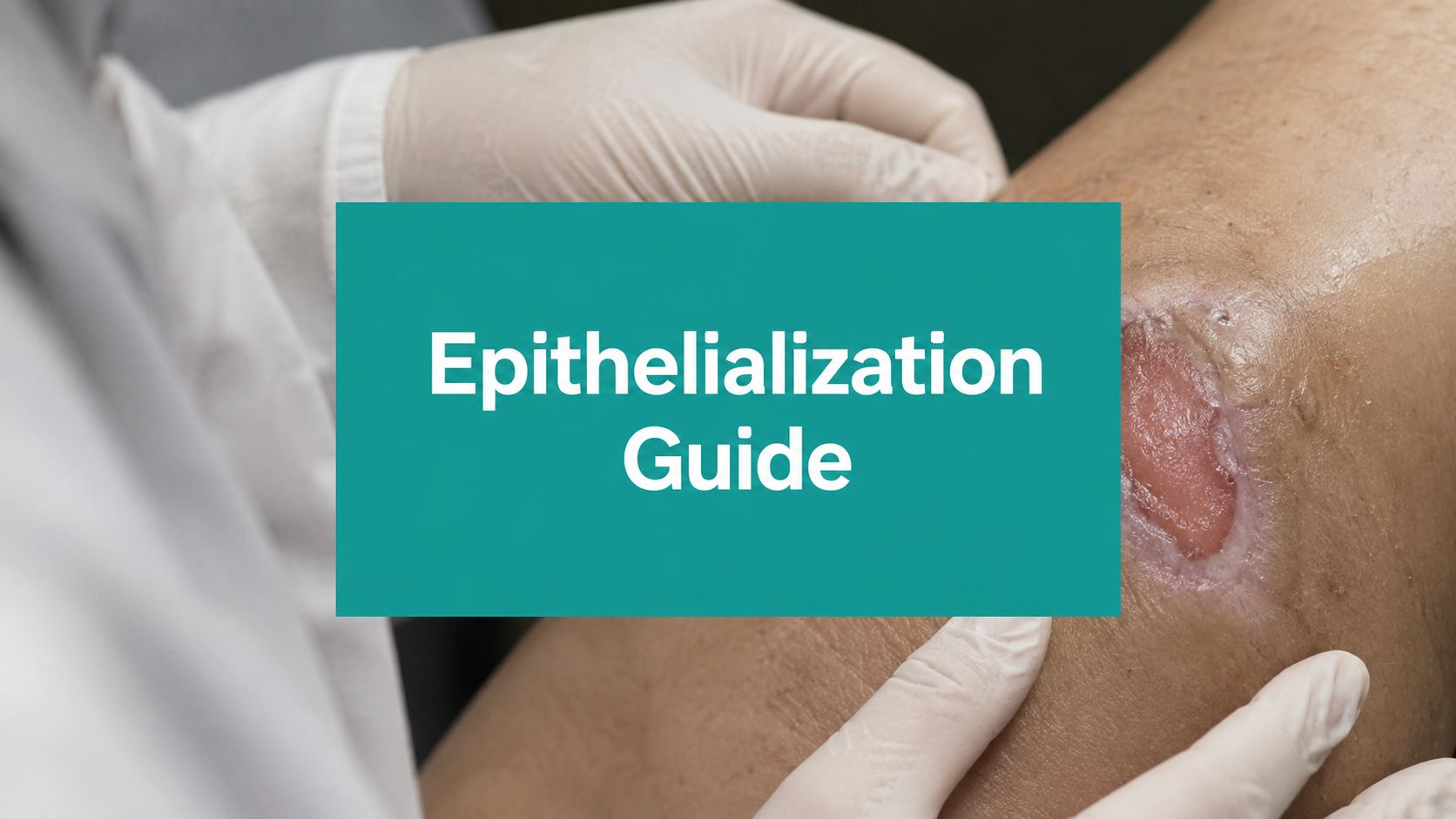
A wound can look busy and still be going nowhere. The base is pink, the exudate has settled down, the edges are cleaner, and everybody in the chart keeps saying “filling in,” but the skin never advances. That's the moment epithelialization matters, because granulation is not closure.
When Granulation Isn't Enough
A diabetic foot ulcer that has been granulating for weeks without visible edge advancement is a common trap. The wound looks healthier than it did at intake, so the team relaxes, the dressing plan stays the same, and the patient keeps coming back for the same visit pattern with no real endpoint. The problem is that a wound can build tissue and still fail to close if the wound bed is not ready for epithelial migration. Granulation tissue and epithelialization are not the same process, and treating them like they are the same leads to wasted time and weak documentation.
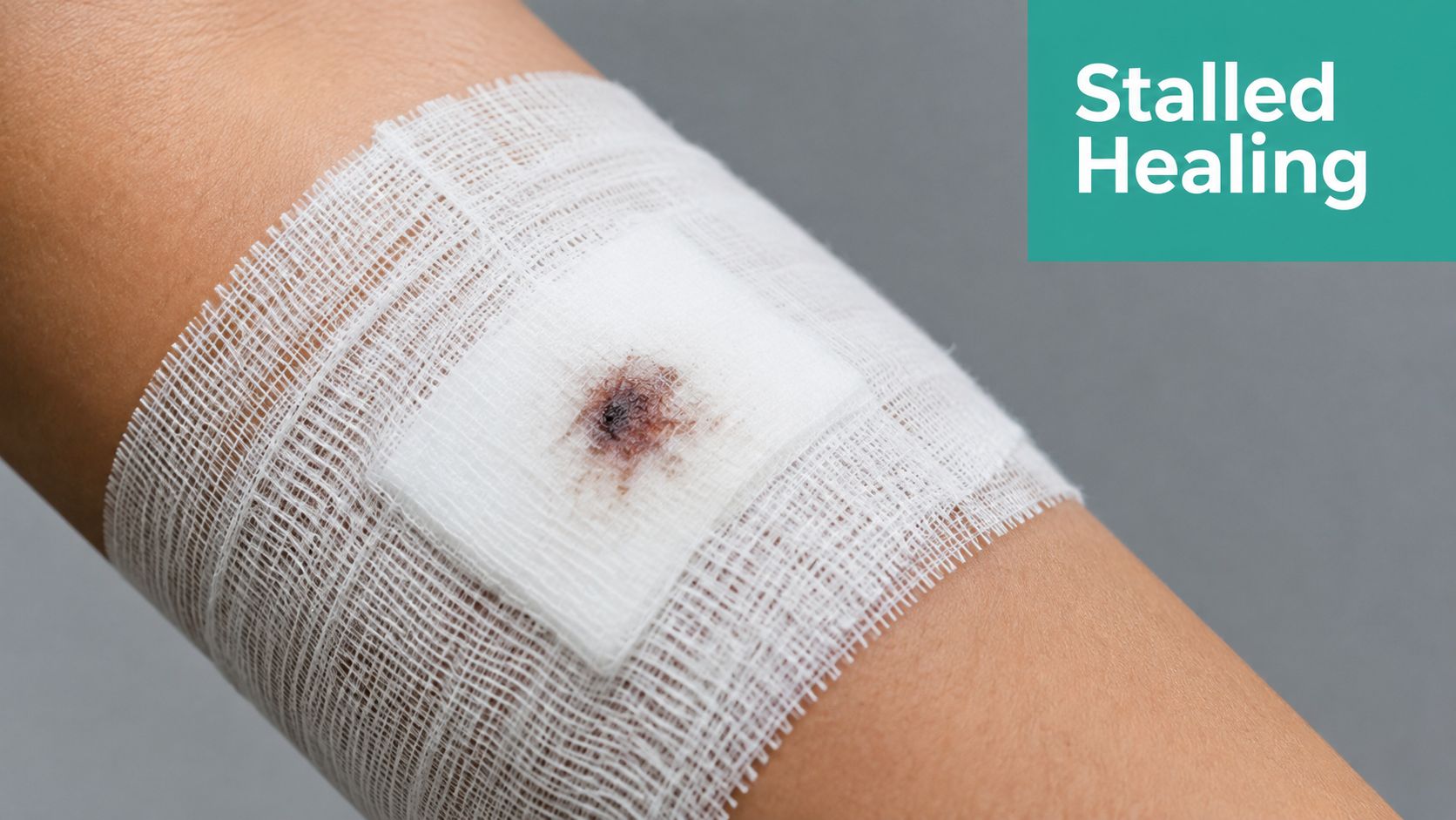
A wound that is filling in has depth change. A wound that is closing has edge migration. Those are not interchangeable, and surveyors know the difference when they read a run of notes that describe “improving granulation” but never mention epithelial tongue, marginal progression, or surface coverage. If the wound is static at the margins, the chart needs to say so plainly.
Practical rule: if the wound base looks healthier but the perimeter hasn't changed, don't call it improvement unless you can show epithelial advancement.
That distinction matters for treatment planning, patient expectations, and payer justification. A stalled Wagner 1 plantar ulcer with good offloading is a very different problem from a sloughy Wagner 2 or 3 wound with edema, biofilm, and heavy exudate. One may need continued protection and close measurement. The other may need a harder reset of the wound bed, source control, and a different escalation path.
Surveyors tend to look for exactly that logic. They want to see whether the team recognized a lack of epithelial progress, whether the treatment changed in response, and whether the documentation supports the medical necessity of continued visits. If the wound keeps “looking better” but the measurements, margins, and closure narrative don't move, the record starts to look repetitive instead of clinically thoughtful.
The Biology of Epithelial Migration
Epithelialization starts fast, but only when the wound environment lets it. Keratinocytes activate within 24 hours after injury, then migrate from the wound edge into the fibrin-fibronectin clot. Basal keratinocyte proliferation begins at 48 to 72 hours, and the surface eventually stratifies into a stable sheet. The practical point is simple, epithelial closure is a coordinated advance, not a random skin crawl across a wound bed. The cellular sequence depends on growth factors, cytokines, proteases, and matrix remodeling.
Think of the epithelial front like a crew laying flooring over scaffolding. The workers at the edge move first, but they can only keep advancing if the surface underneath is stable enough to support them. Human wound models also show that closure isn't only edge migration, because keratinocyte islands in the wound bed can contribute too. That's why viable edge tissue and preserved adnexal structures can speed closure in a way a superficial glance won't capture. Human skin wound models describe that dual mechanism clearly.
What the wound actually looks like
Advancing epithelium is usually fragile, thin, and pink at the margin. It can look almost translucent. It's easy to miss if you don't inspect the edge under good light and compare week to week. Stalled epithelialization tends to show up differently, with rolled edges, maceration, pale borders, or an edge that just keeps sitting there while the base granulates underneath.
Moisture is a real divider here. Epithelial cells migrate more easily on a moist surface, while dry crust and scab formation inhibit epithelialization. Moist wound care became standard for that reason. Clinically, that means a dry, crusted ulcer edge is often a bad sign even when the wound base looks tidy.
The timeline depends on wound type. Well-approximated surgical wounds can re-epithelialize within about 48 hours. Incisional wounds may complete epithelialization in 24 to 38 hours because the migration distance is less than 1 mm. Larger open or chronic wounds may take 2 to 3 weeks or even several weeks, especially if the bed isn't fully granulated. Acute primarily closed wounds are often complete in 1 to 3 days, while larger open wounds behave very differently.
If the edge is rolling over, whitening, or sitting in excess moisture, the wound is telling you the bed is not ready for clean epithelial advance.
What Speeds Up or Stalls Epithelial Closure
The wound microenvironment drives the result more than people like to admit. Growth factors, cytokines, matrix metalloproteinases, extracellular matrix signals, and mechanical cues all shape whether keratinocytes move or stall. That's why a wound can have viable edge tissue and still fail to epithelialize if perfusion is poor, inflammation stays high, or bacteria are controlling the surface. Modern reviews emphasize that re-epithelialization is not just a local skin event.
A Wagner 1 plantar ulcer with good offloading and a moist bed often behaves like a controlled biological environment. A Wagner 2 or 3 wound with edema, biofilm, hypoxia, or heavy exudate is living in the opposite world. The difference isn't cosmetic. It affects whether the epithelial tongue can keep moving or whether it gets trapped by local inhibitors.
| Wound Type | Promoting Factors | Common Inhibitors | Typical Timeline |
|---|---|---|---|
| Well-approximated surgical wound | Edge apposition, moist surface, low bioburden | Dry crust, tension on the incision, contamination | 24 to 48 hours |
| Acute open wound | Clean bed, moist dressings, viable edge tissue | Necrotic debris, desiccation, repeated trauma | Often 1 to 3 days when favorable |
| Plantar diabetic ulcer | Offloading, debridement, controlled moisture | Pressure, callus, biofilm, edema | 2 to 3 weeks or longer if stalled |
| Chronic sloughy ulcer | Source control, moisture balance, perfusion support | Slough, infection, maceration, poor granulation | Several weeks if the bed isn't corrected |
Matching the intervention to the limiter
If the wound is dry, the problem may be moisture balance. If it's wet and white at the edges, the issue may be too much exudate or periwound maceration. If the base is red but the surface never closes, think about persistent inflammation, bioburden, pressure, or vascular limitation. There isn't one fix for all of that, and dressing changes alone won't rescue a wound that still has the wrong biology underneath.
The chart should reflect the limiter, not just the dressing. That's where a lot of notes fail. They document what was placed on the wound, but not why the epithelial front is still stuck. Hypergranulation and excess moisture can interfere with edge advancement, so if the wound is proud, wet, and macerated, you have to say that explicitly.
Measuring and Documenting Epithelial Progress
If you can't measure epithelialization, you can't defend it. Standard wound length, width, and depth matter, but they don't tell the whole story when the surface is creeping inward. Track the distance from the edge to the visible epithelial line in millimeters when you can, and describe which margins are advancing. Use photography consistently, with the same angle, scale, and lighting, so the edge change is obvious from visit to visit. Wound measurement examples should show the perimeter change, not just the base size.
What good SOAP language sounds like
Strong documentation is specific. Weak documentation is lazy.
- Strong: “Periwound margins are intact with a thin, pink epithelial rim advancing from the medial edge. Wound length and width are decreased from prior visit, depth unchanged, moderate serous drainage, no odor, no undermining.”
- Weak: “Wound improving, continue current dressing.”
- Strong: “Epithelialization stalled at the inferior margin, with rolled edge and mild maceration. Base is granular but margin progression has not changed since last visit, despite offloading and moisture control.”
- Weak: “Looks better today.”
Those details matter to payers because they show medical necessity, response to treatment, and the reason the plan changed or stayed the same. They also matter to surveyors because repeated vague language looks like autopilot charting. If the wound hasn't changed, say that. If it has changed only in granulation but not in epithelial coverage, say that too.
Documentation rule: don't bury epithelial progress inside generic wound improvement language. Put it in the assessment and tie it to the treatment decision.
For billing, that precision supports the visit level and the reason for ongoing care. If debridement is performed, the note should support the tissue removed and the wound depth, with the CPT selection aligned to the actual anatomic depth, whether that's 11042 through 11047. If advanced therapy is being considered, the chart needs to show that conservative measures were appropriate, attempted, and not enough to move epithelialization forward.
Interventions That Support Epithelial Advancement
The right dressing is the one that matches the biology, not the one that sounds advanced. Hydrocolloids and other occlusive or semiocclusive options can support moisture balance in cleaner, lower-exudate wounds that need a protected surface. Foams help when exudate needs absorption without drying the bed. Alginates fit wetter wounds. Collagen matrices can be useful when the wound needs a better scaffold, especially after the bed has been cleaned up and the edge is ready to move.
Debridement is usually where bedside reality starts. Sharp debridement is the fastest way to remove slough, callus, and nonviable tissue when the wound can tolerate it. Enzymatic or autolytic approaches may fit a wound that needs a gentler pace, but they won't rescue a wound buried in biofilm and devitalized material if the bedside plan is never reassessed. The point isn't to debride for the sake of the procedure. The point is to clear the barrier so epithelial cells can advance.
Negative-pressure wound therapy, skin grafts, and biologic dressings can all support epithelialization when the wound complexity justifies them. They're most defensible when the wound is clean, the base is ready, and the record shows why simpler care wasn't enough. That is where medical necessity gets won or lost. A big wound with persistent drainage and stalled margins needs a documented rationale, not just a product name in the order set.
One practical option teams use for edge tracking and wound analytics is EkagraHealth AI, which captures wound images, performs objective measurement, and helps classify tissue so progression can be followed over time. That kind of documentation can support the story you're already telling at the bedside, but it still has to match the clinical picture.
When to Escalate and How to Justify It
Stalled epithelialization should trigger a reset, not a habit. If the wound edge isn't advancing, review perfusion, offloading, compression, and infection control before you add another dressing layer and hope for the best. A chronic wound with hypoxia, persistent edema, or ongoing bioburden may have viable keratinocytes at the edge, but those cells won't close anything until the local blockers are addressed.
Escalate when the wound bed looks technically “better” but the perimeter stays static. Escalate when margins are macerated, rolled, or repeatedly undermined by pressure. Escalate when drainage remains heavy, odor persists, or the wound has the kind of slough and inflammation that keeps epithelial cells from advancing. Those are not cosmetic findings. They are the reason the wound is not closing.
Documentation has to connect the finding to the action. If you order vascular workup, the note should say why. If compression is changed, the record should show what changed in the edema and periwound. If the patient is referred, the reason has to be tied to the stalled biology, not to a generic “slow healing” phrase.
For denials, the usual problems are predictable. The note doesn't prove failed conservative care. The measurements jump around without explanation. The wound is described as improving, but the edge never moves. Or debridement is billed without enough detail to support 11042 to 11047. That's the kind of chart that gets pushed back because it reads like procedure capture instead of clinical reasoning.
Surveyors look for consistency. They want to see that the plan changed because the wound changed, or didn't change. If epithelialization is stalled, say it, show what you corrected, and document why escalation was the right move.
Putting It All Together in Practice
At every visit, check three things first, edge, base, and environment. Is the epithelium advancing, is the wound bed ready for it, and is something blocking closure at the margin? Then document the answer in plain language, with the measurements, drainage type, periwound condition, and the specific action you took because of what you saw.
Clean claims start with clean wound language.
A good epithelialization note is short, concrete, and hard to argue with. It tells the reader whether the wound is closing or only granulating, and it shows why the next step makes sense. That's good medicine, and it keeps the chart aligned with the revenue cycle.
If your team wants cleaner epithelialization documentation, tighter SOAP notes, and better support for wound measurements and coding, visit EkagraHealth AI and see how it helps turn bedside wound assessment into usable clinical documentation and cleaner claims.
