
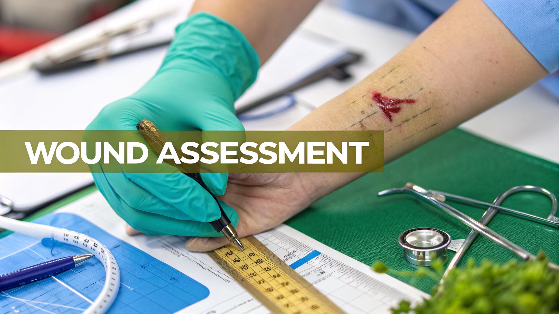
Describing a wound for nursing isn't just about jotting down a few notes. It’s a systematic, objective assessment of its location, size, tissue types, exudate, and the surrounding skin—all coming together to create a clear clinical picture. This detailed documentation is absolutely vital for tracking healing, guiding our treatment decisions, and keeping the entire care team on the same page.
Why Getting Wound Descriptions Right Is So Critical
It’s easy to think of wound documentation as just another box to check on a long to-do list. But I can tell you from experience, a precise and thorough description is one of the most impactful things you can do for your patient. It’s far more than a simple note; it's the very foundation of effective wound care and carries serious weight for patient outcomes, legal protection, and your facility's bottom line.

Think of your detailed observations as the first line of defense against delayed healing and serious complications. A vague note like "wound looks better" is clinically useless. In contrast, an objective entry—"sacral pressure injury now measures 3.2 cm x 2.8 cm with 75% red granulation tissue, up from 50% last week"—gives the whole team a clear, measurable baseline to work from.
This level of detail directly shapes the treatment plan and helps us spot trouble early. Your words paint a picture that guides physicians, physical therapists, and other nurses, ensuring everyone is providing consistent, informed care.
The Financial and Clinical Impact
The scale of this issue is pretty staggering. In the United States, chronic wounds affect an astonishing one in six Medicare beneficiaries, which adds up to about 10.5 million people. This is a silent epidemic that makes precise wound description in nursing an essential skill. The financial toll is just as massive: Medicare spends an estimated $22.5 billion annually on these wounds. As care shifts more toward clinic-based settings, your descriptive skills directly impact reimbursement and outcomes. You can dig deeper into the economic burden of chronic wounds in this study.
This financial reality puts a spotlight on the importance of what you document. Accurate notes ensure that care is properly coded and reimbursed, which is crucial for the facility's financial health.
Beyond reimbursement, remember this: meticulous notes are your best legal defense. If a negative outcome or legal issue ever arises, a well-documented record of consistent, objective assessments provides undeniable proof that you provided diligent and appropriate care.
It's More Than Just Charting
At the end of the day, mastering the art of describing a wound for nursing is about being a powerful advocate for your patient. Your ability to capture the subtle changes in a wound bed, the color of the exudate, or the condition of the peri-wound skin can be the very thing that prevents an infection, avoids a debridement, or even saves a limb.
It transforms a routine task into a powerful clinical tool. When you truly understand the "why" behind the process, you elevate your practice from simply recording data to actively shaping positive patient outcomes.
Your Framework for a Flawless Wound Assessment
Feeling overwhelmed by all the details you need to capture when describing a wound? Let's break it down into a practical, systematic framework you can actually use at the bedside. This isn't about memorizing a textbook list; it’s about building a repeatable process that guarantees you see the whole clinical picture, every single time. Having a structure like this turns a complex task into a confident assessment.

We'll start with the fundamentals: location, type, and dimensions. These initial observations set the stage for a much deeper examination and give you the baseline you'll measure all future progress against.
Starting with Location and Wound Type
Before you even reach for a measuring guide, the very first thing you need to do is pinpoint the wound's exact anatomical location. "Wound on leg" just doesn't cut it in a clinical note. A much better, more defensible description would be something like, "Venous stasis ulcer, 3 cm superior to the medial malleolus, right lower leg." Using specific anatomical landmarks is absolutely critical for consistency, especially when different clinicians are rotating care.
Next, you need to classify the wound type. Is it an acute injury, or are you dealing with a chronic wound? This classification is a foundational piece of your assessment.
- Acute Wounds: Think of these as recent injuries that generally heal in a predictable timeframe—things like a fresh surgical incision or a simple laceration.
- Chronic Wounds: These are the stubborn ones that have failed to move through the normal stages of healing. Common examples include pressure injuries, diabetic foot ulcers, and venous ulcers.
Getting this initial category right helps frame the entire care plan and sets realistic expectations for healing. From here, we can move on to the hard numbers.
Measuring and Documenting Dimensions
Accurate measurements are completely non-negotiable for tracking whether a wound is healing or getting worse. Vague descriptions are useless here; you need concrete data. Always measure the wound’s length, width, and depth in centimeters, as this is the universal standard in healthcare.
To keep your measurements consistent, think of the wound as a clock face. Length is always measured from head-to-toe (12 o'clock to 6 o'clock), and width is measured side-to-side (9 o'clock to 3 o'clock). It doesn't matter how the wound is oriented on the body. For depth, you'll gently insert a sterile, cotton-tipped applicator into the deepest part of the wound bed. These precise measurements become the data-driven core of your note.
Analyzing the All-Important Wound Bed
Now we get to the heart of the assessment: the wound bed itself. The types of tissue you see offer powerful clues about the stage of healing and any barriers that might be slowing things down. Your ability to confidently identify and describe these tissues is a huge part of describing a wound for nursing.
You'll primarily be looking for four types of tissue:
- Granulation Tissue: This is the good stuff—healthy, new connective tissue that fills the wound. It looks beefy red or dark pink and has a bumpy, granular texture. Seeing this is a fantastic sign of healing.
- Epithelial Tissue: As the wound closes, you'll see this new layer of skin migrating in from the edges. It often appears pale pink, white, or even translucent.
- Slough: This is that soft, moist, devitalized tissue that can be yellow, white, tan, or gray. Slough is bad news; it needs to be removed (debrided) because it’s a perfect breeding ground for bacteria and stalls the healing process.
- Eschar: This is dry, thick, and leathery dead tissue, usually black or brown. Like slough, it's a barrier to healing and almost always needs to be debrided.
Pro Tip: Don't just list the tissues you see; document the percentage of each type present in the wound bed. For example: "Wound bed contains 60% red granulation tissue and 40% yellow slough." This quantitative description paints a clear and objective picture for the next person.
To help you get comfortable with this, here's a quick reference table to help you differentiate these tissues at a glance. For a more detailed look at other helpful resources, you might want to explore these wound assessment tools for nurses.
Wound Bed Tissue Identification Guide
| Tissue Type | Visual Characteristics | Clinical Significance |
|---|---|---|
| Granulation | Beefy red or pink, bumpy texture, moist | Indicates healthy healing and tissue regeneration |
| Epithelial | Pale pink, white, or translucent, thin sheet | Signifies wound closure is in progress from the edges |
| Slough | Yellow, tan, white, or gray; soft and stringy or in a sheet | Non-viable tissue that needs debridement; a medium for bacteria |
| Eschar | Black or brown; dry, leathery, and hard | Necrotic tissue that must be removed for healing to occur |
This table serves as a handy cheat sheet when you're at the bedside and need to make a quick, confident identification.
By systematically working through location, type, dimensions, and the wound bed, you create a comprehensive and defensible note every time. This simple framework ensures you never miss a critical detail, making your documentation a powerful tool for driving excellent patient care.
Reading the Clues: Exudate, Edges, and Surrounding Skin
Once you've got a good handle on the wound bed itself, the real detective work begins. The true story of a wound's progress—or lack thereof—is often hidden in the finer details. Honing your clinical eye for the exudate, the condition of the wound edges, and the health of the surrounding skin is what elevates a basic assessment to an expert one. These are the clues that tell you everything about moisture balance, inflammation, and potential roadblocks to healing.

Think of yourself as an investigator at the bedside. Every element provides a piece of the puzzle. Learning to interpret them correctly is a core skill when describing a wound for nursing and allows you to step in before a small issue spirals into a major complication.
Decoding the Message in Wound Exudate
Wound drainage, or exudate, is one of the most revealing signs of what’s happening below the surface. A proper description always covers its amount, color, and consistency.
First, let's talk about the amount. We need to move beyond vague terms like "draining" and use standardized language. It’s simple and effective:
- Scant: The wound bed is moist, but you see no measurable drainage on the old dressing.
- Minimal/Small: Drainage covers less than 25% of the dressing.
- Moderate: Drainage soaks between 25% and 75% of the dressing.
- Copious/Large: The dressing is saturated, with drainage covering more than 75%.
Quantifying the amount this way directly informs your next move, especially dressing selection and change frequency. A wound with copious drainage screams for a highly absorbent dressing, while one with scant exudate might need a product that actually donates moisture to prevent it from drying out.
Next up is the color and consistency, which can signal anything from normal healing to a full-blown infection.
- Serous: This is the clear, thin, watery fluid you see in the early inflammatory phase. Totally normal.
- Sanguineous: Fresh, bright red bleeding. This tells you there’s been new trauma to blood vessels.
- Serosanguineous: A mix of the two—thin, watery, and pale red or pink. This is a very common and reassuring finding in a healthy, healing wound.
- Purulent: This is the classic sign of infection—thick, opaque drainage that can be tan, yellow, green, or brown, and it almost always comes with a foul odor.
A sudden change in the amount or type of exudate is a major red flag. If a wound that was producing minimal serosanguineous fluid yesterday is suddenly putting out moderate purulent drainage today, it’s a clear call to action. It's time to assess for infection.
Examining the Wound Edges for Healing Clues
The wound margins, or edges, tell you if the wound is actively trying to close or if something is getting in the way. In a healthy wound, the edges are attached, well-defined, and you can often see that beautiful, pale pink tissue—new epithelium—creeping in from the sides.
But unhealthy edges tell a different story. Here’s what to look out for:
- Undefined or Diffuse: The edges seem to just blend into the surrounding skin. This can be a subtle sign of swelling or a deeper tissue injury.
- Rolled or Curled (Epibole): This is a huge barrier to healing. The top layer of skin has curled down over the edges, essentially telling the migrating epithelial cells to stop. The wound thinks it’s healed, and all progress stalls.
- Hyperkeratosis: You'll see this as a thick, calloused, scaly rim around the wound. It's common in chronic diabetic or venous ulcers and acts like a tough physical wall, blocking closure.
Noticing and documenting these characteristics is vital. An edge with epibole won't heal on its own; it needs an intervention like debridement or silver nitrate to get back on track. For a deeper dive, our guide on proper wound edge description is a great resource.
Assessing the Critical Peri-Wound Skin
The final piece of this puzzle is the peri-wound skin—the neighborhood the wound lives in. Its health directly impacts the wound's ability to heal and can reveal underlying problems like runaway moisture, infection, or pressure.
When you're describing a wound for nursing, here's what to look for on that surrounding skin:
- Maceration: The skin looks white, wrinkled, and "waterlogged." This is caused by too much moisture, usually from uncontrolled exudate. Macerated skin is incredibly fragile and can break down easily, making the wound even bigger.
- Erythema: Redness is normal during inflammation, but if that redness is spreading, feels warm to the touch, or is accompanied by hardness, you're likely looking at cellulitis.
- Induration: As mentioned, this is a distinct hardening of the tissue. Gently palpate the area around the wound. If it feels firm or dense, that can point to a deeper infection or inflammation.
- Excoriation: This is skin breakdown from scratching or chemical irritation. It often looks raw and can be caused by harsh adhesives or the enzymes present in wound drainage.
These assessment points are more than just documentation details; they are foundational to good care. The global wound care market was valued at USD 24,774.1 million in 2025 and is projected to hit USD 35,562.8 million by 2032, with chronic wounds taking up a massive 65% of that. A nurse’s ability to meticulously describe a wound—not just its size, but the subtleties of its edges and surrounding skin—is what drives evidence-based treatment in this massive and growing field.
By pulling together your observations of the exudate, edges, and peri-wound skin, you create a rich, detailed story. This holistic picture is what empowers you and the entire care team to make smart, timely decisions that actually move the needle on healing.
Turning Your Observations Into Defensible Documentation
You’ve done the hands-on assessment. Now comes the part that’s just as critical: translating what you saw into clear, concise, and legally sound documentation. This is the moment your clinical observations become a permanent part of the patient's record.
Getting this right isn't just about ticking boxes. It ensures the entire care team is on the same page, tracks the wound's true status, and ultimately protects both the patient and your license.

The goal is to paint a picture with words—a picture so clear that another clinician could read your note weeks from now and visualize the wound exactly as you saw it. This means using standardized methods and leaving vague, subjective language at the door.
Standardizing Your Measurements
Consistency is everything when you're tracking progress, and it all starts with how you measure. The "Clock Method" is the gold standard for a reason—it’s simple, repeatable, and eliminates any guesswork.
Think of the wound as a clock face. The patient's head is always 12 o'clock, and their feet are always 6 o'clock.
- Length is always measured from the 12-to-6 axis (head-to-toe).
- Width is always measured from the 3-to-9 axis (side-to-side).
This rule applies no matter the wound's shape or where it is on the body. It ensures every nurse on every shift measures the exact same way, giving you reliable data to track healing over time.
For those hidden wound cavities, you’ll need to gently probe with a sterile, cotton-tipped applicator.
- Depth: Carefully insert the applicator into the deepest part of the wound bed. Mark the applicator at the skin's surface and then measure it.
- Undermining: Gently sweep the applicator around the wound edges to find any "lips" or shelves where the tissue has separated. You'll document this using the clock face, like "undermining noted from 2 o'clock to 5 o'clock, measuring 1.2 cm deep."
- Tunneling: This is a narrow channel extending from the wound into deeper tissue. Document its location and depth in the same way.
From Vague Language to Objective Fact
If there's one mistake that undermines good wound care, it's using subjective or lazy phrasing. Words like "looks better," "less drainage," or "healing well" are clinically meaningless and legally indefensible. They offer no real data and can be interpreted a dozen different ways.
Your charting must be a fortress built on objective facts, not subjective opinions. A note that says "Granulation tissue increased from 50% to 75% of the wound bed" is powerful. A note that says "Looks better" is a liability.
The demand for this kind of precision is only growing. The advanced wound care market is projected to hit USD 27.5 billion by 2034, driven by the need for better outcomes for issues like the 1.5 million diabetic foot ulcers seen annually in the U.S. As you can read in this advanced wound care market analysis from GMI, this growth puts a premium on a nurse's ability to document specific metrics—like classifying a Stage 3 pressure ulcer with full-thickness tissue loss—which is critical for both treatment and reimbursement.
To help you move from vague to valuable, we’ve put together a few examples. You can also get more structured guidance from our complete wound care documentation template.
Weak vs. Strong Wound Documentation Phrases
The words you choose in your charting can mean the difference between a clear clinical picture and a confusing, legally vulnerable note. Here’s a quick comparison of common phrases to avoid and what you should be writing instead.
| Vague Phrase (To Avoid) | Strong Phrase (To Use) | Why It Matters |
|---|---|---|
| Wound looks better. | Wound bed now 75% granulation tissue and 25% slough; length decreased from 4.2 cm to 3.8 cm. | Provides specific, measurable data that demonstrates clear progress or decline. |
| Drainage has decreased. | Exudate changed from moderate serosanguineous to scant serous; dressing changed q48h instead of q24h. | Quantifies the change and links it directly to an actionable intervention. |
| Edges are healing. | Wound edges are well-defined with 0.5 cm of new epithelial tissue migrating from the perimeter. | Offers a precise, measurable sign of re-epithelialization and wound closure. |
| Patient tolerated well. | Patient reported pain level of 2/10 during dressing change, down from 6/10 last week; no grimacing noted. | Uses an objective pain scale and observational data instead of a vague statement. |
By adopting these specific and objective documentation practices, you do more than just complete your charting. You create a clear, accurate, and defensible record that supports continuity of care, justifies treatment decisions, and ultimately protects you, your facility, and your patient. It’s a foundational skill in describing a wound for nursing that reflects true clinical professionalism.
How Technology Is Making Wound Documentation Smarter
Let’s be honest, charting is a huge time suck. It’s the part of the job that pulls you away from your patients. But what if you could slash that documentation time and get more accurate notes in the process? New tools are completely changing the game for wound care documentation, helping us create objective, consistent notes in a fraction of the time it used to take.
This isn't about replacing your clinical judgment—nothing can. It’s about giving it a major boost with smart technology that handles the tedious administrative stuff, so you can focus on your patient.
Freeing You from the Keyboard with Voice Capture
Picture this: you walk into a patient's room, do your wound assessment, and just say what you see out loud. That’s exactly what ambient voice capture technology does. It listens as you describe the wound—the size, tissue types, what the surrounding skin looks like—and turns your spoken words into a perfectly structured note.
No more fumbling with a measuring guide, a penlight, and a keyboard. You can stay focused on your patient, making the whole experience better for them while the system does the heavy lifting in the background. It transforms a clunky, frustrating process into a smooth, conversational one and dramatically cuts down on after-visit charting.
Bringing Objectivity with AI-Powered Image Analysis
One of the toughest parts of describing a wound for nursing is keeping everyone on the same page. What you call "moderate" exudate, the next nurse might call "copious." AI-powered image analysis is stepping in to fix this by bringing hard data into the picture.
Here’s a glimpse of how it works:
- Automated Measurements: You snap a photo of the wound with a specialized device, and the software instantly calculates the length, width, surface area, and even depth. This takes all the guesswork out of manual measurements.
- Tissue Analysis: Smart algorithms can scan the image and pinpoint the different tissue types in the wound bed, giving you exact percentages for granulation, slough, and eschar.
- Visual Progress Tracking: The technology builds a visual timeline of the wound, so the entire care team can see healing trends with just a glance.
Think about the power this gives you. Instead of just relying on subjective descriptions, you have concrete data and visual proof to support your assessment. This leads to much sharper treatment decisions and a clearer story of the wound's progress.
This tech also helps with one of the biggest headaches: coding. By structuring the data correctly from the get-go, these systems can help map your findings to the right CPT and ICD codes. That’s a massive win for accurate billing and reimbursement.
At the end of the day, these tools don’t just make documentation faster. They make it smarter, more precise, and ultimately more valuable for everyone involved in the patient’s care.
Common Questions About Describing a Wound
Even after years on the floor, some wound assessment challenges can still make you second-guess yourself at the bedside. Describing a wound accurately demands a sharp eye and consistent terminology, but tricky situations can leave even the most experienced clinicians scratching their heads. This section is designed to tackle those common sticking points.
Think of it as your go-to reference for those moments when you need a quick, clear answer to a confusing observation. Getting these details right is what makes your documentation a truly reliable reflection of the wound’s status.
How Do I Differentiate Slough from Purulent Drainage?
This is easily one of the most frequent points of confusion, and for good reason—they can look deceptively similar. Both often appear yellow or tan, but their physical properties are completely different.
Slough is dead, non-viable tissue. It’s typically stringy or presents as a sheet-like film that clings firmly to the wound bed. You can't just wipe it away; it's attached.
Purulent drainage, however, is a liquid byproduct of infection. It's thick, opaque, and will be loose in the wound bed or soaked into the dressing. If you gently irrigate the wound, purulent drainage will wash right out, while the slough will stay put. The biggest giveaway is often the company it keeps—purulent drainage rarely shows up alone. Look for other classic signs of infection like increased warmth, redness, and a foul odor.
What Is the Difference Between Undermining and Tunneling?
Both undermining and tunneling describe tissue destruction happening beneath the skin's surface, but they refer to very different shapes of tissue loss.
- Undermining is like a shelf or a lip that runs along the wound's edge. The tissue damage is wider and more spread out, creating a broad area of separation just under the visible wound margin. When you document it, you'll use the clock method: "Undermining noted from 1 o'clock to 4 o'clock, measuring 2.1 cm at its deepest point."
- Tunneling is a much narrower channel or passageway that goes from the wound deeper into the surrounding tissue. Think of a small, distinct "tunnel" with a clear entrance and a single track. Your documentation would pinpoint its location and depth: "A 3.4 cm tunnel is present at the 7 o'clock position."
Here’s a simple way to remember it: Undermining is like a shallow cave with a wide opening, while tunneling is like a deep, narrow mine shaft. Both are critical to document because they create "dead space" where bacteria can thrive, which has to be managed for healing to progress.
How Should I Describe a Mixed-Tissue Wound Bed?
Let's be realistic—it's incredibly rare for a wound bed to be composed of just one type of tissue. Most of the time, especially with chronic wounds, you're going to see a combination of granulation tissue, slough, and sometimes eschar. The gold standard here is to document the percentage of each tissue type you see.
For instance, a rock-solid note would say something like: "Wound bed consists of approximately 60% beefy red granulation tissue, 30% adherent yellow slough, and 10% black eschar along the 3 o'clock margin."
This quantitative approach is far more powerful than just listing what's there. It creates a clear, objective baseline that allows the next nurse to immediately assess if the wound is getting better (more granulation) or worse (more non-viable tissue). It’s the difference between a vague description and a data-driven assessment that truly guides care.
At Ekagra Health AI, we know that precise, efficient documentation is the backbone of excellent wound care. Our AI-powered platform helps you translate complex bedside observations into structured, defensible notes in seconds, using ambient voice capture and automated analysis. You can cut charting time by up to 70% and get back to focusing on your patients. See how our clinician-designed tools can support your practice.
