
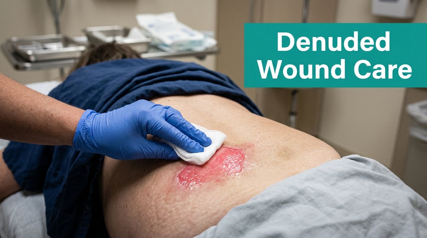
A nurse calls it a “red open area” on the sacrum, charts Stage 2 pressure injury, starts the usual dressing plan, and moves on. Two weeks later the payer rejects the claim, the MDS team is irritated, and the chart doesn't support what was billed. The skin breakdown was real. The staging was wrong.
That scenario shows up all the time in SNFs, home health, wound clinics, and hospital consults. Denuded skin gets treated like a simple surface wound when the primary problem is identification, then documentation, then coding. If you miss the first step, the rest of the chart falls apart.
More Than Just a Rash Identifying the Denuded Wound
A denuded wound is not just “excoriation” and it's not a pressure injury with a softer name. Clinically, it's partial-thickness epidermal loss that leaves a painful, moist, erythematous dermal surface exposed. It often weeps. It often burns more than it “hurts.” And it usually shows up where moisture, friction, irritants, or all three have been working on the skin for days.
Where people go wrong
The most common miss is in the sacrum, buttocks, perineum, gluteal cleft, and skin folds. Staff see open red skin over a bony area and default to pressure staging. That shortcut creates problems fast.
Per Medbridge's review of denuded wounds, denuded wounds are not pressure injuries and should not be staged as such under NHTL guidance. The same source also notes that documentation needs to specify wound bed color, drainage, and periwound maceration, and that 43% of wound care practices report coding errors due to ambiguous tissue descriptors in SOAP notes.
Practical rule: If the cause is moisture, chemical irritation, friction, adhesive trauma, or rubbing, stop reaching for pressure staging language first.
Etiology matters more than appearance alone
A denuded wound can look superficially similar to a Stage 2 pressure injury because both involve partial-thickness skin loss. The difference is cause. Pressure injury comes from pressure and shear. Denudation usually comes from:
- Moisture exposure: urine, stool, sweat, wound drainage, or trapped humidity in skin folds
- Chemical irritation: fecal enzymes and prolonged contact with irritants
- Friction: brief, repeated rubbing from transfers, linens, briefs, or body habitus
- Adhesive trauma: aggressive tape or dressing removal on fragile skin
That distinction isn't academic. It drives treatment, coding, survey defensibility, and whether your claim gets paid.
What surveyors and payers notice
Surveyors will flag inconsistent documentation. If one note says IAD, another says Stage 2, and the photo shows diffuse moisture injury in the gluteal cleft, the chart looks unreliable. Payers do the same thing from a different angle. They compare diagnosis, treatment, and procedure coding. If those don't line up, denials follow.
A denuded wound is often straightforward to heal. It is not straightforward to defend if you document it poorly.
Reading the Wound Bed Accurate Denuded Wound Assessment
The first few seconds of visual assessment usually tell you most of what you need to know. A denuded wound typically looks shiny, moist, pink to bright red, and weeping, with the epidermis gone and the dermis still intact. Borders may be irregular. The patient often reports stinging with cleansing and contact.
Start with what you see and what caused it
Don't begin by forcing the wound into a code bucket. Begin with the tissue and the cause.
Use this sequence at bedside:
- Identify the trigger: incontinence, perspiration, adhesive removal, friction, or mixed causes.
- Describe the bed: shiny, erythematous, moist, weeping, intact dermis, no eschar.
- Check the edges: diffuse and irregular suggests moisture injury more than pressure.
- Assess the surrounding skin: look for maceration, satellite rash, blanching, or diffuse inflammation.
- Quantify drainage: not “some drainage.” Quantify it.
- Document pain quality: burning and stinging are common with exposed dermis.
If your team needs tighter wound-bed language, a structured reference for wound bed descriptions helps standardize terms before they become coding problems.
Differentiate denudation from common mimics
The two big mimics are IAD and pressure injury, with candidal rash sitting in the differential if the fold environment fits.
A simple bedside comparison helps:
| Finding | Denuded wound | IAD | Pressure injury mimic concern |
|---|---|---|---|
| Surface | Open, moist, partial-thickness | Often diffuse inflammation with possible erosion | May show more localized damage over pressure points |
| Cause | Friction, moisture, chemical irritation, adhesive trauma | Moisture and irritants from incontinence | Pressure and shear |
| Borders | Irregular | Patchy or diffuse | More localized to pressure pattern |
| Symptoms | Burning, tenderness, serous weeping | Burning, itching, discomfort | Pain may be present, often linked to loading |
A denuded wound can exist within broader IAD. That's where people get tripped up. The dermatitis is the surrounding moisture-related skin injury. The denuded area is where the epidermis is gone.
If the note says “red open area” and nothing else, the chart is already weak.
Exudate documentation needs actual thresholds
Exudate isn't a side note. It drives dressing choice and shows whether the plan makes sense. According to WCEI guidance on exudate quantities, moderate exudate saturates 25% to 75% of the bandage, while large or copious exudate means more than 75% saturation with wet wound tissues.
That matters because high moisture around denuded tissue quickly leads to more maceration.
Document these pieces every time:
- Amount: scant, small, moderate, large/copious
- Type: serous, serosanguineous, other
- Impact on periwound: none, mild maceration, advancing maceration
- Dressing performance: intact on removal, saturated, leaking, displaced
When newer clinicians skip the periwound, they miss the story. With denuded skin, the periwound often tells you whether your plan is helping or making the wound larger.
Choosing the Right Dressing for Denuded Wounds
The treatment goal is simple. Protect the exposed dermis and control moisture without causing more trauma. That sounds obvious, but a lot of wound plans still fail on one of those two points.
What doesn't work well? Wet-to-dry dressings, harsh adhesives, frequent unnecessary changes, and anything that rips off fragile new epithelium on removal. You may get a cleaner-looking surface for a moment, then you've restarted the injury.
Moisture balance decides whether this improves or spreads
A denuded wound needs a moist healing environment, but not a soaked one. That distinction is where good dressing selection lives.
Wounds International's consensus report on exudate management states that uncontrolled exudate delays healing in 68% of chronic cases and increases infection risk by 2.3x. That same gap shows up in everyday billing, because exudate volume rarely gets tied cleanly to the rest of the note.
For bedside dressing decisions, I'd rather see a simple principle-based choice than a memorized product habit. If the wound is weeping lightly, protect it. If it's soaking the dressing and the periwound is turning white and soggy, absorb more and secure less aggressively.
Dressing Selection for Denuded Wounds by Exudate Level
A practical chart helps. If your clinic wants a quick reference, this wound dressing selection chart is useful for keeping choices consistent.
| Exudate Level | Primary Goal | Recommended Dressing Types |
|---|---|---|
| Scant | Protect exposed dermis and reduce friction | Skin barrier film, zinc oxide paste, transparent film if low-friction location |
| Moderate | Maintain moisture balance and prevent periwound maceration | Hydrocolloid in appropriate cases, bordered or non-bordered foam, barrier paste to surrounding skin |
| Heavy | Absorb drainage, protect surrounding skin, reduce leakage | Absorptive foam dressing, barrier film or zinc-based protection to periwound, more frequent reassessment of fit and saturation |
What I'd avoid first
Not every denuded wound needs an advanced approach. Many need a less aggressive one.
- Avoid strong adhesives: fragile epidermal edges don't tolerate repeated stripping.
- Avoid routine wet-to-dry: it's traumatic and rarely justified for this tissue type.
- Avoid over-occlusion in high-moisture folds: if the dressing traps drainage, maceration wins.
- Avoid “one dressing for all denuded skin” habits: gluteal cleft, abdominal fold, and lower leg all behave differently.
Match the location to the plan
Gluteal cleft and perineal denudation usually need barrier support plus absorbency, not fancy layering that won't stay in place. Skin folds need moisture control and friction reduction. A denuded area from adhesive removal often needs little more than protection and time, provided you stop the offending tape.
The mistake is treating every red open surface the same. Denuded skin heals fast when you remove the cause and stop re-injuring it.
Managing Infection Risk in Denuded Tissue
Denuded tissue has lost its barrier. That doesn't mean every moist red wound is infected. It does mean you need to watch for change, not just appearance.
Early on, many denuded wounds are inflamed because the tissue is irritated and exposed. Warmth and erythema alone don't prove infection. What matters is whether the wound trajectory changes for the worse.
Watch the exudate, not just the color of the skin
A practical sign of trouble is drainage change. Per Wounds International's guidance on wound infection and exudate assessment, a shift from clear, thin, serous exudate to opaque, discolored, thick, purulent drainage in tan, yellow, green, or brown is a primary indicator of developing infection or high bacterial levels. That's why consistency, odor, and color belong in every assessment.
What deserves escalation
Use clinical judgment, but these are the patterns that should get your attention:
- Drainage change: serous becomes thick, opaque, and discolored
- Pain shift: new or increasing pain, especially if the prior complaint was just stinging with contact
- Failure to improve: the denuded area enlarges despite moisture control and protection
- Periwound deterioration: advancing maceration, increased friability, spreading inflammation
- Systemic concern: if the patient's broader picture suggests more than a localized surface issue
Normal inflammation stays relatively stable or improves when you remove the cause. Infection tends to declare itself by changing the wound.
Culture only when it will change management
A superficial swab on every denuded wound isn't good practice. Surface contamination is common. Culture makes more sense when the wound has clear clinical signs of infection and the result will influence antimicrobial treatment.
For localized bioburden concerns, a conservative approach usually starts with better moisture control, reduced trauma, and a topical antimicrobial strategy when clinically appropriate. Systemic antibiotics belong to a higher threshold. If the tissue involvement becomes deeper, the differential changes and the coding changes with it too.
That's especially true in diabetic foot work. Once you're dealing with deeper infection patterns, you're no longer in the denuded-skin lane. You're in ulcer classification, limb threat, and possibly advanced adjunctive care.
Coding Denuded Wounds for Proper Reimbursement
The effectiveness of otherwise solid wound care is frequently undermined. The wound is assessed correctly, treated reasonably, then coded like a pressure injury or billed with a debridement code the tissue doesn't support.
Denuded wounds usually do not support sharp debridement codes 11042 to 11047. Those codes are tied to debridement of deeper tissue levels, and denuded skin typically doesn't have the necrotic burden or depth to justify them. If you bill those codes on a moist superficial denuded area with no slough or eschar, that chart is vulnerable in an audit.
Debridement coding and the common overreach
Most denuded wounds need cleansing, barrier protection, moisture management, offloading of irritants, and time. They do not need you to turn routine surface care into an excisional narrative.
Selective debridement codes such as 97597/97598 may be considered only when there is actual devitalized tissue requiring selective removal and your note clearly supports that work. If the wound bed is shiny, erythematous, and free of slough, don't force a debridement code into the claim.
A strong coding reference for common wound scenarios is this ICD-10 and CPT coding resource, especially when your team is trying to align bedside findings with billable work.
Code the cause, not the visual shortcut
For denuded skin, the ICD-10-CM pathway should follow etiology. If the denudation is from moisture, irritation, or skin-fold breakdown, code from the underlying cause rather than defaulting to a pressure injury family.
Examples of the coding logic clinicians often need:
- Moisture-related irritant pattern: consider an irritant dermatitis pathway when supported by the clinical picture
- Skin-fold denudation: consider an intertriginous or related skin-fold diagnosis when that's what the exam shows
- Incontinence-driven breakdown: support the moisture-associated etiology clearly in the assessment and plan
The exact code selection still depends on the encounter and documentation, but the point stands. Don't code appearance alone. Code cause plus tissue findings.
Don't mix denuded tissue with deeper ulcer frameworks
One mistake I see in multidisciplinary settings is carrying over diabetic foot ulcer language into superficial skin injury documentation. Keep your frameworks separate.
Per WoundReference's review of diabetic foot ulcer classification, Wagner Grade 3 marks deep infection involvement such as abscess, osteitis, osteomyelitis, or tendon sheath infection, and it's the point where hyperbaric oxygen therapy may be considered if the ulcer fails to heal after 30 days of standard wound care. That's a useful reminder of what true deep tissue complexity looks like. A denuded wound is not that.
If your note reads like a superficial moisture injury but your codes read like a deep ulcer service, expect scrutiny.
Phrases that help and phrases that hurt
Better documentation phrases:
- “Partial-thickness denuded skin to gluteal cleft secondary to fecal incontinence and friction.”
- “Wound bed moist, shiny, erythematous, no slough or eschar.”
- “Periwound mild maceration present.”
- “Plan focused on barrier protection, moisture control, and atraumatic coverage.”
Red-flag phrases:
- “Stage 2-looking area.”
- “Excoriation vs pressure.”
- “Debrided wound” when only cleansing was performed
- “Open area noted” with no etiology, bed description, or drainage detail
Documenting for Success A Denuded Wound Example
A good denuded wound note tells one clean story. Cause, tissue, drainage, surrounding skin, intervention, plan. If one of those is missing, coding gets fuzzy and progress tracking gets worse.
Here's a practical example.
SOAP note example
S:
78-year-old patient seen for painful skin breakdown in gluteal cleft. Staff report frequent fecal incontinence. Patient endorses burning pain with cleansing and repositioning. No report of pressure-related pain pattern.
O:
Denuded wound to gluteal cleft. Partial-thickness epidermal loss. Measures 3.2 cm x 2.1 cm x 0.1 cm. Wound bed shiny, erythematous, moist, no slough or eschar. Exudate scant serous. No purulence or malodor. Periwound with mild maceration noted from 3 to 9 o'clock. Borders irregular. Findings consistent with moisture- and friction-related denudation rather than pressure injury.
A:
Denuded wound secondary to fecal incontinence and friction. No evidence of deep tissue involvement. No clinical signs of acute infection at this visit.
P:
Cleansed with NS. Zinc-based skin barrier paste applied to periwound and vulnerable surrounding tissue. Atraumatic absorptive dressing applied to protect site and manage moisture. Incontinence care reinforced with prompt cleansing and moisture barrier after each episode. Reassess wound bed, exudate, and periwound maceration at next visit. Avoid aggressive adhesive products.
What makes that note defensible
Notice what's missing. No pressure stage. No unsupported debridement claim. No vague “excoriation.” The note ties cause to appearance and treatment.
That's what payers want. It's also what surveyors look for when they compare the note to the photo.
For photography, keep it basic and reproducible:
- Use a measuring guide: include a ruler for scale in the same plane as the wound
- Fix the lighting: shadows create false depth and hide weeping surfaces
- Show the periwound: don't crop so tightly that you lose the maceration pattern
- Annotate consistently: location and orientation matter for follow-up comparison
- Protect patient privacy: before sharing records externally, it's smart to remove PDF metadata for privacy so hidden identifiers don't travel with the file
Small details that save time later
Teams get into trouble when the image shows one thing and the note says another. A clean workflow uses the same descriptors every visit, in the same order, with the same photo method. That reduces handoff confusion and makes progression easier to defend.
My advice to newer clinicians is simple. Build your note so a coder, surveyor, and covering clinician can all understand the wound without calling you.
The Clinicians Takeaway on Denuded Wounds
Three things matter with a denuded wound, and none of them are optional.
First, identify it correctly. Don't let location fool you into pressure staging. If the cause is moisture, friction, irritant exposure, or adhesive trauma, document that plainly and keep the diagnosis aligned with the etiology.
Second, treat the tissue gently. The goal is protection and moisture balance. Not aggressive debridement. Not strong adhesive fixation. Not dressing choices that look active but keep re-injuring the surface. Denuded tissue often improves once you stop the insult and stop disturbing the wound bed.
Third, chart the full picture. Bed appearance, exudate amount and type, periwound condition, likely cause, and exact intervention all need to sit in the same note. If those elements don't connect, the claim won't either.
Good denuded wound care is less about doing more and more about naming the problem correctly, protecting the tissue, and writing a note that can survive review.
That's the difference between a common skin injury that heals uneventfully and a common skin injury that creates denials, survey issues, and endless chart corrections.
EkagraHealth AI helps wound care teams document denuded wounds and other complex cases with less back-and-forth. It drafts SOAP notes, maps CPT and ICD-10-CM codes, supports wound image capture and annotation, and helps practices get cleaner claims out the door. If your team is spending too much time translating bedside findings into billable documentation, take a look at EkagraHealth AI.
