
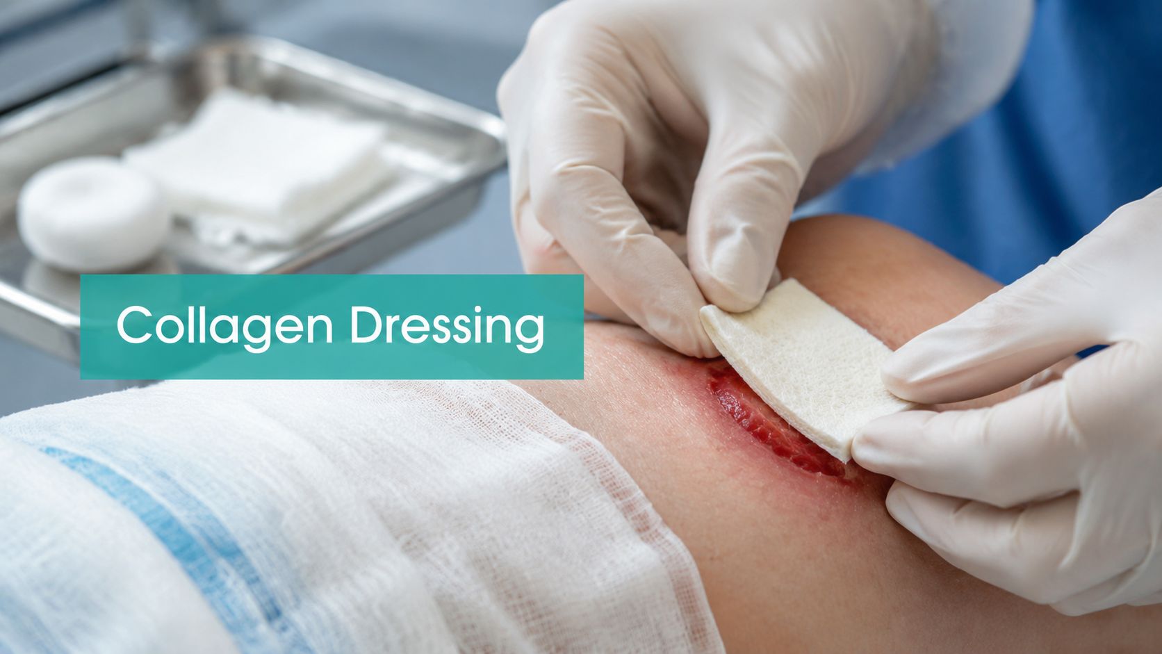
A Wagner 2 diabetic foot ulcer can look deceptively manageable: clean margins, moderate drainage, no obvious spreading infection, and a patient who says offloading is finally happening. Yet after several weeks of sharp debridement, pressure relief, and moisture control, the measurement barely changes. That plateau is where a collagen dressing may earn a place in the plan.
Collagen isn't a substitute for vascular assessment, infection control, compression, offloading, or debridement. It's a wound-bed support tool for a clean, stalled chronic wound. The clinical outcome and the payer outcome depend on the same discipline: select the right wound, apply the dressing correctly, and document why it belongs there.
When Standard Care Hits a Wall
A plantar ulcer under the first metatarsal head had been treated with sharp debridement, an appropriate offloading plan, and a dressing selected for the drainage pattern. The wound bed was viable, the periwound skin was protected, and there was no clinical evidence of spreading infection. Still, serial measurements showed little meaningful change over several weeks.
That pattern matters. A wound that's slowly progressing may need time and continued standard care. A wound that remains clean but repeatedly stalls may need help with the inflammatory environment. In a 2022 meta-analysis of 11 randomized trials involving 961 patients and 961 wounds, complete healing occurred in 53.4% of wounds treated with collagen plus standard care, compared with 34.5% with standard care alone, with a pooled risk ratio of 1.53 (2022 meta-analysis of collagen dressings).
Read the plateau, not just the wound
Before selecting collagen, I look for a credible standard-care foundation:
- Etiology is addressed: Venous ulcers need compression when clinically appropriate. Plantar diabetic foot ulcers need effective offloading. Pressure injuries need pressure redistribution and a plan for nutrition, mobility, and support surfaces.
- The bed is prepared: Adherent slough, necrotic tissue, and suspected biofilm require appropriate cleansing and debridement. Collagen placed over a dirty bed is usually wasted.
- Perfusion is considered: An ischemic ulcer needs vascular evaluation and treatment planning. A collagen dressing can't compensate for inadequate arterial inflow.
- The drainage pattern is understood: Low-to-moderate exudate may allow the matrix to remain in useful contact. Heavy drainage can overwhelm some formulations and damage the periwound.
- The wound has stalled: Serial measurements, tissue description, and photographs should support the conclusion rather than a vague statement that the wound is “nonhealing.”
For patients with diabetic foot disease, prevention remains part of the treatment plan. A consistent daily foot inspection routine can identify new pressure, drainage, or skin breakdown before a small problem becomes a deeper ulcer.
Know when collagen is the wrong move
An infected wound, an ischemic wound, or a wound covered in adherent slough needs its primary problem addressed first. Purulent drainage, expanding erythema, malodor with clinical deterioration, increasing pain, or systemic findings should shift attention toward source control and urgent evaluation.
Collagen is most defensible in a clean, adequately perfused, nonhealing chronic wound with controlled bioburden and a reasonable moisture environment. It's particularly relevant to stalled venous ulcers, diabetic foot ulcers, and other chronic wounds after the fundamentals are in place. The goal isn't to add an advanced dressing because the wound looks difficult. The goal is to correct a wound-bed environment that has stopped progressing.
How Collagen Dressings Work at the Wound Bed
A useful way to think about collagen is as a temporary construction scaffold. It gives cells a surface for attachment and migration while the patient's own tissue rebuilds the wound. The scaffold doesn't replace viable granulation tissue, and it won't create healing where perfusion, pressure control, or infection management is absent.
Chronic wounds often contain excessive protease activity, including matrix metalloproteinases. In practical terms, the wound can degrade newly formed extracellular matrix faster than it can organize new tissue. Collagen-combination products, especially oxidized regenerated cellulose/collagen formulations, are used partly because they can bind proteases while supporting the wound bed.
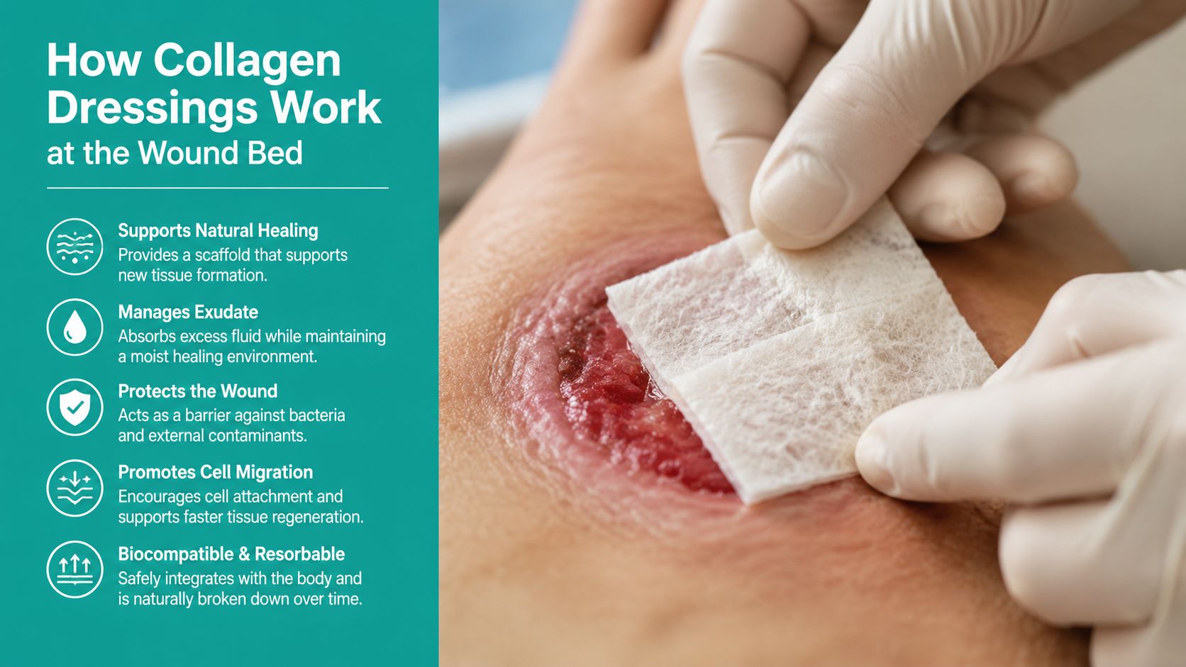
Follow the biology step by step
- Prepare the surface. Cleansing and debridement remove barriers between the dressing and viable tissue. The matrix needs contact with a wound bed that can participate in repair.
- Place the collagen in contact with the bed. The product should conform to the wound rather than sit folded on the surrounding skin. Irregular wounds may require careful trimming.
- Maintain appropriate moisture. Collagen generally needs a moist environment to interact with the wound surface. Excessive fluid, however, can dilute or displace the matrix and macerate the periwound.
- Protect the interface. A secondary dressing manages drainage, prevents trauma, and keeps the primary dressing in place.
- Reassess the response. The wound's measurements, tissue quality, drainage, and periwound condition determine whether the plan is working.
The product's source and formulation matter clinically. Native collagen matrices may provide structural support, while oxidized regenerated cellulose/collagen combinations also target the protease-heavy environment seen in some chronic wounds. The evidence is strongest for combination products rather than treating every collagen dressing as interchangeable.
A meta-analysis of 13 comparative studies found that ORC/collagen dressings improved complete healing, increased relative wound area reduction by 13.5%, and reduced adverse events compared with control dressings. The reported complete-healing odds ratio was 1.74, and the adverse-event odds ratio was 0.63 (meta-analysis of ORC/collagen dressings). Those findings support a targeted use strategy, not automatic use in every chronic wound.
Matching Collagen Type to Wound Characteristics
The correct product depends on more than the word “collagen” on the package. I match the formulation to etiology, depth, exudate, tissue quality, and bioburden. A product that performs well in a moderately draining, clean ulcer may be a poor choice for a heavily exuding wound with unstable periwound skin.
A practical selection framework
For a Wagner 1 or 2 diabetic foot ulcer, the first questions are whether the ulcer is being offloaded, whether perfusion is adequate, and whether the bed is clean. If those conditions are met and the wound has plateaued, a collagen-combination product may be reasonable, particularly when the inflammatory burden appears high.
Venous leg ulcers require the same discipline. Collagen doesn't replace compression. If compression is absent or ineffective, changing the primary dressing won't address the underlying venous hypertension. For a clean venous ulcer with moderate exudate and delayed progress, collagen can support the wound bed while compression addresses the cause.
Pressure injuries require attention to pressure redistribution, shear, moisture, nutrition, and mobility. Collagen may be considered when the wound remains stalled despite those measures, but it shouldn't distract the team from the mechanical cause.
| Wound Type | Exudate Level | Recommended Collagen Type | Key Considerations |
|---|---|---|---|
| Wagner 1 or 2 diabetic foot ulcer | Low to moderate | Collagen or ORC/collagen combination | Confirm offloading, perfusion, depth, and bioburden control |
| Venous leg ulcer | Moderate | Collagen or ORC/collagen combination | Continue clinically appropriate compression and monitor maceration |
| Chronic pressure injury | Low to moderate | Formulation matched to bed and drainage | Correct pressure, shear, moisture, and nutritional factors first |
| Heavily exuding chronic wound | Heavy | Use caution with collagen selection | Absorptive management may need priority before collagen |
| Sloughy, ischemic, or infected wound | Any | Do not use collagen as the primary corrective step | Address debridement, perfusion, and infection concerns first |
A wound dressing selection chart can help standardize this reasoning across clinicians, particularly in mobile programs and facilities where several people document the same wound.
Avoid overgeneralizing the evidence
A systematic review pooling 1,538 wounds across 13 studies found significant improvements in wound closure and wound area reduction with ORC/collagen dressings compared with standard dressings. The reported values were P = 0.027 for wound closure and P = 0.006 for percent wound area reduction (systematic review of ORC/collagen dressings).
Those results don't mean every formulation has identical performance, and they don't remove the need for clinical judgment. Collagen is poorly matched to an uncontrolled wound. It also isn't a replacement for sharp debridement, offloading, compression, or vascular workup.
Application Technique and Exudate Management
Application starts before the package is opened. Confirm the wound's current dimensions, tissue composition, drainage amount and character, odor, pain, and periwound condition. If the wound has changed substantially since the last visit, reassess the diagnosis and the underlying cause rather than applying the same dressing by habit.
Prepare the bed and fit the matrix
Cleanse according to the care plan, then remove loose nonviable tissue and address adherent slough when clinically indicated. The surface should be free of obvious debris and excessive drainage before the collagen is placed. If the wound is infected or deteriorating, pause the collagen plan and escalate treatment.
Cut the dressing to fit the wound bed without unnecessarily overlapping intact periwound skin. Avoid aggressive packing unless the product's instructions and the wound depth support that use. The dressing should contact viable tissue, while the secondary dressing should handle the drainage that the collagen itself cannot safely manage.
For a wound with low-to-moderate exudate, a protective secondary dressing may be enough. For moderate-to-heavy drainage, use an absorptive secondary layer and inspect the edges for maceration. A collagen dressing cleared by the FDA is described as a sterile, single-use, opaque, absorbent collagen membrane matrix for topical use in moderately to heavily exudating wounds and for control of minor bleeding (FDA device documentation). Product labeling still needs to be checked because collagen formulations differ.
Change based on the wound, not the calendar
A dressing change is an assessment opportunity. Look for persistent saturation, strike-through, undermining, odor, periwound whitening, new pain, or a shift from serous to purulent drainage. The exudate classification reference can help staff describe drainage consistently rather than relying on terms such as “a lot” or “bad drainage.”
Negative pressure wound therapy may be used in selected wounds when the wound bed, seal, depth, and overall plan support it. The collagen layer and the negative-pressure interface must be compatible with the treatment protocol. Coordinate with surgical debridement or grafting when the wound's depth, exposed structures, or lack of progress exceeds what a topical matrix can address.
The case series most often cited for native collagen matrix use involved 20 wounds in 19 patients, with a mean wound duration of 66.6 months. It reported a mean surface-area reduction of 29%, a median reduction of 47%, Bates-Jensen score improvement in 85% of cases, and pain reduction in 66.66%, with no adverse events (native collagen matrix case series). Those findings describe a selected clinical experience, not a guarantee. They reinforce the need to pair collagen with correct preparation and moisture management.
Documentation and Coding That Prevents Denials
A collagen claim can fail even when the clinical decision was sound. The usual problem is an incomplete record. “Advanced dressing applied” doesn't establish medical necessity, and “wound improving” doesn't show whether the treatment produced a meaningful response.
Document the decision before documenting the product
At each relevant visit, record the wound's location, etiology, length, width, and depth. Describe the tissue composition, drainage amount and character, odor, undermining or tunneling, periwound condition, pain, and signs of infection. For diabetic foot ulcers, include the Wagner grade when clinically appropriate. Also document the interventions that address the cause, such as offloading, compression, pressure redistribution, or vascular referral.
The rationale should connect the findings to the treatment. A defensible note might state:
Clinical rationale: The Wagner 2 plantar ulcer remains clean and adequately perfused but has plateaued despite sharp debridement, prescribed offloading, and moisture management. Exudate is moderate and serous, with viable granulation and no clinical evidence of spreading infection. ORC/collagen is selected to support the stalled wound bed, with an absorptive secondary dressing and continued offloading.
That language is stronger than “collagen applied for nonhealing wound.” It explains what failed, what remains controlled, and why the selected dressing fits the wound.
Keep debridement and dressing billing distinct
CMS states that CPT 11042–11047 are reported by the depth of tissue removed and by surface area, and that for a single wound the deepest level removed is reported. For multiple wounds, wounds at the same depth are summed, but areas from different depths aren't combined. CMS also states that dressings applied to the wound are part of the service for 11042–11047 and may not be billed separately (CMS debridement policy).
Common denial triggers include:
- Missing measurements: The note doesn't establish baseline size or serial change.
- Unclear depth: The documented tissue removed doesn't support the selected debridement code.
- No failed standard care: The record omits debridement history, offloading, compression, or pressure management.
- Generic wound language: “Moderate drainage” appears without character, periwound findings, or rationale.
- Bundled dressing billing: The dressing is billed separately when CMS treats it as included in the debridement service.
- Unsupported infection statements: The note labels a wound infected without documenting the clinical findings or treatment response.
Use the payer's policy and the patient's diagnosis to select ICD-10-CM codes. Don't choose a code solely because it sounds compatible with collagen. A medical necessity documentation workflow can help clinicians and billing staff use the same required elements, especially when several providers contribute to the chart.
Surveyors and auditors look for internal consistency. The wound description, photograph, measurement, debridement depth, treatment order, and follow-up plan should tell the same story.
Case Examples From Clinical Practice
The stalled plantar ulcer
A patient with a Wagner 2 plantar diabetic foot ulcer had a clean bed and moderate serous exudate. The care team had performed sharp debridement, reinforced offloading, and maintained moisture balance, but serial measurements showed a plateau after an adequate trial of standard care.
The decision to use an ORC/collagen dressing followed the assessment rather than replacing it. The note recorded the ulcer location, dimensions, depth, tissue type, drainage, periwound condition, Wagner grade, offloading plan, prior treatment, and absence of clinical findings requiring infection escalation. The dressing was fitted to the bed, covered with a secondary dressing selected for the drainage, and reassessed at follow-up.
The wound then showed a clear reduction in surface area over the next several weeks. That improvement can't be attributed to collagen alone. Offloading and debridement continued, and the documentation made that relationship explicit.
The wet venous ulcer
The second patient had a venous leg ulcer with heavy drainage, macerated edges, and uncontrolled bioburden. Collagen was applied because the wound was labeled “chronic,” but compression consistency was poor and the exudate quickly overwhelmed the dressing.
The error was sequencing. The wound needed drainage control, periwound protection, compression when appropriate, and assessment of the suspected bioburden before a collagen matrix could be expected to help. The record also failed to explain why collagen was selected or how the team would judge response.
The lesson is straightforward: a difficult wound isn't automatically a collagen wound. If the bed is wet, unstable, infected, ischemic, or mechanically unprotected, correct those barriers first. Advanced dressing selection should follow wound-bed preparation, not substitute for it.
Key Decision Points and Clinical Checklist
Collagen belongs in the plan when the wound is clean, adequately perfused, appropriately offloaded or compressed, and stalled despite optimized standard care. Choose the formulation according to exudate and wound biology, not brand familiarity or habit.
Use this bedside check:
- Cause: Is venous pressure, plantar pressure, immobility, or another cause being treated?
- Bed: Is viable tissue exposed after cleansing and appropriate debridement?
- Bioburden: Are infection concerns and drainage changes addressed?
- Perfusion: Has ischemia been considered?
- Exudate: Can the primary and secondary dressings maintain contact without maceration?
- Documentation: Are measurements, depth, tissue, drainage, periwound findings, prior care, and rationale recorded?
- Billing: Does the debridement code reflect the deepest tissue removed, and is bundled dressing billing avoided?
- Stop signs: Is the wound deteriorating, ischemic, infected, heavily sloughed, or failing despite correct use?
Reassess the wound's trajectory, not just its appearance. If the plan isn't producing progress, step back and revisit diagnosis, perfusion, pressure, compression, debridement, and infection control.
EkagraHealth AI helps wound care teams turn bedside findings into structured SOAP documentation, appropriate CPT and ICD-10-CM workflows, wound measurements, and cleaner claims. Visit EkagraHealth AI to see how the platform can support collagen dressing documentation and reduce the gaps that lead to denials.
