
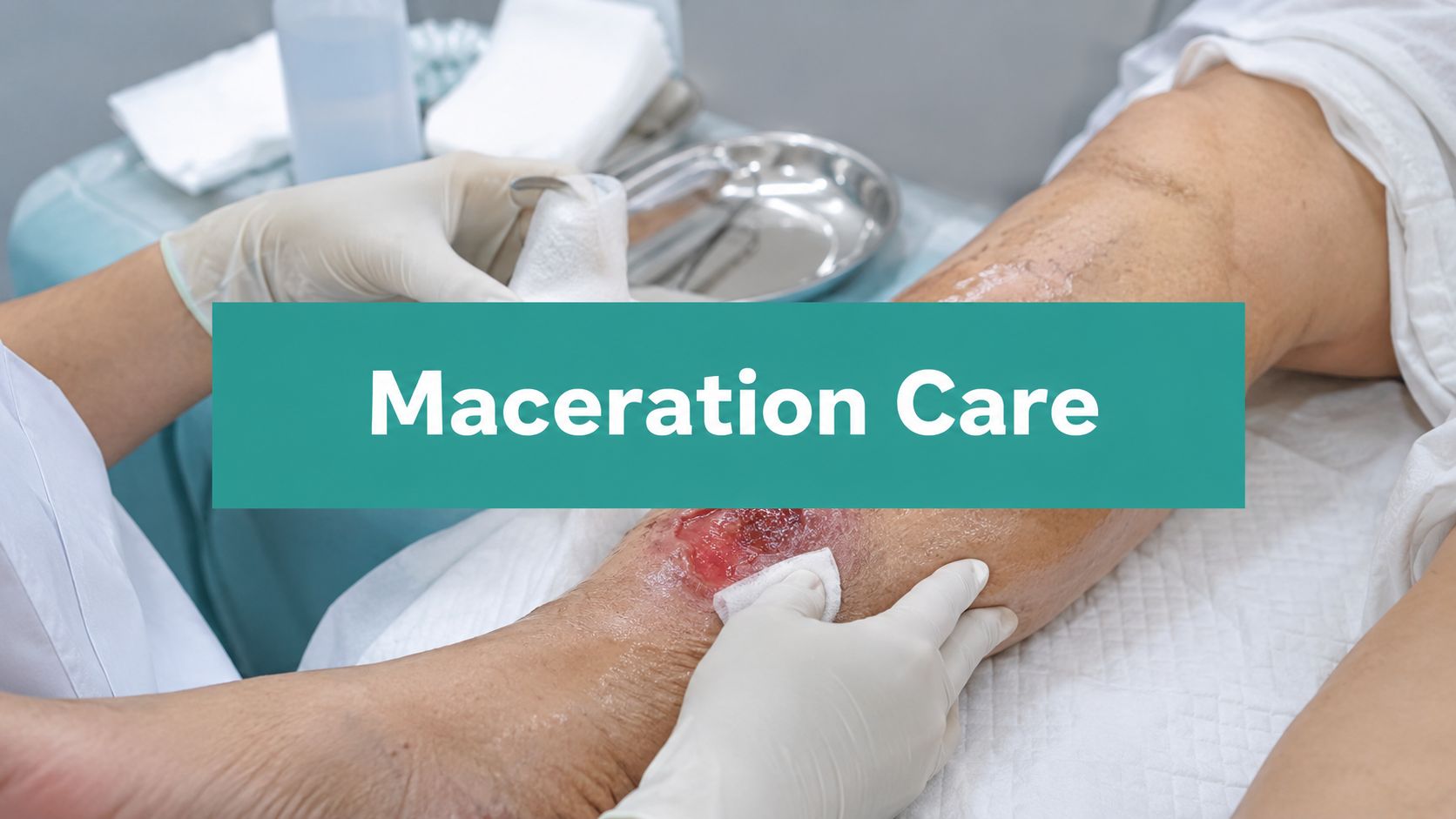
The ulcer looked cleaner than the chart suggested. The wound bed had decent granulation, but the periwound skin was white, wrinkled, and starting to lift at the edges. That's the kind of maceration wound that turns a manageable visit into a stalled plan, because the drainage is telling you the dressing system is already losing the moisture battle.
A draining venous leg ulcer is a classic setup. The fluid isn't just sitting there, it's sitting against skin that was never meant to stay wet for hours or days at a time. In chronic wounds, that persistent contact with exudate swells the stratum corneum, weakens the skin barrier, and makes the edge more likely to break down, leak, and invite infection rather than contract and close (Wounds International).
One practical point gets missed all the time. A dry edge around a draining ulcer usually means the dressing is matching the exudate load. A white, soggy rim usually means the current plan is failing. That distinction matters clinically, and it matters in the chart.
Recognizing Maceration Before It Derails Healing
The patient usually tells the story before the skin does. “It's draining more,” “the bandage slips,” or “the edge feels sore” often comes up before the wound bed looks worse. By the time the rim is white and wrinkled, the periwound has already been overhydrated long enough for the tissue to lose strength and start breaking down.
What the edge is really telling you
Periwound maceration is a form of moisture-associated skin damage. It happens when exudate, urine, sweat, or other fluids sit on the skin long enough to overhydrate it. In chronic wounds, the problem gets worse when the underlying driver of heavy drainage is not controlled, such as venous hypertension in a venous leg ulcer.
That is why I do not treat the edge as a cosmetic finding. A prospective study of diabetic foot ulcers found maceration was an independent predictor of healing failure, with an adjusted hazard ratio of 0.324 (Wiley). In plain terms, ulcers with maceration healed less favorably than ulcers without it.
Practical rule: if the wound bed looks acceptable but the periwound is whitening, softening, or wrinkling, the moisture plan is already off.
The mechanism is predictable. Exudate from chronic wounds can contain proteases, and prolonged contact overhydrates the skin while breaking down protein structure. Occlusive or nonbreathable cover dressings can make it worse by trapping moisture at the surface. A useful next step is to document the type of exudate clearly, because serous, serosanguinous, and purulent drainage do not create the same dressing problem. For a concise reference on drainage patterns, see this guide to wound exudate types.
Systematic Periwound Assessment Techniques
A quick look at the base of the wound isn't enough. Maceration often starts at the margins, and those margins can be assessed in under a minute if you use the same sequence every time. I teach staff to start with color, then texture, then extent, then cause.
The edge first, then the bed
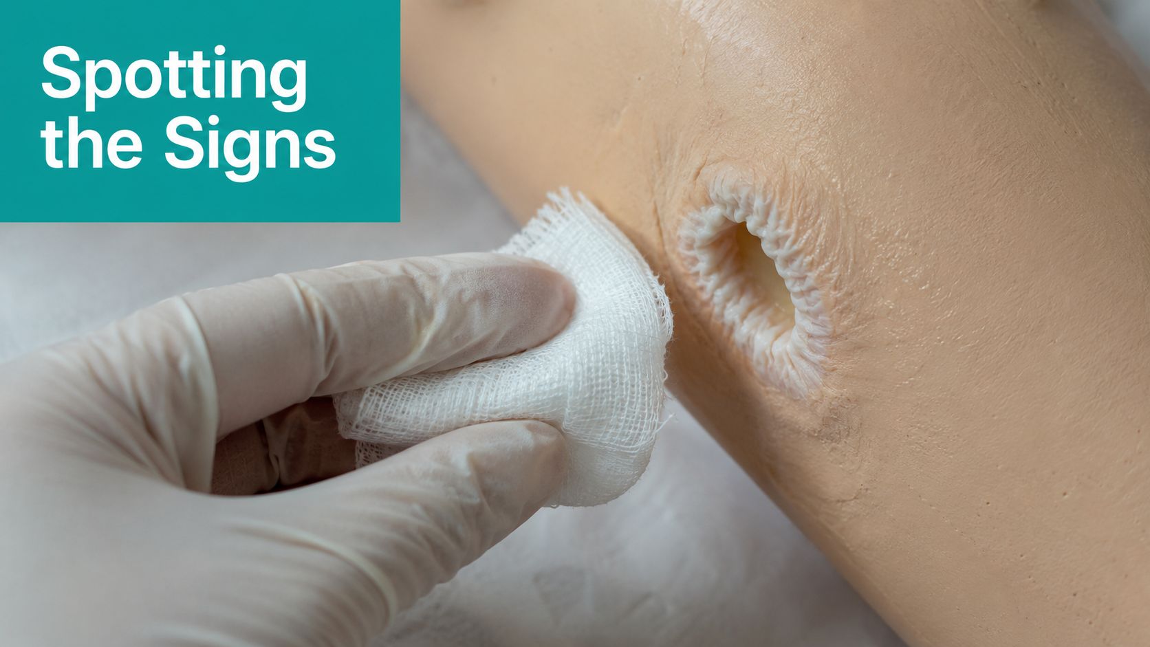
Look for whitening, a translucent rim, wrinkling, bogginess, or denudement extending from the wound edge. The skin may feel softer than the surrounding tissue, and in some cases it will look slightly raised or waterlogged. That's different from healthy moisture, which should not leave the tissue swollen or fragile.
Measure the periwound the same way you measure the wound. Document how far the changes extend from the edge, and be consistent from visit to visit. If the wound is irregular, map the vulnerable zone by clock face and note which quadrants are affected.
The surveyor wants to see a pattern, not a guess. “Periwound maceration improved” is weak. “White, wrinkled periwound extending 0.5 cm from the superior and lateral margins, improved with increased dressing frequency” is defensible.
What to document so the note holds up
Record the type of exudate, amount, odor if present, and whether drainage is strike-through, pooling, or saturating the primary layer. If the exudate is moderate to heavy, say so. If the drainage is serous, serosanguinous, or purulent, name it. Many notes fail at this point, because they describe “drainage present” and leave the reviewer to fill in the rest.
Differential diagnosis matters too. Maceration can resemble fungal intertrigo or irritant/contact dermatitis, especially in moist skin folds. Fungal rashes tend to have more surface inflammation and satellite lesions, while contact dermatitis often tracks with adhesive exposure or a product pattern. Maceration is usually more directly tied to fluid contact at the wound edge.
For difficult-to-see locations, use palpation with a gloved finger and inspect after dressing removal, before cleansing blurs the tissue findings. In tunneled or undermined wounds, don't assume the edge is stable just because the bed looks clean. Edges fail first in a lot of these wounds.
The internal references on exudate classification and charting help here, especially when the drainage pattern changes from visit to visit: exudate types and what they mean. In practice, that kind of structured language keeps the note aligned with what happened at the bedside.
Dressing Selection for Moisture Balance
The mistake I see most often is treating all exudate as if a single absorbent pad can solve it. It can't. The right dressing depends on how much fluid the wound is producing, whether the periwound is already damaged, and whether the dressing is helping maintain a moist environment without flooding the edge.
Comparing dressing choices by drainage burden
| Exudate Level | Primary Dressing | Secondary Layer | Expected Wear Time |
|---|---|---|---|
| Light | Nonadherent contact layer or thin absorbent dressing | Light protective cover if needed | Longer, if the edge stays intact |
| Moderate | Absorbent foam or hydrofiber | Secondary cover to secure and contain drainage | Variable, based on saturation |
| Heavy | Hydrofiber or alginate in direct contact with the wound, often layered | Absorbent secondary cover and edge protection | Shorter, with more frequent changes |
Foam alone often fails on highly exudative wounds. It can be useful, but once it saturates, the fluid has nowhere to go except into the periwound. That's when the rim turns white and the dressing starts leaking at the tape line.
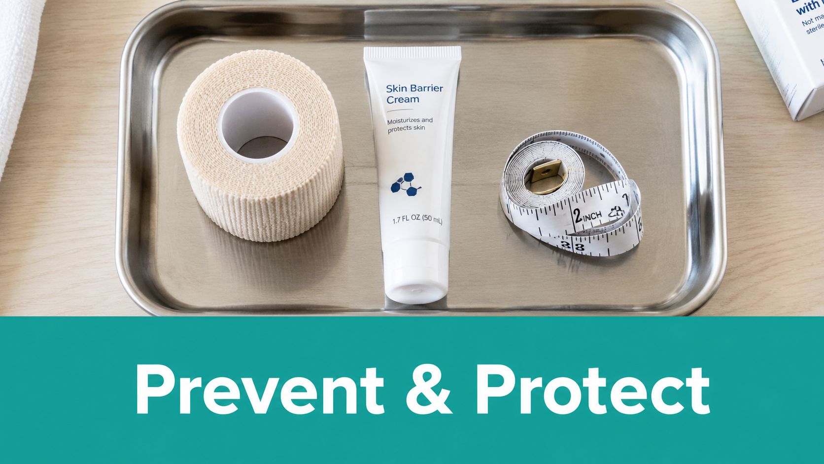
Hydrofiber or alginate does better when the wound is producing a lot of drainage, because the primary layer can manage fluid at the wound surface before it reaches the surrounding skin. Barrier creams, liquid barriers, or polymer skin protectants belong on intact periwound skin when the edge is at risk. Don't smear them into the wound bed.
Occlusive cover dressings are the wrong answer when the problem is excess moisture. They trap the moisture you're trying to control.
The decision point is whether you need a dressing change or a plan change. If drainage is heavy because the underlying cause is still active, like venous hypertension or ongoing pressure, dressing selection alone won't fix it. That's when you tighten compression, improve offloading, or address another driver instead of just changing the outer layer.
The dressing selection framework here pairs well with a structured chart review tool such as EkagraHealth AI wound dressing selection chart, especially when multiple clinicians are rotating through the same patient and need the same moisture-balance logic documented consistently.
Documentation and Coding That Passes Audits
Good care can still be denied if the note doesn't show why the service was needed. Auditors look for the clinical reason the dressing changed, the specific moisture problem, and the response to prior treatment. If the chart reads like routine maintenance, the claim gets treated like routine maintenance.
Coding the condition and the work
For a maceration wound, the billing story starts with the underlying diagnosis, then documents the periwound complication. Maceration itself is often documented as a complication or moisture-associated skin damage finding rather than a standalone disease. The exact ICD-10 choice depends on the wound etiology, such as venous ulcer, diabetic ulcer, or pressure injury, plus the associated skin breakdown.
If maceration has progressed to tissue breakdown and you're performing debridement, the CPT range 11042–11047 becomes relevant. Those codes need depth, tissue type, and the documented area debrided. If the note doesn't support the tissue level, the code won't hold.
A solid SOAP note usually includes the following:
- Subjective: drainage increased, dressing saturation, pain at the edge, or slipping dressings.
- Objective: wound dimensions, periwound appearance, exudate type and amount, odor, undermining, and whether the edge is intact or denuded.
- Assessment: maceration linked to exudate burden and the underlying wound cause.
- Plan: dressing change frequency, barrier application, compression or offloading changes, and follow-up interval.
Documentation pitfall: saying “continue same dressing” while also describing worsening maceration reads like no skilled adjustment happened.
CMS review often hinges on whether the note shows active management of a complication rather than passive observation. If maceration worsened despite prior intervention, say exactly what changed and why. For example, increased drainage saturation, more frequent dressing replacement, and a switch to a higher-absorbency primary layer are all skilled decisions when they're tied to the exam findings.
The medical necessity language that tends to survive audit is concrete: worsening periwound breakdown, increased exudate burden, need for barrier protection, need for increased dressing frequency, or need for a different absorbent category because the current regimen failed. The documentation guidance at EkagraHealth AI medical necessity documentation aligns with that same logic, because the note has to show the problem, the intervention, and the response.
Prevention Protocols and Offloading Strategies
Once the edge is stable, prevention is mostly about not letting moisture, pressure, or friction recreate the same problem. The plan has to match the wound location. Plantar wounds need offloading. Venous wounds need compression. Skin folds need moisture control. Incontinence-related breakdown needs a different containment strategy altogether.
What actually prevents recurrence
For venous insufficiency, don't under-dose compression if it's indicated and tolerated. In practice, too little compression lets exudate keep leaking, and the periwound gets soft again. For plantar wounds, pressure relief has to be real, not theoretical. If the patient keeps walking on the ulcerated foot, the edge will stay inflamed and moist.
For intertriginous areas, use moisture-wicking fabric, keep the area clean and dry, and avoid thick products that trap sweat in the fold. If incontinence is part of the picture, barrier protection and scheduled hygiene matter more than the brand of the dressing. The skin needs protection from continuous wetness, not just a prettier cover.
Patient teaching only works if it's short and specific. Tell patients when to change the dressing, how to keep it dry during showers, and what the warning signs are, such as leakage, odor, edge whitening, or pain at the margin. I've had better adherence when instructions are simple enough to repeat back without reading from a handout.
A practical learning aid like VideoLearningAI workplace learning tools can be useful for staff onboarding when you need quick refreshers on dressing changes, barrier use, and edge assessment during busy clinic days. That kind of point-of-care reinforcement is often more realistic than expecting everyone to remember every protocol from a quarterly meeting.
Case Examples From Clinical Practice
A venous leg ulcer with heavy drainage came in with a clean bed and a soggy rim. The initial plan had been foam alone, and the wound kept striking through by the next day. The fix was a hydrofiber primary layer with a more absorbent secondary cover, plus compression to address the venous driver. Once the edge stayed dry, the wound started contracting instead of spreading outward.
A diabetic foot ulcer on the plantar surface looked deceptively quiet until the periwound turned white and soft. The problem was not the granulation tissue, it was the dressing failure and ongoing pressure during ambulation. After offloading was tightened and the moisture plan changed, the edge stabilized. The chart also supported the wound's risk profile with the appropriate Wagner grading language and a clean record of serial measurements.
A pressure injury with periwound breakdown needed more than a routine dressing change. The note described denudement, exudate amount, pain at the border, and the intervention that reduced saturation. Debridement was billed only when the tissue findings supported it, with the correct 11042–11047 code selection and depth documented clearly.
In all three cases, the biggest difference wasn't a clever product. It was whether the note showed that the clinician recognized moisture failure, linked it to the underlying cause, and changed the plan in a way that made sense to a reviewer.
EkagraHealth AI helps wound teams capture those details at the point of care, map them to the right CPT and ICD-10 logic, and keep the documentation tied to the actual periwound findings. If you're trying to reduce denials around maceration, exudate, and dressing necessity, visit EkagraHealth AI and see how its workflow fits wound care charting.
