
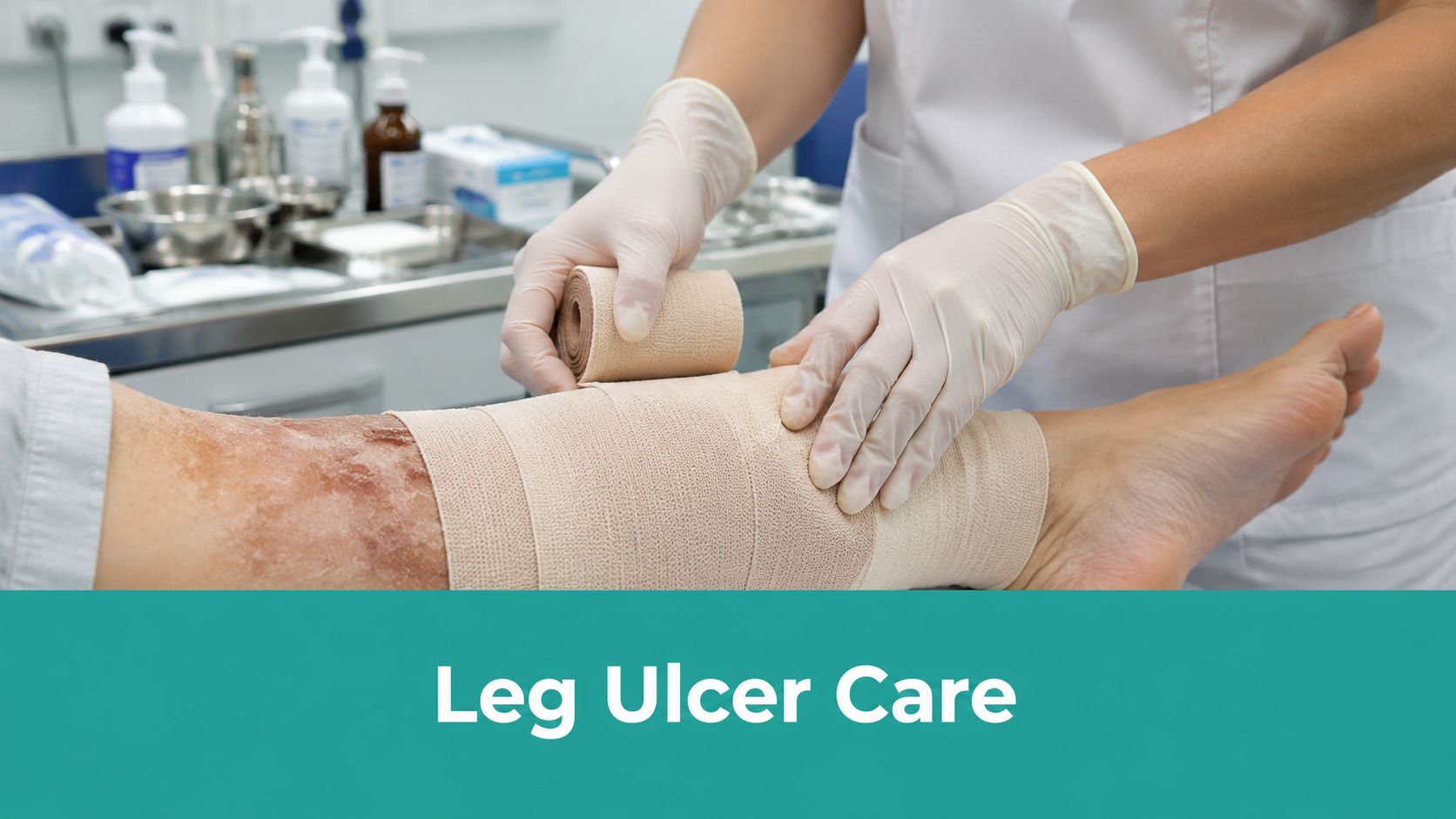
A leg ulcer patient rarely arrives as a clean, single-problem visit. The chart may say “nonhealing lower leg wound,” but what walks into the room is usually edema, venous disease, pain, drainage, skin damage, missed follow-up, and documentation gaps that are already setting up the next denial.
That's why wound care for leg ulcers has to run as a workflow. Assessment, compression decision, debridement choice, dressing logic, reassessment timing, and chart language all have to line up on the same day. If they don't, you may still be busy, but you won't be effective.
Your Next Leg Ulcer Case Is Not Standard
The patient I want you to picture is the one you'll probably see this week. Older adult. Medial gaiter area ulcer. Heavy drainage. Brown staining around the lower leg. “It's been there for months.” Compression was tried “at some point,” but nobody can tell you what level, how consistently, or whether arterial supply was checked first.
That is not a simple skin defect. It's a lower-limb physiology problem with a wound sitting on top of it.

Prognosis starts early
If you remember one prognostic fact, make it this one. A prognostic model indicates that VLUs older than one year and larger than 10 cm² have a 78% probability of closure failure by the sixth month, whereas fresher, smaller wounds (<10 cm², <1 year old) only face a 29% failure rate according to the VLU review in PMC.
That changes how you triage. It should also change how urgently you correct bad care plans.
A wound that has been allowed to drift for months without a structured reassessment is already telling you something. Chronicity matters. Size matters. Sloppy reassessment matters too, because once the wound enters the hard-to-heal lane, every avoidable mistake costs the patient time.
Practical rule: The first few weeks are not “watch and wait.” They're when you decide whether standard care still has a chance.
Treat the limb, not just the ulcer
The novice mistake is focusing on the wound bed and ignoring the leg. The wound bed matters, but if edema is uncontrolled, venous hypertension is still active, and the periwound skin is macerated, your dressing choice alone won't rescue the case.
What works in real practice is a disciplined sequence:
- Define the likely ulcer type: Don't assume venous just because the wound is on the lower leg.
- Establish vascular safety: Compression without arterial assessment is how people get hurt.
- Measure the wound correctly: Baseline dimensions and tissue description are the backbone of both treatment and reimbursement.
- Set a reassessment deadline: If you don't define what “progress” means at the first visit, you'll miss failure when it arrives.
The clinicians who get the best results in wound care for leg ulcers aren't the ones who know the most dressings by brand. They're the ones who run the same disciplined process every time.
The First 15 Minutes Assessment and Triage
A new patient is on your schedule with a “leg ulcer follow-up,” but nobody can tell you whether the wound is venous, arterial, mixed, or neuropathic. The dressing bag is full. The chart is thin. That is how bad plans get repeated, bad compression orders get written, and denials get invited.
Set the room up around triage first. Supplies come after you answer two questions. What is driving this ulcer, and is the limb safe for the treatment you are about to order?
Read the leg before you touch the wound
The ulcer sits on a limb with a history. Start there.
Venous disease usually shows itself with edema, hemosiderin staining, varicosities, lipodermatosclerosis, and a wound in the gaiter region. Drainage is often moderate to heavy. Pain often tracks with swelling and prolonged dependency. If the wound bed gets all your attention and the edema does not, healing slows and the dressing budget climbs for no good reason.
Arterial ulcers usually look more discrete and more ischemic. Expect a punched-out appearance, a pale or fibrotic base, cooler skin, reduced hair growth, delayed capillary refill, and diminished pulses. Pain at rest, nocturnal pain, and tenderness out of proportion to the surface appearance should change your tempo. Compression decisions made too early in this group can injure tissue fast.
Neuropathic ulcers follow pressure and repetitive trauma. Look for callus, deformity, sensory loss, poor footwear, and a pattern that matches loading rather than venous hypertension. A lower-leg wound does not rule out neuropathy. A patient can have edema, venous disease, and loss of protective sensation at the same time.
Mixed disease is common. Treating every lower-leg ulcer as venous is one of the expensive mistakes in wound care. It delays the right workup, exposes the patient to avoidable risk, and leaves you with a chart that will not defend the treatment plan later.
Leg Ulcer Differentiation at a Glance
| Characteristic | Venous Ulcer | Arterial Ulcer | Neuropathic Ulcer |
|---|---|---|---|
| Typical location | Gaiter area, often medial lower leg | Distal foot or areas with poor perfusion | Pressure-bearing or trauma-prone areas |
| Borders | Often irregular | Often punched out | Often callused |
| Drainage | Often moderate to heavy | Often less exudative | Variable |
| Pain pattern | Often worse with swelling or dependency | Often severe, can occur at rest | May be reduced if sensation is impaired |
| Skin clues | Edema, staining, chronic venous skin change | Cool limb, trophic change, diminished perfusion clues | Callus, deformity, sensory loss |
| Pulses | Often present | May be reduced | Variable |
| Immediate concern | Compression planning | Perfusion safety | Offloading and pressure control |
Arterial screening is a treatment decision
Before you order meaningful compression, document perfusion assessment. Palpate pulses. Compare temperature. Check capillary refill, but do not overvalue it. If pulses are weak, absent, or difficult to interpret because of edema, the chart should say that plainly.
An ankle brachial pressure index helps decide whether standard compression is appropriate. The Society for Vascular Surgery and the American Venous Forum clinical practice guideline for venous leg ulcers02152-0/fulltext) supports arterial evaluation as part of the workup before compression is applied. In practice, the number is only useful if it matches the limb in front of you. A reassuring value with ischemic pain, cool skin, or poor pulses should push you to recheck the assumptions, not push harder with compression.
One sentence in the note can protect both the patient and the claim: “Compression level deferred pending arterial assessment because exam raised concern for mixed venous and arterial disease.” That is clinical reasoning. It is also audit-proof reasoning.
What has to be in the first note
A weak first note creates work for everyone after you. It also costs money. If the diagnosis is vague, measurements are incomplete, and the rationale for testing is missing, treatment authorizations stall and debridement claims become harder to defend.
Capture the findings that support both care and coding:
- Location and laterality: Right or left leg, exact site, and whether there are multiple ulcers.
- Measurements in centimeters: Length, width, depth, and method used. If there is undermining or tunneling, measure that too.
- Tissue description: Granulation, slough, eschar, exposed structures, epithelial edge, and percentage estimates if your workflow uses them consistently.
- Drainage and odor: Amount, character, and whether odor persists after cleansing.
- Periwound and limb findings: Edema, maceration, erythema, dermatitis, fibrosis, hemosiderin staining, temperature, and tenderness.
- Vascular exam: Pulses, refill, skin quality, pain pattern, and any noninvasive testing obtained or ordered.
- Working etiology: Venous, arterial, neuropathic, pressure-related, vasculitic, malignant, or mixed.
- Reason for the plan: Why compression was started, modified, or held. Why vascular referral, imaging, or urgent escalation was needed.
Do not write “wound stable” on a first visit. Stable compared with what?
The first 15 minutes should produce a defensible differential, a limb safety decision, and a baseline note that another clinician can trust. If you do that well, the treatment plan has a fair chance to work, and the documentation can survive payer review instead of collapsing the moment someone asks why you chose that path.
Debridement and Dressing Selection That Heals
A lot of stalled leg ulcers are not failing because the patient needs something exotic. They're failing because the wound bed is dirty, the exudate plan is wrong, or the chart never explained why the intervention was medically necessary.

Start with TIME, not habit
The simplest discipline is still the TIME framework. Tissue, Infection, Moisture, Edge. It forces you to look at what is blocking closure instead of repeating the same supply order every week.
Debridement is the cornerstone of the Tissue phase in the TIME framework, and systemic antibiotics are indicated only for overt clinical infection with classical signs such as increased pain, erythema, and purulence, not for colonization, as summarized in the chronic wound treatment review in PMC.
That point matters clinically and financially. Overprescribing antibiotics for colonization doesn't move the wound forward, and it muddies the chart. If you're treating infection, document the clinical signs. If those signs aren't there, don't write as if they were.
Choose debridement on purpose
Not every nonviable surface needs the same modality.
- Sharp or surgical debridement: Use when necrotic tissue, adherent slough, or obvious bioburden barrier is preventing progress and the tissue can be safely removed in clinic.
- Enzymatic debridement: Useful when the patient can't tolerate aggressive sharp work, or when serial softer debridement fits the tissue condition better.
- Autolytic support: Reasonable for selected wounds, but too passive for a stagnant wound that's collecting debris and exudate week after week.
For coding, don't let the procedure note stop at “debrided wound today.” That's a denial waiting to happen. If you bill debridement CPT codes 11042–11047, document the tissue removed, depth reached, instruments used, pain control if used, pre- and post-debridement measurements, and why debridement was necessary that day.
Dressing choice should follow exudate logic
Supply closets tempt clinicians into dressing by habit. Don't.
A dressing is doing a job. If the wound is dry, you need moisture donation. If the wound is heavily draining, you need absorption and periwound protection. If the exudate amount and the dressing's handling capacity don't match, the wound tells on you fast.
Use a simple decision pattern:
- Dry wound bed: Consider a moisture-donating approach such as a hydrogel when the goal is to prevent desiccation.
- Moderate exudate: Foam dressings are often a practical fit when you need absorption without stripping the bed.
- Heavy drainage: Alginates or other high-handling absorptive options make sense when exudate control is the main problem.
- Fragile periwound: Protect the surrounding skin early. Maceration is not a minor side issue. It enlarges the problem.
For a quick framework on matching wound characteristics to dressing categories, I like a simple visual crosswalk such as this wound dressing selection chart. The point isn't the chart itself. The point is consistency.
If the periwound is white, soggy, and breaking down, your dressing plan is underpowered no matter how good the primary dressing sounded in the note.
Mastering Compression and Offloading Protocols
A leg ulcer stalls for a predictable reason more often than clinicians want to admit. The wrap was applied without a vascular safety check, the pressure delivered was too low to matter, or the patient left with no plan for keeping compression in place between visits.
If the ulcer is venous and the limb can tolerate compression, compression treats the underlying venous hypertension. The dressing supports the wound bed. It does not replace compression.

Compression has to match both physiology and execution
Use the vascular assessment you already gathered. As noted earlier, high compression belongs on a limb with adequate arterial inflow. If the ABI is reassuring and the clinical picture fits venous disease, apply true therapeutic compression. If arterial disease is suspected, mixed disease is possible, or the patient reports ischemic rest pain, stop and sort that out before you wrap aggressively.
Technique decides whether the order on paper becomes treatment on the leg. A loose wrap, uneven tension, padding that creates pressure ridges, or a bandage that slides by day two will fail even if the diagnosis is correct. Newer clinicians often document "compression applied" and assume this fulfills the documentation needs. It does not. Record the system used, intended pressure level, laterality, patient tolerance, and whether capillary refill, toe color, pain, and distal sensation were reassessed after application.
That documentation protects the patient and the claim. If an auditor cannot tell why this patient was appropriate for compression and what level was delivered, payment becomes harder to defend.
Common points of failure
Compression plans break down in a few familiar ways:
- Vascular shortcuts: No ABI on file, no pulse exam documented, or no explanation for why compression was safe that day.
- Application errors: Bunching at the ankle, slippage at the calf, or inconsistent tension that creates discomfort without producing therapeutic benefit.
- Poor fluid handling under the wrap: The primary dressing saturates early, exudate tracks into the periwound, and the patient blames the wrap when the underlying problem is dressing capacity.
- No maintenance plan: The patient leaves without replacement supplies, laundering instructions, wear schedule, or a clear transition plan for long-term edema control.
When patients are ready to step down from wraps or need maintenance therapy between flare-ups, use patient education tools that show what gradient support stockings for long-term edema management look like and how they are worn. That reduces the usual confusion between medical compression and ordinary socks.
Offloading still applies when force is part of the problem
A lower-leg ulcer can be venous and still fail because of pressure, friction, gait abnormality, ankle deformity, or neuropathy. I see this missed in mixed-mechanism wounds all the time. The edema gets attention. The repeated trauma does not.
Offloading does not need fancy language. It needs a clear reason and a specific intervention. Document the pressure source, the device or footwear change recommended, whether the patient can use it safely, and what happened when you trialed it. If the patient refused, write that down. If balance, work demands, or cost made the plan unrealistic, write that down too.
Bad notes create expensive problems. "Continue compression, advised offloading" is weak clinical communication and weak billing support. A defensible note states why compression was indicated, what was applied, what offloading barrier was identified, and how the treatment plan was adjusted to fit the patient's actual life.
Recognizing Failure and Escalating Care
Some ulcers don't respond even when the basics are done well. That is not the moment to repeat the same treatment and hope your note sounds more optimistic next week.
Know the failure trigger
For venous pathways, a 40% reduction in wound size at 6 weeks is the marker for normal progression, and when a wound fails to meet that 40% threshold or achieves only 0–20% reduction by the 6-week interval, the protocol calls for full reassessment for infection, inflamed tissue, or improper compression application, often with transfer to a complex pathway or referral to tissue viability specialists, according to Oxford Health's leg ulcer management training material.
That means your six-week review is not just another dressing change visit. It is a decision point.
What to reassess when progress stalls
Start with the obvious, because the obvious is often the problem.
- Compression delivery: Was therapeutic compression applied and tolerated?
- Diagnosis drift: Is this venous, or are you dealing with mixed disease?
- Bioburden and tissue status: Does the wound still need more aggressive debridement?
- Exudate plan: Is maceration or dressing failure undermining the edges?
- Patient adherence: Is the wrap staying on, and is elevation or offloading realistic?
Then think about escalation. Non-improving venous wounds may need vascular imaging and referral when reflux correction is in play. Some may need adjunctive tissue-based treatment, but only after you've documented failed standard care correctly.
Build the chart for escalation from day one
Coverage for advanced tissue products doesn't start when you decide you want one. The chart has to prove standard care was attempted and failed.
For Medicare coverage of skin substitutes or CTPs in venous leg ulcers, the ulcer must have failed to respond to documented standard of care for at least four weeks, defined as 30 consecutive days, with documented compliance, and it must show that a 50% area reduction was not achieved by week 4, as described in this coverage summary on skin substitutes.
If you didn't document baseline area, interval measurements, compression compliance, and the standard care delivered, you haven't built a case. You've built an appeal.
A stalled wound is not asking for more optimism. It's asking for a different plan and a better record.
Document to Treat and to Get Paid
Poor documentation is one of the easiest ways to sabotage good wound care for leg ulcers. The patient may have needed the service. You may have performed the service well. If the note doesn't prove medical necessity, tissue depth, wound status, and treatment rationale, the claim is weak.
What an audit-proof wound note includes
For bone debridement billed under CPT 11044, the note must explicitly state that bone was “exposed and tangibly debrided to healthy bleeding margins,” and the wound must be documented with length × width × depth in centimeters plus tissue types by percentage to avoid denials, per wound care billing guidance for debridement documentation.
That level of specificity shouldn't be reserved for bone cases. Use the same discipline across your wound notes.
Include these elements every time:
- Measurements in cm: Length, width, depth. Not approximate. Not “small.”
- Tissue percentages: Granulation, slough, eschar, epithelial tissue where present.
- Exudate description: Amount and type. Serous, sanguineous, purulent.
- Periwound assessment: Maceration, erythema, edema, induration, callus if present.
- Procedure detail: Tissue level debrided, instruments, anesthesia, hemostasis, tolerance.
- Medical necessity statement: Why that intervention was needed at this visit.
Denials usually start with vague language
“Selective debridement performed” is weak. “Wound cleaned and dressed” is weaker. Those phrases don't tell a payer what tissue was present, what tissue was removed, or why the intervention could not be deferred.
Your note should sound like a clinician who observed the wound directly. If you removed subcutaneous nonviable tissue, say that. If the wound had moderate serous drainage with periwound maceration, say that. If pain control was required to complete debridement, say that too.
For practices trying to standardize those elements at the point of care, a structured workflow such as medical necessity documentation support can help keep the note aligned with CPT and ICD-10-CM requirements. EkagraHealth AI is one example of a platform built around that documentation and billing backbone.
Don't forget transmission and referral hygiene
The chart isn't finished when you sign it. Leg ulcer care often depends on moving records, photos, orders, and referral packets quickly between clinic, home health, vascular, and facility teams. If your staff still handles that inconsistently, it's worth tightening the process with resources on SendItFax for compliant document transmission, especially for referral packets that include wound measurements and treatment history.
Documentation is clinical care. In this field, it's also revenue protection. When the note clearly links assessment, intervention, and medical necessity, both the patient and the claim are more likely to move forward.
If your team is tired of losing time to wound charting, coding cleanup, and claim rework, EkagraHealth AI is built for that exact problem. It helps wound practices capture structured visit documentation, support coding, and keep the clinical record aligned with what payers expect.
