
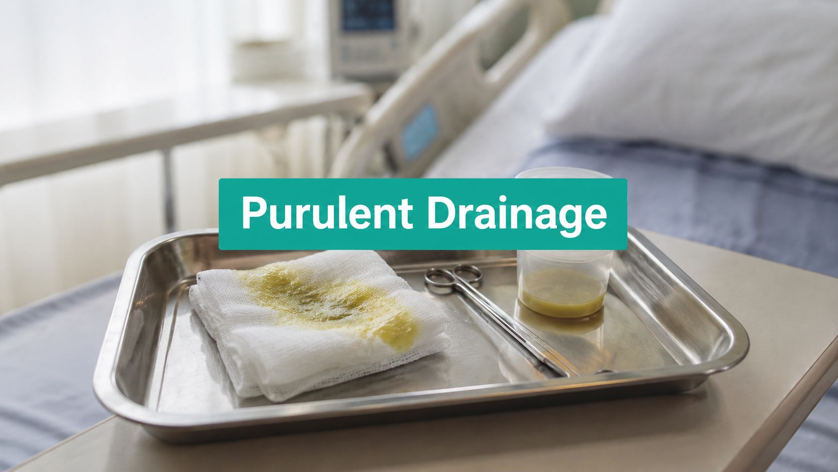
You're standing at the bedside with a dressing in your hand and a note still open. The wound has gone from “draining” to clearly infected, but the hard part isn't spotting that something's wrong. The hard part is describing it precisely enough that treatment makes sense, the culture is worth sending, and the claim doesn't come back denied.
That's where newer clinicians often get tripped up. They write “purulent drainage present,” order an antibiotic, and move on. Clinically, that's thin. From a billing standpoint, it's worse. Purulent drainage changes the whole picture of the wound, from infection workup to dressing choice to whether debridement can be defended.
Identifying Purulent Drainage vs Other Exudate Types
Purulent drainage is a thick, opaque exudate composed of necrotic white blood cells, primarily neutrophils, viable and non-viable bacteria, tissue debris, and inflammatory proteins. It's a definitive biomarker for active wound infection rather than normal healing progression, as described in this clinical review of purulent drainage characteristics and treatment considerations.
That distinction matters. A lot of wound fluid looks concerning to a novice eye, but not all exudate signals infection. Purulent drainage is never part of routine wound closure. Serous and serosanguineous fluid may be expected in healing tissue. Purulence is different.
What it looks like in practice
At the bedside, purulent drainage is usually milky, viscous, opaque, and often foul-smelling. The color may be yellow, green, brown, or cream. If the periwound is getting redder, more tender, more swollen, and the dressing is coming off with thick opaque drainage, stop calling it “just drainage.”
For a quick refresher on the broader exudate family, this breakdown of types of exudates is useful, especially when staff are documenting inconsistently across settings.
Wound Exudate Type Comparison
| Exudate Type | Color | Consistency | Clinical Significance |
|---|---|---|---|
| Purulent | Yellow, green, brown, cream, opaque | Thick, viscous, milky | Suggests active wound infection |
| Serous | Clear to pale yellow | Thin, watery | Often consistent with normal healing |
| Sanguineous | Red | Thin to slightly thicker fluid containing blood | Indicates bleeding |
| Serosanguineous | Pink to light red | Thin, watery | Common in healing wounds, especially early on |
What people mislabel
The most common error is calling cloudy fluid “purulent” without describing it. The second most common error is missing purulence because the wound has been heavily irrigated right before assessment. If you only document “moderate drainage,” you've left out the most clinically important part.
Practical rule: If the exudate is opaque and thick, document the exact color and consistency, then assess the wound for infection. Don't chart it as a generic increase in moisture.
There's also a difference between a wound with some slough and a wound producing purulent exudate. Slough is tissue in the wound bed. Purulence is drainage. They often appear together, but they aren't interchangeable terms, and using them loosely causes confusion in both treatment planning and coding review.
Bedside Assessment and Microbiology
Before you order anything, get disciplined about how you assess the drainage. I use a simple bedside frame: color, odor, consistency, amount. If you want your note to hold up later, those four descriptors need to be there every time.
Here's the visual cue that helps most.
Use a COCA bedside check
When I assess suspected purulence, I want a note that another clinician can act on without seeing the patient.
Color
Yellow may be straightforward purulence. Green or brown deserves closer attention because color shifts can reflect pathogen burden, and green discharge is clinically significant when bacterial colonization is suspected.Odor
Don't stop at “malodorous.” Describe whether the odor is slight or foul and whether it persists after cleansing. A dirty dressing smells. An infected wound often still smells after the wound is cleaned.Consistency
“Thick,” “viscous,” or “milky” says much more than “draining.” Thin fluid behaves differently in the dressing and often points you toward a different differential.Amount
Light, moderate, or heavy matters for both treatment and documentation. It also helps explain periwound maceration, dressing failure, and frequency of change.
When to culture and how to do it correctly
Purulent drainage warrants prompt wound assessment for associated infection findings such as erythema, edema, and increased pain. If you're going to culture, cleanse the wound first. Then obtain the sample from deep tissue, avoiding necrotic tissue and surface drainage, because superficial contamination doesn't guide useful antibiotic selection.
A bad swab gives you bad treatment. That's the point.
Surface drainage often reflects what's sitting on top of the wound, not what's driving the infection in viable tissue. If you send a superficial sample from dried drainage or necrotic debris, you may end up treating colonizers instead of the actual pathogen burden. That's how patients stay on the wrong antibiotic while the wound keeps worsening.
Cleanse first. Then sample viable wound tissue with intent. If you culture the surface sludge, don't trust the result.
The wound has to be assessed as a whole while you do this. Drainage alone doesn't tell the entire story. Look at the edge, undermining, warmth, pain escalation, and whether the periwound is intact or breaking down. If the patient has a diabetic foot ulcer, pressure injury, or another chronic wound that suddenly changes drainage character, assume the biology has changed until proven otherwise.
What Purulent Drainage Really Signals Clinically
A lot of clinicians still think in a straight line. Pus means infection. Infection means antibiotics. Sometimes that's true. Often it isn't enough.
The more useful clinical question is this: why is the wound still producing purulent drainage despite treatment? In chronic wounds, the answer may not be a simple planktonic infection. It may be biofilm.
Purulence isn't always a simple antibiotic problem
Recent findings summarized in this discussion of chronic wound drainage and biofilm-associated infection report that up to 65% of chronic wounds with purulent exudate harbor non-viable bacterial biofilms that don't respond to standard oral antibiotics alone. The same source notes treatment failure in 30 to 40% of diabetic foot ulcers within 14 days when this is missed.
That tracks with what many of us see in practice. The drainage improves briefly, then returns. Odor drops for a few days, then comes back. The wound bed stays stalled. The patient has “already had antibiotics,” but the wound still looks infected.
If you want a patient-facing explanation of the basic infection signal, this overview on whether pus is a sign of infection is fine. For clinical decision-making, though, the key point is that persistent purulence in a chronic wound should make you think beyond oral therapy alone.
What changes when biofilm is involved
Biofilm changes the treatment sequence. If the wound is carrying mature biofilm, antibiotics may have limited effect until you disrupt the barrier mechanically. That usually means debridement, and not symbolic debridement. Real removal of non-viable tissue and biofilm burden.
A chronic wound with ongoing purulent drainage that “fails antibiotics” often hasn't failed pharmacology. It has failed source control.
Debridement planning involves both clinical and operational considerations. If a diabetic foot ulcer is Wagner grade progression risk, or a pressure injury has depth and undermining with persistent purulence, waiting too long on definitive tissue management is a mistake. You can't medicate your way through every infected cavity or stalled wound bed.
Purulent drainage also raises concern for deeper involvement. In chronic wounds, persistence can signal deep tissue infection or abscess formation, which is why some wounds need imaging, more aggressive debridement, or escalation to negative pressure therapy after the wound is adequately prepared.
Documentation That Justifies Treatment and Gets Paid
“Purulent drainage present” is not a billable story. It's a vague observation.
Payers want enough detail to understand why the wound required infection-specific treatment, more frequent dressing management, debridement, and in some cases advanced therapies. If the note doesn't connect the bedside findings to medical necessity, the claim is exposed.
Here's the workspace side of that problem.
What has to be in the note
Per these documentation standards for purulent drainage, coding, and claim defense, you need more than the presence of drainage. Color, consistency, volume, odor intensity, onset timing, and associated symptoms such as fever or localized tenderness directly influence CPT coding for debridement, including 11042 to 11047, and ICD-10 diagnosis coding. If those details are missing, you risk undercoding and denials.
A second source focused on payer requirements says documentation should capture five specific data points for purulent drainage audits: color, consistency, amount, odor, and onset, with associated symptoms like increased pain, warmth, or spreading redness, because those details distinguish purulent from serous drainage and help justify higher-cost infection treatment and debridement in payer review. That summary appears in this payer-focused overview of purulent drainage documentation expectations.
What auditors and denials commonly hinge on
If the chart says “drainage increased,” that doesn't tell the reviewer whether the wound worsened, remained moist, or crossed into infection. If the note says “debrided due to infection” but never describes thick yellow or green viscous drainage, associated periwound erythema, or pain escalation, you've made the procedure look unsupported.
Use language that links findings to action:
For selective debridement
Document non-viable tissue or biofilm burden, wound bed appearance, and why tissue removal was needed to control bioburden and support healing. If you bill 97597/97598, the note should show exactly what was removed and why selective work was medically necessary.For excisional debridement
If you're reporting 11042 to 11047, depth and tissue type must be clear. “Sharp debridement performed” isn't enough. State whether you removed necrotic subcutaneous tissue and what wound findings supported excision.For diagnosis coding
Link the drainage findings to the infected wound diagnosis you're treating. Generic wound documentation with no infection descriptors often weakens the ICD-10-CM picture and invites downcoding.
Coding reality: The payer isn't in the room. Your note has to recreate the room.
One thing that helps multidisciplinary teams is standardizing the drainage language itself. If nurses, APPs, and physicians all use different descriptors, coding review becomes messy fast. A simple internal workflow document can fix that. Teams building one from scratch may find this SOP guide for operations managers useful because the same operational logic applies to wound documentation consistency.
If your practice is trying to tighten note quality around infection findings, medical necessity, and procedure support, this resource on medical necessity documentation is worth reviewing with both clinicians and billers.
Treatment Implications From Dressings to Debridement
Once you've identified true purulent drainage and documented it correctly, treatment gets more straightforward. Not easy. Straightforward. The wound needs bacterial burden control, exudate management, tissue assessment, and often source control.
Match the dressing to the drainage
Dressings fail when clinicians choose based on habit instead of exudate volume. For purulent drainage, the hierarchy matters. This overview of exudate-driven dressing selection notes that light drainage is managed with thin foam or hydrocolloid dressings, moderate drainage calls for medium-absorbency foam or alginate, and heavy drainage requires thick foam, super-absorbent dressings, or negative pressure wound therapy (NPWT).
That isn't just a product preference issue. It's wound preservation.
Practical dressing decisions
Light purulent drainage
If the wound is infected but the drainage volume is limited, a thinner absorbent option may be enough. The goal is to manage moisture without sealing in thick material that needs to leave the wound.Moderate drainage
Foam or alginate often justifies its use. You need enough absorption to prevent strike-through and enough structure to protect the periwound from maceration.Heavy drainage
If dressings are saturating quickly, periwound skin is whitening or breaking down, and the cavity is deep, escalate. Thick absorptive coverage or NPWT may be more appropriate, assuming the wound is adequately assessed and prepared.
Purulent exudate that sits against the skin will macerate the periwound and make an already infected wound harder to control. When staff keep using low-absorbency dressings on thick infected drainage, they create two problems at once. The infection remains active, and the surrounding skin starts to fail.
Topical control vs systemic treatment
Purulent drainage usually pushes the clinician to think about antibiotics. Fair enough. But treatment shouldn't default to oral therapy in every case without looking at tissue quality, wound depth, and whether the wound can be penetrated by the therapy you choose.
Silver-impregnated antimicrobial dressings can help reduce bacterial burden locally. Hypochlorite cleansing may also be useful in selected infected wounds. Those measures are adjuncts, not magic. If there's deep necrotic tissue, undermining packed with devitalized debris, or a cavity that's effectively holding infected material, local antimicrobial support won't replace debridement.
When debridement stops being optional
If the wound has persistent purulence, non-viable tissue, stalled healing, or suspected biofilm burden, debridement often becomes the hinge point. Mechanical or sharp debridement can convert an infected stagnant wound into one that can respond to the rest of your plan.
That's especially true in chronic wounds such as diabetic foot ulcers and pressure injuries. Ongoing purulent drainage in those wounds raises the possibility of deeper infection and abscess formation. Some patients need imaging. Some need surgical debridement. Some need cavity management with NPWT after tissue removal.
A common mistake is under-debriding because the wound “doesn't look that bad” at the surface. Purulent drainage can be the visible clue to a much worse problem underneath. If the wound tunnels, undermines, probes deeper than expected, or keeps producing thick exudate despite care, broaden your concern.
A treatment sequence that usually works better
I teach newer clinicians to think in this order:
Cleanse the wound well
Remove loose surface material so you can see the bed and assess the true odor and exudate.Assess the whole infection picture
Drainage characteristics matter, but so do pain, warmth, erythema, edema, and systemic symptoms.Decide whether the wound needs culture
If yes, obtain it correctly from viable tissue after cleansing.Choose the dressing based on drainage volume
Don't underdress a heavily draining wound.Address non-viable tissue and suspected biofilm
If the wound is stalled and purulent, debridement may be what makes everything else start working.Escalate when depth or persistence suggests more
Chronic wounds with ongoing purulence may need imaging, surgical involvement, or advanced cavity management.
The dressing controls the consequences of the drainage. It does not fix the cause of the drainage.
There's another trade-off worth mentioning. Aggressive wound programs generate a lot of sensitive documentation, images, and communication between field staff, clinics, and billing teams. If your operation is tightening workflows around infected wound management, it's worth reviewing this expert guide on preventing healthcare breaches so the clinical side and the operational side mature together.
At the bedside, the core principle stays simple. Purulent drainage is not a normal healing finding. It's a sign that the wound has changed and your plan has to change with it. Good clinicians recognize it. Better clinicians describe it well, treat the underlying cause, and leave a note strong enough to defend every step they took.
EkagraHealth AI helps wound care teams turn bedside findings into clean, defensible documentation without losing time to manual charting. If your clinicians are managing infected wounds, documenting debridement, and trying to keep CPT and ICD-10-CM coding aligned with what happened in the room, EkagraHealth AI is built for that workflow.
