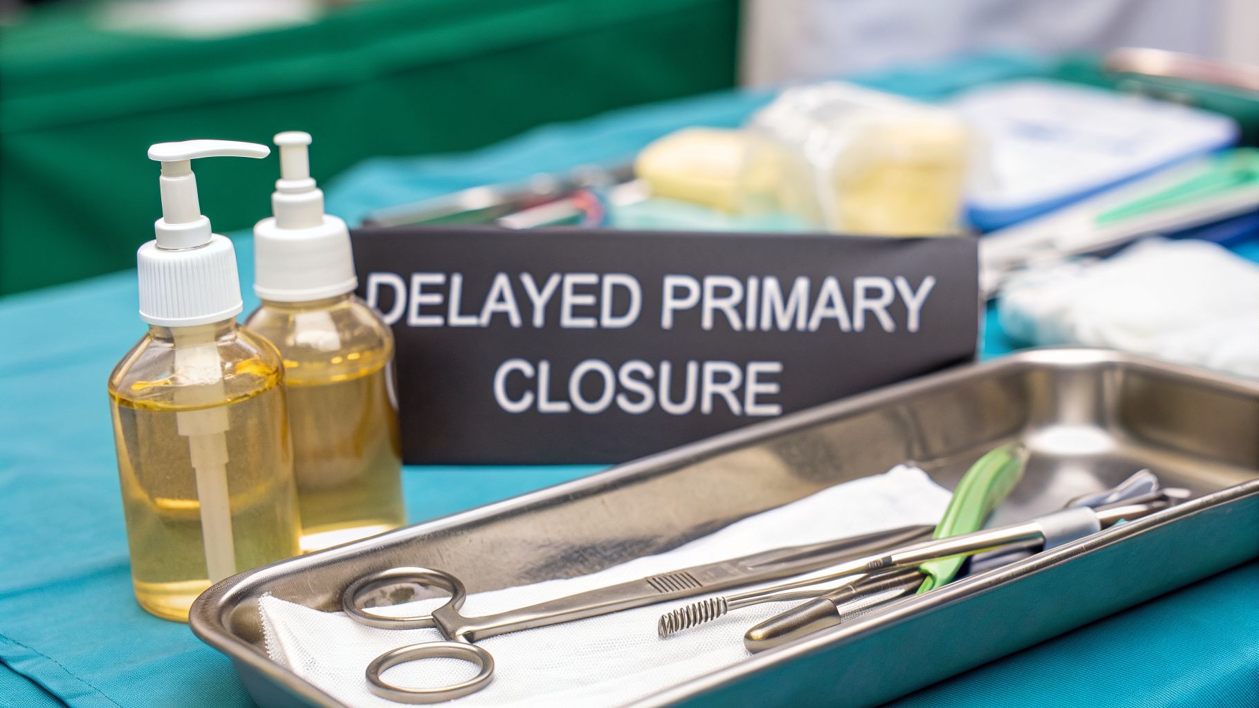
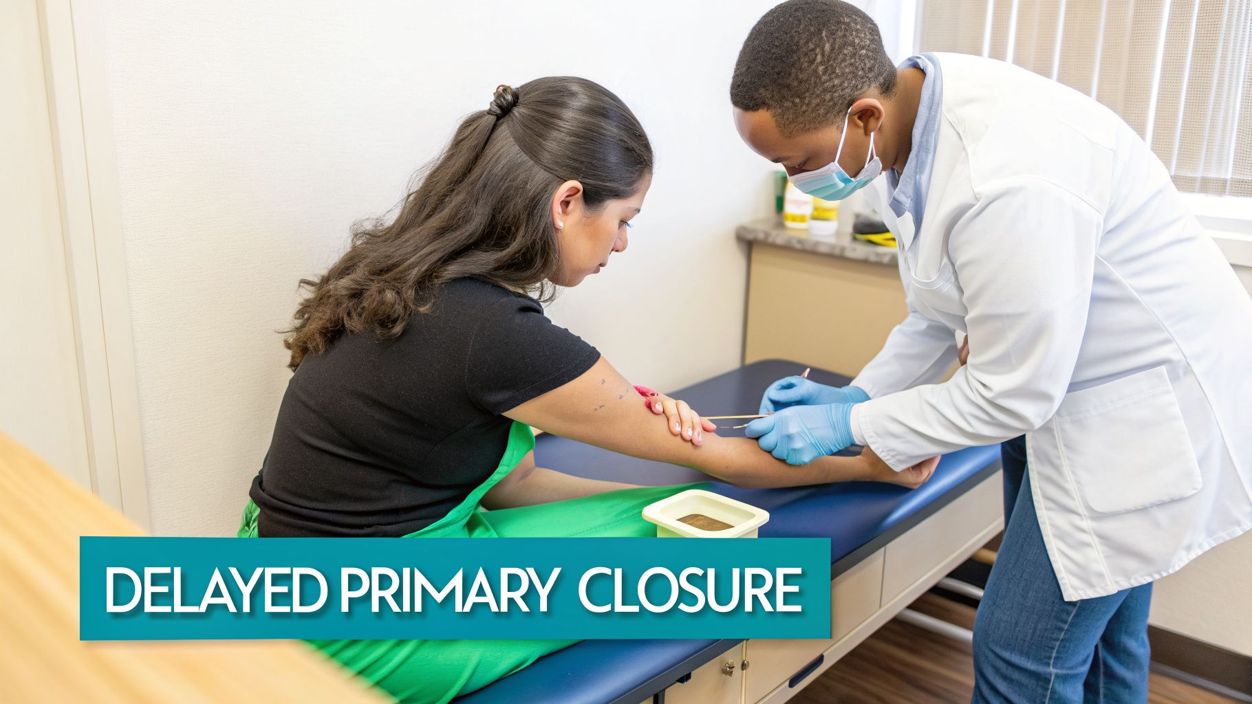
Tertiary intention healing, often referred to by clinicians as delayed primary closure, represents a calculated and strategic approach to managing complex wounds. It's fundamentally a hybrid method designed for messy, high-risk injuries that are too contaminated to be sutured shut immediately. By employing this technique, medical professionals deliberately pause the closure process. This pause creates a crucial window to meticulously clean the wound bed, which dramatically reduces the risk of developing a severe and debilitating infection. Think of it as a tactical retreat to ensure a future victory in the healing process.
This guide will delve into the nuances of tertiary intention wound healing, exploring when it is the appropriate choice, how it is managed in practice, and what realistic outcomes patients and clinicians can expect. Understanding this method is essential for any healthcare provider involved in acute or chronic wound care, as it represents a cornerstone of modern, evidence-based practice.
Why Some Wounds Need a Delayed Start
Imagine the scenario of a deep animal bite or a traumatic injury filled with gravel, soil, and other debris. If a clinician were to suture that wound shut immediately—a process known as primary intention—they would effectively be trapping a dangerous payload of bacteria and foreign material beneath the skin. This action would inadvertently create a perfect, warm, and moist incubator for an abscess or, even worse, a severe deep-tissue infection like cellulitis or osteomyelitis. Such complications can lead to systemic illness, prolonged hospital stays, and even limb loss.
This is precisely where tertiary intention wound healing proves its value. It serves as a critical intervention to prevent this worst-case scenario from unfolding. By intentionally leaving the wound open for a period of approximately 4 to 7 days, clinicians buy themselves a critical window of time to gain control over the wound environment. This delay is not passive; it is an active phase of intensive wound management.
You can think of tertiary intention as the best of both worlds—it borrows from both primary and secondary healing. The payoff is a much lower infection risk and a better, faster healing outcome than just letting a contaminated wound granulate on its own.
During this initial open phase, the entire clinical focus is on aggressive cleaning and decontamination. This is when the medical team performs meticulous debridement to remove all non-viable (necrotic) tissue, which serves as a food source for bacteria. The wound is irrigated, often with copious amounts of sterile solution, to physically flush out contaminants. Furthermore, antimicrobial dressings and systemic antibiotics are frequently employed to bring the bacterial load under control. The ultimate objective is to transform a dirty, dangerous wound into a clean, healthy one that is primed and ready for definitive surgical closure.
Understanding the Three Types of Healing
To truly appreciate the strategic advantage of tertiary intention, it is helpful to understand its place within the broader spectrum of wound healing pathways. Each method represents a specific tool in the clinician's toolkit, chosen based on the unique characteristics of the wound.
- Primary Intention: This is the standard procedure for clean, well-approximated wounds, such as a surgical incision made in a sterile environment. The wound edges are brought together and secured right away with sutures, staples, or adhesive strips. Healing is typically rapid, and the resulting scar is minimal.
- Secondary Intention: This method is employed for wounds with significant tissue loss, like a deep pressure ulcer or a large avulsion, where the wound edges cannot be pulled together. The wound is left open to heal from the base upwards through the natural processes of granulation, contraction, and epithelialization. This is a slow process that results in more significant scarring and a higher risk of infection if not managed properly.
- Tertiary Intention: This is our hybrid model, a bridge between the other two. The wound management begins as if it were to heal by secondary intention—left open and packed—but it concludes with a surgical closure akin to primary intention. It is the ideal strategy for a wound that is too contaminated for immediate closure but does not have so much tissue loss that it must be left open indefinitely to granulate.
Choosing tertiary intention is a deliberate clinical judgment call. It is a proactive measure to manage the immediate and significant risk of infection without sacrificing the long-term goals of a strong, functional, and cosmetically acceptable repair. A solid grasp of the underlying principles is crucial for success. You can learn more about the wound healing process to understand how these physiological stages play out, which is essential knowledge for managing complex cases effectively.
When to Choose Delayed Primary Closure
The decision to leave a wound open is a significant clinical judgment. It is a strategic maneuver where the immediacy of a quick closure is intentionally traded for the enhanced safety of a dramatically lower infection risk. This requires careful assessment and a clear understanding of the wound's history and presentation.
At its core, tertiary intention wound healing, also known as delayed primary closure, is an exercise in patience and proactive risk management. Clinicians wait until a wound is demonstrably clean and stable enough to be closed safely. This makes it the perfect and often necessary choice for a specific subset of high-risk injuries where primary closure would be reckless.
Of course, this approach is entirely inappropriate for clean wounds where the risk of infection is inherently low. In those situations, promoting rapid healing and minimizing scarring through primary closure is the priority. Knowing when to utilize tertiary intention—and, just as importantly, when to avoid it—is a fundamental skill that directly impacts patient outcomes and safety.
Prime Candidates for a "Wait and See" Approach
Delayed primary closure is reserved for wounds where the initial bacterial load or the degree of tissue damage is simply too great to risk immediate suturing. It's akin to hitting the pause button on the final step of wound repair. This deliberate delay provides a crucial interval for the body’s innate immune defenses, augmented by our clinical interventions, to prepare the wound bed for a successful and lasting repair.
This method becomes the standard of care in several well-defined clinical scenarios:
- Grossly Contaminated Traumatic Wounds: Any injury that involves significant contamination with dirt, debris, or organic matter is a ticking time bomb for infection. A classic example is a farmer whose arm gets caught on a rusty piece of farm machinery. Closing such a wound immediately would trap a host of bacteria and foreign material, making a severe abscess almost inevitable.
- Wounds with Significant Dead Tissue: Crush injuries or wounds with a substantial amount of devitalized (dead) tissue create a perfect, nutrient-rich breeding ground for bacteria. Leaving the wound open allows for thorough and often repeated surgical debridement, giving the clinical team a chance to clear out all non-viable tissue before even considering closure.
- Deep Animal and Human Bites: Bites, particularly from dogs, cats, or other humans, are notorious for injecting a high concentration of pathogenic bacteria deep into the tissue. The standard of care often involves delaying closure to properly irrigate the wound, initiate antibiotic therapy, and ensure no signs of infection emerge before suturing.
- Infected Surgical Sites: If a surgical incision becomes infected and subsequently opens back up (a process known as dehiscence), it is immediately converted to management by tertiary intention. The site is debrided, packed, and dressed until the infection is completely resolved. Only then is it re-closed surgically.
The decision to use tertiary intention is all about proactive risk management. It’s an admission that some wounds are just too compromised from the start for primary closure and need a stabilization period to prevent a much more serious problem.
To help simplify this critical decision-making process, here's a quick reference guide.
When to Use Tertiary Intention A Quick Reference
This table provides a clear, at-a-glance comparison of wound characteristics that are ideal for tertiary intention versus those where it's the wrong call.
| Wound Characteristic | Ideal for Tertiary Intention | Contraindicated for Tertiary Intention |
|---|---|---|
| Contamination Level | High bacterial load, visible debris, soil, or organic matter. | Clean, minimal contamination. |
| Time Since Injury | Typically >6-8 hours old, allowing for bacterial proliferation. | Fresh, recent injury (typically <6 hours old). |
| Tissue Viability | Significant devitalized (dead) or crushed tissue present. | Healthy, well-perfused tissue edges. |
| Cause of Injury | Deep animal/human bites, crush injuries, high-velocity projectiles. | Clean surgical incisions, simple lacerations from a sharp, clean object. |
| Signs of Infection | Existing signs of infection (purulence, erythema, warmth). | No signs of established infection. |
Ultimately, choosing the right closure method comes down to a careful and comprehensive assessment of the wound itself, the mechanism of injury, and the overall health status of the patient.
When Tertiary Intention Is Not the Right Choice
While tertiary intention is a powerful tool in the arsenal of wound management, it is not a one-size-fits-all solution. Applying this method when it is not indicated can unnecessarily prolong the healing process, increase healthcare costs, and subject the patient to additional procedures without any discernible clinical benefit.
Contraindications for tertiary intention are quite clear and are essentially the inverse of the indications:
- Clean Surgical Wounds: A sterile incision from a planned surgery has an extremely low risk of infection. These wounds should always be closed via primary intention to achieve the fastest healing and the best possible cosmetic result.
- Minor, Clean Lacerations: A simple, clean cut from a kitchen knife, for example, can be safely and immediately sutured, stapled, or glued with tissue adhesive.
- Wounds with Severe Vascular Compromise: In patients with severe peripheral artery disease, blood flow to the affected area might be so poor that the wound is incapable of healing, regardless of how it is closed. In such cases, leaving a wound open for tertiary closure might just increase the infection risk without any realistic chance of eventual successful closure, necessitating a different vascular or reconstructive approach.
Making these clinical judgments requires a sharp eye for distinguishing between normal post-injury inflammation and the early signs of a brewing infection. For clinicians seeking to refine their assessment skills, our article on whether pus is always a sign of infection can offer valuable clarity. By correctly identifying which wounds necessitate a delayed approach, we can effectively balance the risk of infection with the goal of achieving a strong, well-healed wound.
The Two Phases of Tertiary Intention Management
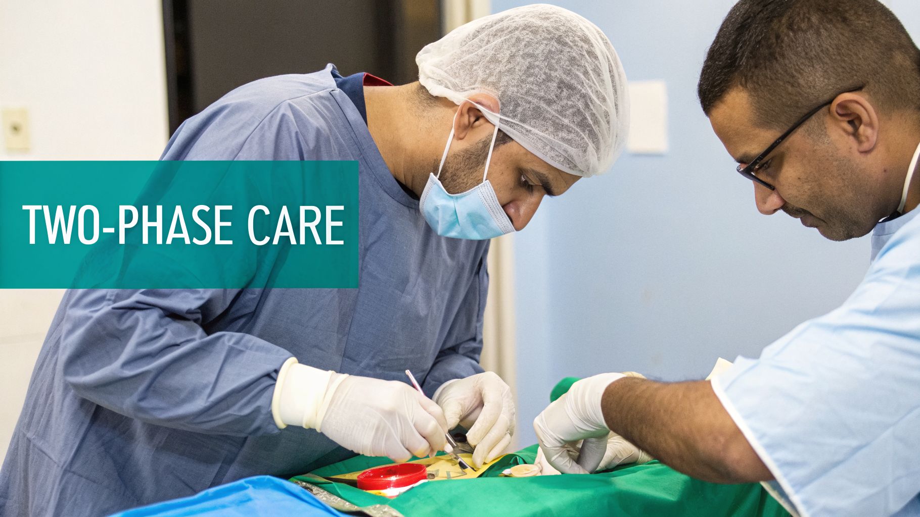
Successfully managing a wound with tertiary intention is not a passive waiting game. It is a highly active, hands-on, and strategic process that is meticulously broken down into two distinct and equally important phases. I often like to use the analogy of preparing a garden plot before planting. First, one must diligently clear all the weeds, rocks, and debris, and then enrich the soil. Only then can the seeds be planted with a reasonable expectation that they will grow into healthy plants.
This deliberate, two-step approach is what enables clinicians to transform a high-risk, contaminated wound into one that can be safely and permanently closed. Getting both phases right is the absolute key to preventing serious complications and achieving a successful long-term outcome.
Phase One: The Open Phase of Decontamination
This critical first phase kicks off the very moment the clinical team decides that immediate closure would be unsafe. For the next 4 to 7 days, the entire focus is singular: cleaning house. The overarching goal is to aggressively decontaminate the wound, reduce the bacterial burden, and create a healthy, well-vascularized foundation for new tissue to grow.
The absolute cornerstone of this phase, and one which cannot be overstated, is debridement. This step is non-negotiable. The clinical team must meticulously remove every last bit of dead (necrotic) tissue, foreign material, and bacterial biofilm from the wound bed. Necrotic tissue is not only a buffet for bacteria but also acts as a physical barrier to the body's natural healing processes. Following debridement, the wound is thoroughly flushed with irrigation fluid to wash away any remaining contaminants and cellular debris.
With the wound cleaned, the next step involves selecting the appropriate dressing. This is not a one-size-fits-all decision; it must be tailored to the specific needs of the wound at that moment, which are assessed at every dressing change.
- Antimicrobial Dressings: If there is a high bacterial load or early clinical signs of infection, clinicians will often choose dressings containing agents like silver, iodine, or PHMB to combat microbes directly at the wound surface.
- Highly Absorbent Dressings: In these early inflammatory stages, wounds can produce a significant amount of fluid (exudate). Using highly absorbent dressings, such as calcium alginates or hydrofibers and foams, is crucial for managing this moisture, preventing maceration of the surrounding skin, and maintaining a clean wound environment.
- Systemic Antibiotics: When there is evidence of a spreading or systemic infection (such as cellulitis or fever), systemic antibiotics are essential. Administered orally or intravenously, these medications work from the inside out, complementing the topical work of the dressings.
Throughout this open phase, the wound is under constant surveillance. The clinical team is looking for positive signs that the strategy is working—a decrease in drainage, reduction in surrounding redness and swelling, and, most importantly, the appearance of healthy, bright red granulation tissue. This bumpy, cobblestone-like tissue is the definitive sign of a clean, healing wound bed that is ready for the next step.
The open phase is a dynamic battleground. The wound is essentially auditioning for closure, and it has to prove it’s clean and ready. We're constantly monitoring and adjusting our plan based on what we see each day.
Phase Two: The Closure Phase
Once the wound bed is deemed clean, displays good granulation tissue formation, and is free of any clinical signs of infection, the team can finally prepare for the main event: surgical closure. This is a critical judgment call that carries significant weight. If closure is performed too early, there is a high risk of trapping residual bacteria and creating a painful abscess. Conversely, if one waits too long, it can unnecessarily prolong the patient's healing journey and increase the overall cost of care.
The decision to proceed is based on clear clinical evidence: a healthy-looking wound, minimal serous drainage, and no lingering signs of infection like purulence or erythema. This is an area where newer technologies are becoming incredibly helpful. AI-powered wound assessment tools, for instance, can analyze a digital image to provide objective, quantifiable data on tissue types, surface area, and volume. This helps to turn a subjective assessment of "it looks good" into a decision backed by concrete, defensible evidence.
Once everyone on the care team agrees that the time is right, closure is performed in a surgical setting. The specific technique employed will depend on the wound's size, depth, and location.
- Simple Suturing: For smaller wounds where the edges can be brought together without excessive tension, the surgeon can use sutures or staples, just as would be done for a primary closure.
- Skin Grafts or Flaps: For larger defects with significant tissue loss, more complex reconstructive surgery may be necessary. This can involve using a split-thickness skin graft or a local or free tissue flap to cover the exposed area and achieve a durable closure.
By carefully navigating these two distinct phases, tertiary intention wound healing accomplishes precisely what it is designed to do: create a stable, fully healed wound with the lowest possible risk of infection and a strong, functional, and cosmetically acceptable long-term result.
Realistic Healing Timelines and Success Rates

One of the most challenging and crucial conversations in wound care revolves around time. When a patient, often anxious and in pain, asks, "How long will this take to heal?" they deserve an honest, transparent, and evidence-based answer. This is particularly true when we are discussing the management of wounds through tertiary intention wound healing. Setting realistic expectations from the very first day is fundamental to building a therapeutic alliance, fostering patient trust, and ensuring adherence to what is often a long and arduous treatment plan.
When patients have a clear understanding of the road ahead, including potential challenges and realistic milestones, they are far more likely to remain engaged and committed to the plan. This isn't just about managing their expectations; it’s about creating a defensible and transparent care plan that is built on a foundation of honesty and real-world data.
The Problem with Advertised Success Rates
Many have likely seen advertisements for wound care centers or specific products that tout impressive healing rates, often in the range of 90% or higher. It is critical for both clinicians and patients to approach these numbers with a healthy dose of skepticism. Such figures are almost always misleading. They often originate from studies that cherry-pick the healthiest patients, exclude difficult cases, or use reporting methodologies that conveniently leave out anyone who dropped out or did not complete the full course of treatment. This practice creates an unrealistic fantasy benchmark that sets everyone—patients, clinicians, and administrators—up for disappointment and frustration.
A far more transparent, ethical, and clinically responsible metric is the modified intent-to-treat (mITT) analysis. This statistical approach includes every patient who received at least one treatment, regardless of whether they completed the full protocol. This methodology provides a much clearer and more honest view of what is actually achievable in the real-world clinical setting. When we look at large-scale data from respected sources like the US Wound Registry (USWR), the picture of success changes dramatically.
Big data analysis reveals a national healing rate of approximately 66% when all patients are reported transparently. This figure starkly contrasts with the often-touted 90%+ success rates and provides a more grounded perspective for clinical practice.
Knowing these real-world benchmarks is an incredibly powerful tool for any clinician. It allows for frank and honest conversations with patients about their prognosis. It also helps to justify the chosen care plan to payers and administrators, which is invaluable when fighting claim denials or advocating for necessary resources.
Real-World Timelines for Complex Wounds
The clock of healing ticks differently for every wound. For complex wounds that are managed with tertiary intention, the overall timeline is heavily dependent on a multitude of factors, including the wound type, its severity, its location, and the patient's overall health and comorbidities. Chronic wounds such as diabetic foot ulcers (DFUs), venous leg ulcers (VLUs), and pressure ulcers (PUs) each present their own unique set of challenges that dictate the pace and probability of healing.
Real-world registry data provides a sobering but essential look at these timelines. For instance, after a standard 12-week period of care, the healing rates for some of the most common and challenging chronic wounds are nowhere near 100%.
- Diabetic Foot Ulcers (DFUs): Only about 30% of these wounds achieve complete closure within 12 weeks. The insidious triple threat of neuropathy (loss of sensation), peripheral artery disease (poor circulation), and a high susceptibility to infection creates significant and persistent roadblocks to healing.
- Pressure Ulcers (PUs): The story is unfortunately similar here, with roughly 30% of pressure ulcers healing by the 12-week mark. The deep tissue damage, often extending to muscle or bone, and the persistent pressure that caused the injury in the first place make recovery a slow and challenging process.
- Venous Leg Ulcers (VLUs): These wounds tend to have a slightly better short-term outlook, with nearly 45% achieving healing in 12 weeks. However, this success is almost entirely contingent on the patient’s unwavering commitment to consistent, adequate, and effective compression therapy to manage the underlying venous hypertension.
These statistics are not meant to be discouraging; they are a vital dose of reality. When you can sit with a patient and explain that their VLU has a nearly 50/50 chance of healing in three months with diligent care, you frame their journey in truth and shared responsibility. You can explore more about these figures in the comprehensive research on real-world healing benchmarks to better inform your practice and patient counseling.
Documentation and Coding for Better Reimbursement
A harsh reality in modern healthcare is that you can do everything right clinically, but if your documentation doesn't tell the right story, you simply will not get paid. It is a frustrating but unavoidable truth that even the most skillful and successful management of tertiary intention wounds can get bogged down in claim denials, payment delays, and endless appeals. This creates a huge administrative drag on any practice and diverts resources away from patient care.
Think of your charting as the financial and legal backbone that supports your clinical decisions. It must clearly, convincingly, and meticulously justify why you chose a two-stage closure and why an immediate primary closure would have been not just suboptimal, but potentially dangerous. This written narrative is your single best defense against reimbursement challenges and your most powerful tool in demonstrating medical necessity.
Justifying Medical Necessity Through Documentation
Your documentation needs to paint a crystal-clear picture for the payer, leaving no room for ambiguity or negative interpretation. A chart note that simply says "dirty wound" is a major red flag for an auditor and is practically an invitation for a denial. You must be specific, objective, and detailed in your descriptions.
Using a structured wound care documentation template is an excellent strategy to ensure you consistently capture all the essential details. Your chart notes must meticulously detail the specific factors that forced your hand—things like gross contamination from a farm injury, embedded foreign material like glass or gravel, or a significant amount of dead, devitalized tissue that required extensive debridement.
Key Elements for Defensible Charting:
- Initial Assessment: Clearly document the wound's state at presentation. Use precise language. Mention the high bacterial load, the traumatic origin (e.g., animal bite, crush injury), the time elapsed since injury, and the presence of necrotic tissue or foreign bodies that made delayed closure the only safe and prudent option.
- The "Why": This is the most important part of your note. Explicitly state your clinical reasoning in a dedicated section. For instance, write something like, "Primary closure was deferred due to the high risk of abscess formation and deep-tissue infection secondary to heavy contamination with organic matter and significant tissue devitalization."
- Ongoing Monitoring: Each follow-up note during the open phase is a new chapter in the story of healing. Document every dressing change, the wound's response, the signs of decreasing bioburden (e.g., less exudate, reduced odor), and the first appearance of healthy granulation tissue. This proves you are actively managing the wound toward a planned closure, not just passively waiting.
Your documentation is more than a clinical record; it’s a legal and financial argument. It must prove that delaying closure wasn’t just a preference, but a medical necessity to prevent a much worse outcome for the patient.
Navigating ICD-10 and CPT Codes
Accurate coding is the process of translating your hard clinical work into a language the billing system understands. Because tertiary intention happens in distinct stages, each step must be coded correctly to reflect the full scope of care provided. Miscoding, or under-coding, is a fast track to a denied claim and lost revenue.
It all starts with the ICD-10 codes, which explain the "why" of the treatment by identifying the specific wound, its location, its cause, and any complicating factors like infection or foreign body.
Next, you use Current Procedural Terminology (CPT) codes to describe the "what"—the actual procedures and services you performed. For tertiary intention, this is never a single code but a series of codes over time.
Coding the Two-Stage Process:
- Initial Debridement (Phase One): You will code for the initial surgical debridement required to clean out the contaminated and necrotic tissue. This is its own distinct procedure, completely separate from the final closure. The specific code will depend on the depth of the debridement (e.g., skin, subcutaneous tissue, muscle).
- Interim Wound Care (Phase One): All those follow-up visits and dressing changes during the open-wound phase should be captured with the appropriate evaluation and management (E/M) codes, reflecting the complexity of the ongoing assessment and management.
- Delayed Primary Closure (Phase Two): Once the wound is clean and ready, you'll use the specific CPT code for delayed primary closure of a wound. Here's the critical step: you must attach modifier -58. This special modifier tells the payer that the closure was a planned, staged procedure that was prospectively related to the initial treatment, not an unexpected return to the operating room for a complication. This is crucial for proper payment.
This complex, multi-stage process adds to the financial weight of wound care. The economic burden of these wounds highlights the need for more efficient care models, especially as the global wound healing market, valued at USD 28.1 billion in 2023, is projected to grow to an astonishing USD 42.1 billion by 2032. For busy clinical teams, tracking these staged procedures is a major administrative challenge. This is precisely where innovative tools like Ekagra Health AI can make a difference, using a voice-first system to capture notes, automatically generate structured charts, and map the correct codes. This helps slash denials and provides the data needed for evidence-based decisions on when to finally close the wound.
You can explore the full wound healing market analysis to learn more about this growing field. Also, check out our guide on the ideal wound care documentation template to see how structured data can simplify this entire process.
How to Prevent Common Complications

While tertiary intention wound healing is an incredibly effective and often life-saving tool for managing difficult wounds, it is not a foolproof plan. The very concept of intentionally delaying closure means that we are walking a clinical tightrope, and we must be exceptionally vigilant to avoid potential complications. The best strategy is always proactive prevention—identifying and mitigating risks before they have a chance to escalate into full-blown crises.
Ultimately, a good outcome depends on a deep understanding of the common pitfalls and a systematic approach to stepping in before they can derail the healing process. This requires a well-coordinated game plan involving the entire clinical team and, crucially, the patient.
Avoiding Premature Closure and Abscess Formation
Arguably the single biggest mistake a clinician can make when managing a wound by tertiary intention is closing it too early. If you seal in lingering bacteria or residual non-viable tissue, you are practically guaranteeing the formation of a deep infection or a painful, walled-off abscess. This would completely undermine the entire rationale for delaying closure in the first place and would likely necessitate another, more invasive surgery to drain the infection.
This is why the decision to finally close the wound cannot be based on a gut feeling or a calendar date. It must be driven by clear, objective, and defensible proof that the wound is physiologically ready for closure.
- Objective Assessment: The wound bed should be covered in a healthy, robust layer of bright red granulation tissue. You should not see any lingering yellow slough, black eschar, or pockets of purulent material (pus).
- Minimal Exudate: A noticeable and sustained drop in the volume of wound drainage is an excellent sign. It indicates that the intense inflammatory phase is winding down and the local bacterial load is well under control.
- No Clinical Signs of Infection: A thorough examination of the surrounding skin is essential. It should not be red (erythematous), hot to the touch, or hard and swollen (indurated). The patient should not have systemic signs like fever or malaise.
This is where meticulous, consistent documentation, including serial photography and automated wound measurements, really pays dividends. Having a visual record of healing provides an objective way for the entire team to track the growth of healthy tissue and makes the final decision to close both evidence-based and unequivocally safe.
The most critical step in preventing an abscess is patience. Waiting an extra day or two for the wound to declare itself clean is far better than rushing to closure and facing a major setback a week later.
Managing Infection and Dehiscence
Even after a perfectly timed and executed closure, the patient is not entirely out of the woods. A post-closure infection is still a risk, as is the mechanical failure of the closure, known as dehiscence (wound separation). Dehiscence is often triggered by an underlying, subclinical infection that was not apparent at the time of closure or by excessive tension on the suture line, often from patient activity or swelling.
We know this staged approach works. A landmark 2017 study that utilized a modified intent-to-treat (mITT) framework on a massive dataset of over 667,000 wounds found a robust overall healing rate of 74.6%. For wounds that are notoriously prone to issues, like infected surgical sites, using tertiary intention can slash the rate of recurring infection by up to 50%. These are the kinds of impressive outcomes that make platforms capable of reducing documentation time by 70% so critically important for busy clinicians who want to adopt best practices. You can explore the findings on wound healing benchmarks for a deeper dive into this data.
With these post-closure risks in mind, patient education becomes a critical piece of the complication prevention puzzle. Patients need to understand that they have an active role to play in protecting the newly closed wound.
Patient Education Essentials:
- Activity Restrictions: Clearly explain what activities (e.g., lifting, bending, stretching) they need to avoid so they don’t put undue strain on the fresh closure site.
- Symptom Reporting: Teach them the specific red-flag symptoms to look for—fever, new or worsening pain, spreading redness, or any drainage from the incision line—and instruct them to call the clinic immediately if any of these occur.
- Adherence to Follow-Up: Stress how absolutely vital it is for them to make it to every single scheduled follow-up appointment. There is no substitute for a trained professional laying eyes on that wound to catch early signs of trouble.
When you combine these proactive clinical steps with clear, consistent patient communication, tertiary intention stops being a high-risk gamble. It becomes what it is meant to be: a predictable and highly successful pathway to a stable, lasting wound closure.
Frequently Asked Questions
When you are dealing with a complex process like tertiary intention, a few common questions always seem to come up from both patients and fellow healthcare professionals. Let's walk through some of the practical answers to clear up the most important points about this vital two-stage closure process.
How Long Should a Wound Be Left Open Before Closure?
While there is no single magic number that applies to every case, the typical window that clinicians work with is 4 to 7 days. It's helpful to think of this time as a controlled "cooling off" period. This delay gives the body’s own powerful inflammatory response a chance to begin the cleanup process from the inside out, while we assist from the outside with interventions like debridement and antimicrobial dressings to manage any overt infection.
Ultimately, however, the calendar does not decide when it is time to close the wound—the wound itself does. The decision must be based on clinical observation. You are looking for clear, unmistakable signs that the wound is ready. This includes a wound bed that is covered with healthy, bright red granulation tissue, drainage that is minimal and serous (clear or pale yellow), and the absence of any lingering signs of infection like pus, significant surrounding redness, or warmth.
Can Any Wound Be Treated with Tertiary Intention?
Definitely not. This is a very specific tool for a specific job, and using it inappropriately can cause more harm than good. You would never use delayed closure on a clean, routine surgical incision or a simple cut from a clean object. In those cases, closing the wound right away (primary intention) is always the faster, safer, and more effective route to healing.
Tertiary intention is a specialized strategy that is reserved for wounds that are deemed too high-risk to seal up immediately. It is our go-to strategy for a well-defined set of circumstances:
- Heavily contaminated wounds, such as those resulting from a farm or industrial accident where there is exposure to soil or organic matter.
- Wounds that contain a lot of foreign material or debris that cannot be completely removed in the initial assessment.
- Deep animal or human bites, which are notorious for their high bacterial load and risk of deep-space infection.
- Surgical wounds that have dehisced (opened back up) as a result of a post-operative infection.
The entire point of the technique is to use it only when the clear and present danger of sealing in bacteria is greater than the benefit of a quick and cosmetically appealing closure.
What Are the Main Differences in Dressing Choices?
Your dressing strategy completely flips between the two distinct phases of tertiary intention healing, because the job you need the dressing to do changes entirely from one phase to the next.
In the open phase, dressings are active tools for decontamination and moisture management. After closure, their role shifts to simple protection of the intact incision.
- Open Phase (Decontamination): Here, we are on the offensive against bacteria and excess moisture. This is when we bring out antimicrobial dressings (such as those containing silver, iodine, or honey) to actively knock down the bioburden on the wound surface. We also rely heavily on highly absorbent dressings, such as calcium alginates or hydrofibers and foams, to manage the heavy drainage that is common in this inflammatory stage and protect the surrounding skin.
- Closed Phase (Protection): Once the wound has been successfully sutured or stapled shut, the mission changes from offense to defense. The goal is now simply to protect the new, fragile incision line from mechanical friction and external contaminants. A simple, low-adherent protective dressing, or even just sterile adhesive strips (like Steri-Strips), is usually all that is needed to shield the site and keep it clean as it gains tensile strength.
How Does Documentation for Tertiary Intention Differ?
Documenting for tertiary intention is significantly more involved and complex than documenting for a wound healing by secondary intention. The reason for this is that you are justifying a planned, two-part surgical procedure, not just tracking the slow, continuous, and non-procedural process of granulation.
Your notes need to tell a clear and compelling story from start to finish. You must begin by explaining why the delay in closure was a medical necessity—detailing the initial contamination, infection, or tissue damage. From that point on, every subsequent entry should document the wound's positive progression toward becoming clean enough for closure. Finally, you must document the closure itself as a staged procedure. This is absolutely crucial for billing and reimbursement, where you will often use a specific modifier, like -58, to communicate to the payer that this second surgery was a planned and integral part of the initial treatment plan, not a complication.
Managing the complex documentation, coding, and clinical tracking required for tertiary intention wound healing can be a significant administrative burden. Ekagra Health AI streamlines this entire workflow. Our voice-first, AI-powered platform captures patient encounter notes, automatically generates structured charts, suggests accurate codes, and submits clean claims in minutes—reducing documentation time by up to 70%. Transform your wound care practice from "voice to claim" with a single, modern solution. Discover the future of efficient wound management at https://ekagrahealth.ai.
