When a wound starts to heal, you'll often see a new, pinkish-red tissue begin to fill the space. This is granulation tissue, and it's essentially the biological scaffolding your body constructs to repair the damage. It's a temporary but absolutely critical structure made of new blood vessels, connective tissue, and immune cells all working together. Understanding its role is fundamental for anyone involved in wound care, as its appearance provides direct insight into the body's repair process.
What Is Granulation Tissue? A Definitive Answer
Think of healing a wound like rebuilding a house after a storm. First comes the demolition and cleanup crew—that’s the inflammatory phase. Next, the construction team arrives to lay the foundation and erect the framework. That framework is granulation tissue. It provides the essential structure that new skin (epithelium) will eventually migrate across to close the wound, serving as a bed for the final stages of healing.
For clinicians, granulation tissue is one of the most important visual cues we have. Its presence, color, and texture tell a detailed story about how well the healing process is progressing. It is the visible sign that the body has successfully transitioned from a state of defense to one of active rebuilding.
This new tissue typically has a vibrant, slightly bumpy, "beefy red" look. That classic appearance comes from a dense, brand-new network of capillaries, a process called angiogenesis. These tiny blood vessels are the supply lines, delivering the oxygen and nutrients that fuel cellular repair and rebuilding. Without this robust blood supply, the cells responsible for creating new tissue cannot function, and the healing process will stall.
The Building Blocks of Healing
Granulation tissue isn't just one thing; it's a dynamic mix of several key components working in concert:
- New Capillaries: These fragile vessels form the lifeline, giving the tissue its bright red color and feeding the healing process. They branch out to create a dense network that ensures every part of the new tissue receives adequate perfusion.
- Fibroblasts: These are the workhorse cells. They migrate into the wound and start producing collagen, the protein that builds the new connective tissue matrix, also known as the extracellular matrix (ECM). This matrix provides structural integrity to the wound bed.
- Immune Cells: Macrophages are the on-site cleanup crew, continuing to clear away debris and dead cells while defending against infection. They also play a crucial role in signaling, releasing growth factors that orchestrate the entire rebuilding effort.
This biological foundation usually starts to appear between days 7 and 30 after the initial injury. In fact, research shows a strong positive link between the thickness of this tissue and the wound's healing trajectory, confirming its role as a vital progress marker. You can find more details about the timeline of granulation tissue formation on the National Center for Biotechnology Information's site.
For wound care professionals, the quality and quantity of granulation tissue are direct visual cues. Its appearance provides immediate feedback on whether the healing process is on track or if we need to intervene to address issues like poor circulation or infection.
Being able to quickly assess this tissue is a fundamental clinical skill. A quick glance can tell you a lot, but knowing exactly what to look for is key. The table below offers a straightforward guide to help you differentiate healthy from unhealthy tissue in seconds. This rapid assessment is crucial for timely clinical decisions.
Healthy vs Unhealthy Granulation Tissue At a Glance
Here is a quick reference table for clinicians to differentiate between healthy and unhealthy granulation tissue during a visual wound assessment.
| Characteristic | Healthy Granulation Tissue | Unhealthy Granulation Tissue |
|---|---|---|
| Color | Bright, beefy red or pink | Dark red, dusky, pale pink, or greyish/yellow |
| Texture | Bumpy, granular, "cobblestone" appearance | Smooth, gelatinous, fragile, or overly shiny |
| Moisture | Moist but not overly wet | Can be very dry (desiccated) or excessively wet (macerated) |
| Bleeding | Bleeds easily with minor contact (due to fragile new capillaries) | May bleed poorly or excessively; may be friable and crumble |
| Growth | Level with or just below the surrounding skin | May be recessed (stalled healing) or overgrown, rising above the wound edges (hypergranulation) |
| Odor | Typically no odor | May have a foul or sweet odor, often indicating infection |
This at-a-glance comparison is a great starting point for any wound assessment. Recognizing these differences early allows for faster intervention, which can make all the difference in achieving a positive healing outcome for the patient. Early detection of unhealthy tissue can prevent complications and accelerate the overall healing timeline.
How Does Granulation Tissue Actually Form?
To really get what granulation tissue is, we have to look at the bigger picture of how a wound heals. Think of wound repair as a carefully choreographed process, happening in four overlapping stages. Granulation tissue isn't just something that shows up; it's the main event of the third stage, creating the very foundation for recovery. It represents the pinnacle of the body's regenerative efforts.
This whole cascade kicks off the second an injury happens. Each step perfectly sets the stage for the one that follows, all working toward the final goal: closing the wound with new, healthy tissue. This biological sequence is a testament to the body's remarkable ability to repair itself.
The Four Phases of Healing
Seeing where granulation tissue fits into the full timeline is key to understanding its job. While healing is a continuous flow, clinicians usually break it down into four distinct phases:
- Hemostasis: This is the body's emergency response team. Right away, blood vessels tighten up to slow bleeding, and platelets swarm the area to form a clot, essentially plugging the hole. This initial step prevents further blood loss and provides a temporary matrix for invading cells.
- Inflammation: With the bleeding stopped, the cleanup crew arrives. Specialized immune cells, like neutrophils and macrophages, move in to clear out bacteria, dead cells, and other debris. This is a critical step for preventing infection and preparing the wound bed for new tissue growth.
- Proliferation: Now we get to the main event. Starting around 3-5 days post-injury, the body gets serious about rebuilding. This is the phase where granulation tissue is formed, actively filling the wound defect. It is characterized by angiogenesis, collagen deposition, and wound contraction.
- Maturation (Remodeling): The final stage can go on for months, sometimes even years. The collagen framework built during proliferation is reorganized and strengthened, eventually becoming a scar. The tissue's tensile strength increases significantly during this phase.
This sequence is non-negotiable. Without proper hemostasis and inflammation, the proliferative phase simply can't get off the ground. If you want a more detailed look at these stages, you can learn more about the complete wound healing process and see how every step builds on the last.
The Cellular Construction Crew
During the proliferative phase, the wound bed is buzzing with activity. It’s like a construction site run by a highly specialized team of cells, all working together to build that new granulation tissue. This symphony of cellular action is orchestrated by a complex array of growth factors and cytokines.
Here are the key players:
- Fibroblasts: These are your master builders. They move into the wound and start laying down collagen, which is the primary protein that creates a scaffold, giving the new tissue its strength and structure. They are responsible for creating the extracellular matrix that fills the wound space.
- Endothelial Cells: Think of these as the utility workers laying down new pipes and wires. Through a process called angiogenesis, they sprout new, tiny blood vessels (capillaries) that grow into the wound bed. This rich new blood supply is what gives healthy granulation its signature bright, beefy-red look.
- Macrophages: These cells are the ultimate multitaskers. They start as the cleanup crew in the inflammation phase but then switch hats to become site foremen. They continue clearing away debris while also releasing growth factors that direct the fibroblasts and endothelial cells to start building.
The classic look and feel of granulation tissue is a direct result of these cells working in harmony. That bumpy, granular texture comes from the new capillary loops pushing up. The vibrant red color is from all the fresh blood flowing through them. It’s a visible sign that the body is actively, and successfully, rebuilding itself from the inside out.
Assessing Granulation Tissue in Clinical Practice
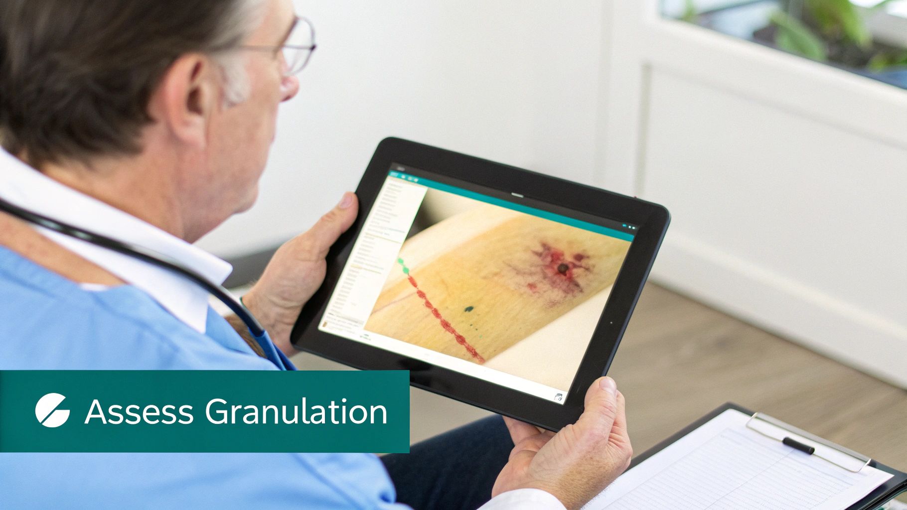
This is where the rubber meets the road. Translating the biology of wound healing into practical bedside skills is what separates good wound care from great wound care. When you look into a wound bed, a systematic assessment of the granulation tissue gives you immediate, valuable feedback on how well the body is healing. It's about moving beyond a quick glance and developing a consistent eye for what you're seeing.
The whole process starts with observing three key characteristics. Each one tells a part of the healing story, from the quality of blood flow to the moisture balance in the wound environment. A thorough assessment is the first step toward effective intervention.
Key Visual Indicators of Healthy Granulation
A trained eye can spot the signs of healthy granulation tissue from across the room. These are the green flags that tell you the proliferative phase is chugging along nicely and the wound has everything it needs to rebuild. Consistent, accurate assessment ensures that any deviations from the normal healing trajectory are caught early.
Here’s what you should be looking for every single time you assess a wound:
- Color: You want to see a vibrant, beefy red or bright pink—think of the color of fresh raspberry flesh. This brilliant color tells you there’s a rich supply of new capillaries feeding the area with oxygen and nutrients. If you see pale, dusky, or dark red tissue instead, that's a warning sign for poor perfusion or a potential infection.
- Texture: Healthy tissue has a distinct bumpy or granular feel, almost like cobblestones. This texture is literally the loops of new capillaries pushing up toward the surface. A surface that looks smooth, shiny, or gelatinous should make you think about other culprits, like biofilm or stalled healing.
- Moisture Level: The wound bed needs to be moist, but not sopping wet. A properly balanced moisture level is crucial for cells to migrate and do their jobs. Tissue that’s too dry (desiccated) or too wet (macerated) will bring healing to a screeching halt.
Consistently checking these three things—color, texture, and moisture—gives you a solid baseline to track progress. Any deviation from this healthy picture is your cue that the treatment plan might need a tweak to get healing back on track.
Measuring and Documenting Progress
Beyond a simple visual check, it's critical to quantify how much of the wound bed is actually covered in granulation tissue. We typically document this as a percentage of the total wound surface area. This objective data is crucial for tracking healing over time and for communicating effectively with other members of the care team.
For instance, a good clinical note might read, "75% of the wound bed is covered with bright red, granular tissue." This objective measurement provides a clear, data-driven way to watch the wound heal over time. It helps prove that your interventions are working and justifies the medical necessity for continued treatment. To really nail this down, you can explore our full guide on how to standardize wound bed descriptions for clearer team communication.
Let’s be honest, though—this manual process can be subjective. What one clinician calls 75% another might call 65%. This is where modern AI-powered tools are making a huge difference. By analyzing a simple photo of the wound, this technology automates the measurement, giving you highly accurate and consistent data. It eliminates the guesswork, reduces those inter-clinician discrepancies, and helps build a far more reliable patient record. That level of precision ultimately helps the entire wound care team make better-informed decisions, leading to more consistent and improved patient outcomes.
Recognizing and Managing Problematic Granulation Tissue
While healthy granulation tissue is a fantastic sign, the process can sometimes go off the rails, throwing up serious roadblocks to healing. As clinicians, we need to be just as skilled at spotting these problematic variations as we are at identifying the good stuff. Early recognition of abnormal tissue is key to preventing long-term healing complications.
The two most common challenges you'll encounter are tissue overgrowth and, conversely, its failure to appear at all. Each requires a completely different game plan.
When Good Tissue Goes Bad: Hypergranulation
When the healing process kicks into overdrive, you get hypergranulation—often called "proud flesh." This is what happens when granulation tissue grows excessively, mounding up and rising above the level of the surrounding skin.
This overgrowth creates a very real physical barrier. It stops the new skin cells, the epithelium, from migrating across the wound bed to close the gap. Think of it like a lawn that’s grown so thick and tall it completely smothers the delicate flowers trying to sprout at its edges. The epithelial cells simply can't climb over that mountain of overgrown tissue. This process, called epibole, effectively stalls the final closure of the wound.
Hypergranulation often looks like a dark red, spongy, or friable mass that bleeds at the slightest touch. Its presence is a clear signal that something is disrupting the delicate balance of healing.
What's causing this overgrowth? It's usually one of a few culprits:
- Prolonged Inflammation: An inflammatory response that just won't quit, often due to an underlying infection or a foreign body, can keep tissue production in high gear.
- Excessive Moisture: A wound environment that’s too wet can encourage cells to proliferate out of control. This is often linked to inappropriate dressing choices that trap exudate.
- Friction or Trauma: Constant irritation from dressings or repeated pressure on the wound site can trigger this over-the-top tissue response.
Managing hypergranulation means getting to the root of the problem. That might involve treating an infection or swapping out the dressing to better handle moisture. In some cases, we might need to use topical treatments like silver nitrate or corticosteroids to cauterize or shrink the excess tissue. You're essentially "mowing the lawn" back down to the right level so healing can get back on track.
Recognizing hypergranulation is more than a simple observation; it is a diagnostic clue. It tells the clinician to investigate deeper for underlying issues like a hidden infection or mechanical irritation that are actively sabotaging the healing process.
The Other Side of the Coin: Insufficient Granulation
On the opposite end of the spectrum is hypogranulation, where the wound just fails to produce enough tissue to fill the defect. This leaves you with a wound that looks completely stalled, with a recessed or hollowed-out bed that never builds up to meet the surrounding skin. The wound edges may appear rolled or thickened, further indicating a chronic, non-healing state.
Seeing this is often a red flag for a significant systemic or local problem that's starving the wound of what it needs to rebuild.
Poor blood flow (ischemia) can deprive the area of oxygen, and an out-of-control infection can completely overwhelm the body's repair mechanisms. When you see hypogranulation, it’s a sign that the wound is missing the fundamental building blocks for repair. Getting things moving again depends entirely on identifying and fixing these underlying issues to restart the healing engine. This may involve advanced therapies like negative pressure wound therapy (NPWT) or specialized growth factor products to stimulate cell activity.
Factors That Influence Tissue Development
Healthy granulation tissue doesn't just appear on its own; it's the result of a complex biological process that’s incredibly sensitive to both the patient's overall health and the specific conditions at the wound site. Think of it like trying to grow a garden. You can have the best seeds in the world (the body's healing potential), but if the soil is poor or there's not enough water, nothing will grow. The same exact principle applies to wound healing.
Both systemic (whole-body) and local (wound-specific) factors can make or break the quality and speed of tissue formation. As a clinician, getting a handle on these influences is everything. It's how you pinpoint and fix the root causes when healing stalls out.
Systemic Influences on Healing
Systemic factors are the patient-wide conditions that impact the body’s ability to kickstart a strong healing response. Honestly, these are often the biggest roadblocks to forming the healthy, beefy-red granulation tissue we want to see. Addressing these is crucial for a successful outcome.
Here are the big ones:
- Patient Age: Let's face it, healing capacity slows down as we get older. The cellular response in older adults is often less vigorous, which can put the brakes on the entire wound repair timeline.
- Nutritional Status: The body can't build something from nothing. It needs the right raw materials—especially protein, vitamin C, and zinc—to construct new tissue. Malnutrition literally starves the healing process at the cellular level. A nutritional assessment should be part of any chronic wound evaluation.
- Comorbidities: Chronic diseases like diabetes and vascular disease throw a major wrench in the works. Diabetes messes with immune function and circulation, while vascular problems choke off the supply of oxygen and nutrients that growing tissue desperately needs.
- Smoking: Nicotine is a powerful vasoconstrictor, meaning it clamps down on blood vessels. This dramatically slashes blood flow to the wound, depriving the delicate new tissue of oxygen and delaying every phase of healing.
Patient age is a particularly powerful variable. It's not just that healing slows down; the entire inflammatory response that kicks off repair can be altered. This underscores the need for patient-specific care plans that account for these age-related biological shifts.
Local Factors in the Wound Environment
While a patient's systemic health sets the stage, the conditions right there at the wound bed play an equally critical role. These local factors can either nurture granulation tissue or completely sabotage its development. Managing the local environment is a key responsibility of the wound care clinician.
The age-related delay in healing is a perfect example of this. One observational study showed that elderly patients heal more slowly because of reduced cell activity and a weaker immune response. Researchers identified that inflammatory markers like C-reactive protein (CRP) and interleukin-6 (IL-6) were significantly different in older adults, pointing to a changed inflammatory pattern that directly gets in the way of tissue formation. You can dig deeper into these findings on healing delays in elderly patients.
Common local barriers we see all the time include:
- Infection: When the bacterial load in a wound is too high, the body shifts its focus from rebuilding to fighting off the invaders. This battle consumes precious resources and brings granulation to a screeching halt. The presence of biofilm is a particularly stubborn barrier.
- Moisture Imbalance: A wound bed needs to be just right—not too dry (desiccated) and not too wet (macerated). Either extreme creates a hostile environment for the cells trying to rebuild. Dressing selection must be tailored to the amount of exudate.
- Pressure: Constant or heavy pressure on the wound site is a killer for new tissue. It can easily crush the fragile new capillaries, cutting off blood supply and causing the very tissue we're trying to grow to die. Offloading pressure is non-negotiable for healing pressure injuries.
Getting Paid: Why Your Documentation Is Your Bottom Line
In wound care, the old saying "if you didn't document it, it didn't happen" has a financial twist: if you don't document it well, you won't get paid for it. How you describe and quantify granulation tissue isn't just a clinical exercise; it's the foundation of your practice's financial health. Payers aren't in the room with you—they rely on your notes to justify the treatments and advanced therapies you’re providing.
Think about it from their perspective. When a claim gets denied, it’s usually because the documentation was too vague. A chart note that simply says "granulation tissue present" tells them almost nothing. It doesn't prove the wound is complex enough for the level of care you’re billing for.
From the Bedside to the Bank: Closing the Loop
Your clinical notes are the bridge between your assessment and getting reimbursed. This means ditching subjective, lazy descriptions and embracing a consistent, data-driven method for describing the wound bed. Accurate documentation is as crucial as the treatment itself.
When documenting granulation tissue, you need to nail two key details every single time:
- The Percentage: Be specific. Instead of "some granulation," write "60% granulation tissue." This gives payers a concrete number they can use to track progress (or lack thereof) from one visit to the next.
- The Quality: Use descriptive, standard terms that paint a clear picture. Is the tissue "beefy red and granular"? That's a sign of healthy healing. Is it "pale, friable, and bleeds easily"? That points to a problem and justifies a different treatment plan.
This level of precision creates a rock-solid patient record. It directly supports the ICD-10 and CPT codes you select, proving medical necessity and dramatically cutting down on frustrating denials and payment delays. For a closer look at building these notes, check out our guide on creating a comprehensive wound care documentation template.
Your best defense against a claim denial is clear, quantifiable documentation. It translates what you see at the bedside into the language of billing and reimbursement, ensuring you're paid fairly for your expertise.
Let's be honest—this whole process, from seeing the patient to submitting a clean claim, can be a huge time sink. It’s a major source of burnout. This is where AI-powered tools are really starting to make a difference. Imagine simply speaking your observations while you work, and having a platform automatically generate a perfectly structured note, calculate tissue percentages from a photo, and suggest the right codes. This isn't just about saving time; it's about creating consistent, clean claims that fly through the revenue cycle.
Common Questions About Granulation Tissue, Answered
Even when you've seen it a thousand times, granulation tissue can still spark questions in the clinic. Let's tackle some of the most common ones that come up during daily practice, clearing up any confusion and reinforcing what matters most.
Does Healthy Granulation Tissue Need to Be Removed?
Absolutely not. Healthy, beefy-red granulation tissue is the biological scaffold for healing and should never be removed. Think of it as the foundation being poured for a new house—you wouldn't tear it out halfway through construction. It is a sign of successful progression through the proliferative phase.
The only time you'll intervene is when it becomes hypergranulation (often called proud flesh). This is when the tissue grows above the level of the surrounding skin, creating a physical roadblock that stops new skin cells (epithelial cells) from migrating across the wound to close it. In those specific cases, the excess tissue needs to be managed so the edges can finally meet.
How Can I Promote Healthy Granulation Tissue Growth?
Getting that perfect, cobblestone-like wound bed isn't about a single magic bullet; it's about creating the ideal environment for the body to do its work. It's a holistic approach that considers both local and systemic factors.
Here’s what I focus on:
- Keep it Clean: Regular, gentle cleansing is non-negotiable. It keeps the bacterial bioburden down and prevents infection from derailing the whole process. Debridement of non-viable tissue is also crucial to create a clean slate.
- Get the Moisture Right: The goal is a moist wound bed, not a soggy swamp. Choosing the right dressing is key to hitting that perfect balance. Alginates might be used for heavy exudate, while hydrogels can donate moisture to a dry bed.
- Protect, Protect, Protect: This new tissue is incredibly fragile. You have to shield the wound from any pressure, friction, or accidental bumps that could damage the delicate new blood vessels.
- Look at the Big Picture: The body can't build new tissue from nothing. Make sure your patient is getting enough protein, vitamin C, and zinc—the essential building blocks for cellular repair. A nutritional consult can be invaluable.
At its core, promoting healthy granulation is all about removing the roadblocks and giving the body the raw materials it needs. Create the right conditions, and you allow the body’s incredible healing capacity to take over.
What Is the Difference Between Granulation and Scar Tissue?
It’s easy to mix these up, but they represent two very different chapters in the healing story. I like to think of granulation tissue as the temporary scaffolding used during construction, while scar tissue is the final, permanent patch on the wall. They are distinct in both composition and function.
Granulation tissue is the active, bustling worksite. It's temporary, packed with new blood vessels and cells, and its whole purpose is to fill the wound during the proliferative phase. Its primary component is Type III collagen, which is relatively weak.
Scar tissue, on the other hand, is what’s left after the construction crew has gone home. It forms during the final maturation phase, is mostly made of reorganized collagen (stronger Type I collagen), has far fewer blood vessels, and will never quite have the strength or flexibility of the original skin. It is avascular and acellular compared to granulation tissue.
Streamline your entire wound care workflow, from voice-powered documentation to clean claim submission, with Ekagra Health AI. Our end-to-end platform reduces administrative burdens by up to 70%, allowing your team to focus on what matters most—patient care. Discover a more efficient way to document, code, and get reimbursed at https://ekagrahealth.ai.
