When you see pus in a wound, the first question that probably pops into your head is: is this an infection? It’s a natural assumption, but the answer isn't a simple yes or no. While pus is a clear signal of an intense inflammatory response where bacteria are present, it doesn't automatically mean you're dealing with a clinically significant infection that needs antibiotics. Understanding this distinction is fundamental for effective wound care, as it guides everything from local treatment to the potential need for systemic intervention. This guide will walk you through the nuances of assessing purulent drainage, helping you move beyond a simple visual cue to a comprehensive clinical diagnosis.
Rethinking the Role of Pus in Wound Assessment
For clinicians on the front lines, seeing purulent drainage is an immediate red flag for infection. That’s a good instinct—after all, pus is literally the aftermath of a battle, made up of dead white blood cells, bacteria, and tissue debris. It’s evidence that the body is fighting something. It signifies that neutrophils, the first responders of the immune system, have been dispatched to the site to combat microbial invaders.
But here’s the crucial part: relying on the presence of pus alone can lead you down the wrong path. It's better to think of pus not as the final verdict, but as a single, important clue in a much larger clinical investigation. The real challenge for any healthcare provider is to differentiate between simple bacterial colonization, which may not require intervention, and a true, invasive infection that threatens tissue and patient health. The real challenge is to figure out if the bacteria have tipped the scales and overwhelmed the patient's defenses, causing a true infection that requires targeted treatment.
When Your Eyes Might Deceive You
The sight of pus confirms bacteria are in the mix, but it doesn’t tell you if those bacteria are causing a problem that requires systemic treatment. You might be surprised to learn how often pus samples come back from the lab with no active infection to report. This disconnect between visual assessment and microbiological findings is a key reason why a holistic approach is so vital.
A 2023 study drove this point home. Researchers analyzed 4,378 pus samples from wounds, and only 43.9% actually showed bacterial growth. That means more than half of the time, the pus wasn't indicative of a culture-positive infection. This really highlights why we can't judge a wound by its drainage alone. You can read the full research about these findings to dig deeper into the data. The study underscores that what appears purulent may sometimes be sterile inflammation or contain non-viable organisms.
This is exactly why a comprehensive assessment is non-negotiable. Always keep these points in mind:
- Pus signals inflammation, which isn't always the same thing as infection. Inflammation is the body's standard response to injury, while infection involves the invasion and multiplication of pathogenic microorganisms.
- The color and consistency of the pus can offer hints, but they are not reliable diagnostic tools on their own. For example, greenish pus is often associated with Pseudomonas aeruginosa, but this is not a definitive diagnosis without a culture.
- It must be evaluated alongside the other classic signs of wound infection. These are often referred to as the cardinal signs of inflammation and infection: redness (rubor), heat (calor), swelling (tumor), and pain (dolor).
A much better approach is to see pus as one piece of the puzzle. The full picture only becomes clear when you combine that visual cue with other clinical signs—like increasing pain, localized warmth, spreading redness (erythema), and new or worsening swelling (edema). This holistic view helps you distinguish between normal inflammation, harmless bacterial colonization, and a genuine infection that needs intervention.
To help simplify this, here is a quick table summarizing how to approach the presence of pus.
Pus as an Infection Indicator at a Glance
| Clinical Question | Key Takeaway |
|---|---|
| Does pus always mean infection? | No. It confirms an inflammatory response to bacteria but doesn't automatically confirm a clinically significant infection requiring antibiotics. |
| What does a negative culture from a pus sample mean? | It means that visual cues alone are not enough. Over 50% of pus samples in one study were culture-negative, highlighting the need for a broader clinical assessment. |
| What should I look for besides pus? | Assess the entire clinical picture: increasing pain, warmth, spreading erythema (redness), and edema (swelling). These are the classic signs that point toward true infection. |
| Is the color or smell of pus a reliable indicator? | It can provide clues about the type of bacteria present but is not a definitive diagnostic tool on its own. It's just one part of the overall assessment. |
Ultimately, the key is to move beyond a simple "pus equals infection" mindset and embrace a more nuanced, evidence-based assessment of the entire clinical situation. This requires a shift from reactive observation to proactive, critical analysis of all available patient data.
Decoding What's Inside Wound Drainage
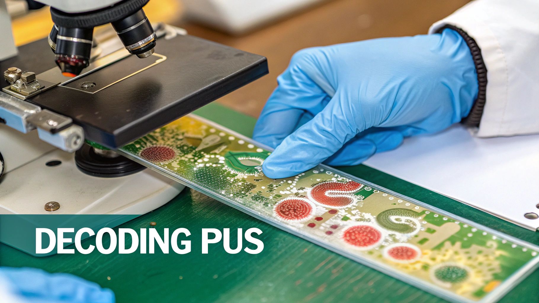
To figure out if pus means infection, you have to know what it is in the first place. I like to think of it as the battlefield debris left over after our body goes to war. When bacteria show up in a wound, the immune system sends in the cavalry—in this case, an army of white blood cells called neutrophils. These cells are phagocytic, meaning they engulf and digest foreign invaders like bacteria.
Pus, which we clinically call purulent drainage, is the thick, often colorful aftermath of that battle. It's a messy cocktail of dead neutrophils, the bacteria they fought (both dead and alive), and bits of broken-down tissue. That’s what gives it that classic opaque, viscous look, setting it apart from other fluids a wound might produce. The specific characteristics of the pus—its color, viscosity, and odor—can sometimes provide clues about the type of microorganisms present.
Differentiating Drainage Types
It's a rookie mistake to assume all fluid leaking from a wound is pus. Being able to accurately identify the type of drainage, or exudate, is one of the most fundamental skills in wound assessment. Each fluid tells a different part of the story about what's happening beneath the surface, reflecting the specific physiological state of the wound bed and the phase of healing.
In the clinic, you'll run into these four main types of wound drainage all the time:
- Serous Fluid: This is the thin, clear, or pale-yellow watery stuff. Seeing it is completely normal, especially in the early inflammatory stage of healing. It’s mostly just protein and water (plasma filtrate) that has leaked from capillaries.
- Sanguineous Fluid: Basically, this is fresh blood. It's bright red and signals active bleeding, which you'd expect to see in deeper, full-thickness wounds or immediately after trauma or debridement. It indicates damage to capillaries.
- Serosanguinous Fluid: When you see a thin, watery fluid that’s pinkish or pale red, you're looking at a mix of serous fluid and blood. It's a very common finding in wounds that are healing properly, particularly after a dressing change.
- Purulent Fluid (Pus): This is the one we're focused on. It’s thick, opaque, and can range in color from yellow and green to tan or brown. Its presence is a major red flag for bacterial activity and a significant inflammatory response.
Telling these apart is critical for getting your documentation right. Accurate description of exudate is essential for communicating the wound's status to other members of the care team and for tracking its progress over time. If you want to sharpen your eye, our guide on wound bed descriptions has more clinical examples to look at.
There's an old, outdated medical idea of "laudable pus," where its presence was thought to be a good sign of the body fighting off disease. We know better now. Unchecked purulent drainage creates the perfect breeding ground for biofilms—organized communities of bacteria encased in a protective matrix—which can completely stall healing or even send it backward.
Recognizing that pus is just one piece of the puzzle is step one. The real skill comes in combining that observation with other clinical signs to build a full, accurate picture of what's going on with your patient. This comprehensive evaluation forms the basis of a sound clinical judgment and an effective treatment plan.
Looking Beyond Pus for Signs of Infection

While purulent drainage is a major clue, you can't stop there. Diagnosing a wound infection with confidence means putting on your detective hat. Think of pus as just one piece of evidence at the scene; your job is to build the full clinical case. It's the combination and progression of signs that truly tell the story, not just the presence of pus. A single sign in isolation can be misleading.
A wound that's genuinely infected will almost always show a cluster of classic symptoms. These are the body's alarm bells, warning you that the bacterial load has tipped the scales and is starting to damage local tissue. A systematic check for these cardinal signs is essential for an accurate diagnosis and timely intervention, preventing the infection from becoming more severe or systemic.
The Classic Signs of a Spreading Problem
The most reliable indicators often have less to do with the drainage itself and more to do with the wound bed and the skin around it (the periwound skin). Taking a methodical approach ensures you won't miss the subtle (and not-so-subtle) clues that point to a bigger problem. This systematic evaluation should be a routine part of every wound assessment.
A great way to organize your thoughts at the bedside is to focus on these four key indicators.
- Pain: Is the pain new, or is it getting worse? Of course wounds can be painful, but a steady increase in pain, especially pain that seems disproportionate to the wound's size, is a huge red flag for infection.
- Redness (Erythema): Look closely at the skin around the wound. Is the redness spreading beyond the original border? Some localized redness is normal during healing, but when it starts to creep outward and feels warm to the touch, you should be thinking cellulitis.
- Exudate: Has the amount of drainage suddenly ramped up? A shift from a small amount of purulent fluid to a copious, continuous flow often signals a worsening bacterial burden. The character might also change, becoming thicker or more malodorous.
- Odor: Has the wound developed a foul or unusual smell? A distinctly bad odor can be a tell-tale sign of certain pathogenic bacteria taking hold, particularly anaerobes. Some describe the smell as sweet, foul, or putrid, which can sometimes hint at the specific organism.
When you see these signs appearing together—say, escalating pain plus spreading redness and more purulent drainage—your confidence in diagnosing a clinical infection should skyrocket. This combination is what justifies escalating care, whether that means starting antimicrobial therapy or heading for debridement.
Differentiating Local vs Spreading Infection Signs
One of the most critical judgments you'll make is determining if an infection is contained or if it's starting to spread. This distinction dictates your entire plan of care. A localized infection might respond well to topical treatments and enhanced local wound care, whereas a spreading infection is a clear signal for systemic antibiotics and may require more aggressive intervention.
This table breaks down the differences you should be looking for.
Differentiating Local vs Spreading Infection Signs
| Clinical Sign | Localized Infection (Superficial) | Spreading Infection (Deep Tissue/Systemic) |
|---|---|---|
| Pain | New or increasing localized pain at the wound site. | Spreading pain beyond the wound margin; deep, throbbing pain. |
| Erythema (Redness) | Well-defined redness limited to the wound border. | Diffuse, spreading redness (cellulitis); red streaks (lymphangitis). |
| Temperature | Localized warmth around the wound. | Systemic fever, chills; significant warmth in the surrounding limb. |
| Edema (Swelling) | Localized swelling at the wound edges. | Pitting edema, induration, or swelling extending far from the wound. |
| Drainage | Increase in volume or change in character of purulent drainage. | Crepitus (gas in tissue), abscess formation, significant purulence. |
By systematically walking through these signs, you shift the conversation from a simple "Is there pus?" to the much more important question: "What is the full clinical picture telling me about this wound?" This involves integrating all findings to form a coherent diagnosis.
This comprehensive approach is the very foundation of effective, defensible, and patient-centered wound care. It's how we move from simply reacting to what we see to proactively managing the wound's trajectory based on a complete understanding of its pathophysiology.
Why Pus Can Interfere with Antibiotics
So, you've confirmed the wound is infected and purulent. The next move is usually an antibiotic, right? Not so fast. Just starting a patient on an antimicrobial might not be enough to turn the tide. The pus itself can create a formidable shield, physically blocking the medication from ever reaching the bacteria it’s supposed to destroy. This is a critical concept in managing infections, especially those involving abscesses.
Think of an abscess as a fortress. The thick, dense collection of pus forms a very real physical barrier. This environment is often acidic and has poor blood supply, which makes it incredibly difficult for systemic antibiotics—the ones circulating in the patient's bloodstream—to actually get inside and reach the core of the infection in high enough concentrations to be effective. This reality is the basis for a surgical principle that every clinician should have etched into their memory.
Ubi pus, ibi evacua—it's a Latin phrase that simply means, "Where there is pus, evacuate it." This isn't just some dusty old saying; it’s a fundamental tenet of modern wound care that holds as true today as it did centuries ago. It’s a powerful reminder that to treat the infection, you first have to dismantle its stronghold. Source control is paramount.
This principle really drives home why source control and antimicrobial therapy have to go hand-in-hand when you're dealing with an infected, purulent wound. Relying on antibiotics alone in the presence of a significant pus collection is often a recipe for treatment failure.
The Science Behind the Barrier Effect
The difficulty of getting antibiotics into a pocket of pus isn't just a theory; it's a measurable pharmacokinetic problem. The actual amount of an antibiotic that reaches the infection site can swing wildly from one patient to the next, even if they're on the exact same dose. The local environment of the abscess—low pH, enzymatic degradation, and binding of drugs to cellular debris—all contribute to reduced antibiotic efficacy.
One pharmacokinetic study looking at how fosfomycin penetrates abscesses found that the drug concentrations inside the pus varied by more than 10-fold between individuals. That kind of massive variability shows just how unpredictable antibiotic delivery can be when a dense wall of pus is standing in the way. You can discover more insights about these drug concentration findings and see the data for yourself.
The Role of Drainage and Debridement
This is precisely why procedures like incision and drainage (I&D) or sharp debridement are so critical. These aren't just about "cleaning up" the wound. They are essential therapeutic interventions designed to break down that fortress and let the antimicrobial treatments do their job. They are the cornerstone of source control.
When you physically remove the purulent material, you're achieving several key things at once:
- Reduces Bacterial Load: You’re instantly lowering the sheer number of bacteria that the patient’s immune system and the antibiotics have to fight. This decreases the overall bioburden.
- Improves Antibiotic Penetration: With that physical barrier gone, systemic antibiotics can finally get to the remaining bacteria in the wound bed and reach therapeutic concentrations.
- Disrupts Biofilm: Purulent, low-oxygen environments are a perfect breeding ground for biofilm. Debridement is a direct, mechanical way to break up these stubborn bacterial communities that are inherently resistant to antibiotics.
Ultimately, successfully managing an infected, purulent wound requires a two-pronged attack. The antibiotics handle the systemic fight, while drainage and debridement clear the battlefield so they can be effective. Trying to do one without the other is a common reason for treatment failure and unnecessarily long healing times.
Bridging Clinical Insights and Documentation with AI

As clinicians, we know the real work doesn’t end after the wound assessment. The moment you determine that pus is indeed a sign of infection, a whole new set of tasks begins—starting with meticulous documentation. This isn't just about keeping a record; it's the critical link that justifies treatments, proves medical necessity, and secures proper reimbursement. Without clear documentation, your clinical reasoning and interventions are invisible to payers and other providers.
Your notes are the bedrock of defensible wound care. Describing purulent drainage with specifics—its amount, color, consistency, and odor—paints a clear picture for anyone who looks at that chart. But let's be honest, capturing that level of detail while juggling a packed schedule is a major source of administrative burnout. The time spent on EHRs is a significant challenge in modern healthcare.
From Spoken Words to Structured Data
This is where modern tech can genuinely make a difference. Imagine you're at the bedside, assessing a wound, and you simply say, "Moderate amount of thick, yellow, non-odorous purulent drainage noted in the wound bed." What if those words could instantly become a structured, coded entry in the patient's record? This is the promise of ambient clinical intelligence.
That’s exactly what voice-first, ambient AI systems are designed for. They act as a clinical assistant, listening to your natural conversation during an exam and pulling out the key details. It’s a simple but powerful way to close the gap between your expert clinical judgment and the rigid data fields of an EHR, translating your narrative into actionable data.
The real win here is capturing the subtleties of your assessment in the moment. It gets you away from the keyboard and frees you up to spend more time thinking, observing, and caring for your patient, rather than focusing on data entry.
Cutting Down on Clicks and Improving Accuracy
The payoff of bringing AI into your documentation workflow goes well beyond just saving a few minutes. When you automate the process of turning clinical observations into coded data, you dramatically reduce the chance for human error. It ensures the specific details you see—the very ones that help you answer "is pus a sign of infection" for this patient—are recorded accurately every single time.
This approach creates a smarter, more efficient workflow with some tangible benefits:
- Faster Charting: Your notes get drafted almost in real-time, helping you leave work on time instead of spending hours catching up on documentation. This combats clinician burnout.
- Better Coding Accuracy: The system can help connect your narrative descriptions to the right CPT and ICD-10 codes, which means fewer claim denials and faster payments. This improves the revenue cycle.
- Stronger Care Continuity: With detailed, consistent documentation, the entire care team has a complete and unambiguous view of the wound’s progress, leading to better-coordinated care.
Tools like Ekagra Health AI are built specifically for this. They give clinicians a much more natural way to handle the administrative side of wound care. And for teams looking to sharpen their initial assessments, pairing this technology with robust wound assessment tools for nurses can elevate the quality of care even further. By embracing this approach, you can create a smooth path from bedside assessment to a fully coded, billable claim, all without compromising the care your patients depend on.
A Practical Framework for Wound Assessment
Seeing pus in a wound should trigger a structured response, not a snap judgment. To figure out if that pus actually signals an infection, you need a reliable framework—one that takes you from simple observation to decisive action. A systematic approach is your best friend here, ensuring you collect all the critical clues before piecing together a diagnosis and a care plan you can stand behind. This methodical process reduces the risk of misdiagnosis and ensures all relevant factors are considered.
The key is to remember that pus is just one piece of the puzzle. Its true meaning only becomes clear when you see it in the context of the whole clinical picture. In the end, sharp assessment skills and precise documentation are the two pillars that support effective wound management. These pillars ensure that care is both clinically sound and administratively defensible.
A Step-by-Step Clinical Approach
So, you're looking at a wound with purulent drainage. Here's a methodical way to think through your next steps. This process helps you cover all your bases before jumping to interventions like culturing, debridement, or starting antibiotics.
-
Observe the Drainage: First things first, get a good look at the pus itself. You'll want to document its amount (is it scant, moderate, or copious?), its color (yellow, green, tan?), its consistency (thick or thin?), and any odor (is it malodorous or is there none?). Getting these details down creates a baseline you can use to track any changes over time.
-
Assess the Periwound Skin: Next, shift your focus to the tissue surrounding the wound. Are you seeing spreading erythema (redness)? Does the area feel warm to the touch? Is there any edema (swelling)? These are the classic signs that a local inflammatory process is starting to ramp up into a full-blown infection. Check for induration (hardness) as well.
-
Evaluate for Systemic Signs: Now, zoom out and look at the bigger picture. Talk to your patient. Are they experiencing new or worsening pain? Check for systemic red flags like a fever or chills, which tell you the body is mounting a system-wide response. Also consider malaise, confusion (especially in older adults), or elevated white blood cell counts on lab tests.
Ultimately, your goal is to synthesize these findings. A wound with a scant amount of non-odorous pus and no other local or systemic signs is managed very differently from a wound with copious, malodorous pus accompanied by spreading cellulitis and fever. The first may require only local care, while the second demands aggressive, systemic treatment.
This kind of comprehensive evaluation is the bedrock for every decision that follows. It reinforces the central message: while pus is a critical signal, its diagnostic power is unlocked only through a complete and thoughtful clinical assessment. For clinicians wanting to sharpen their methods, exploring different evidence-based wound care practices can offer more strategies to improve patient outcomes.
Common Questions from the Field
Even with a solid assessment framework, you're bound to run into tricky situations at the bedside. Let's tackle some of the most common questions that come up when you're dealing with purulent drainage and trying to connect the dots to a potential infection. These real-world scenarios often require nuanced clinical judgment.
What’s the Real Difference Between Purulent and Non-Purulent Drainage?
The simplest way to think about it is that purulent drainage is the evidence of a battle. Non-purulent drainage is just part of the normal construction process of healing.
Purulent drainage, which we all know as pus, is that thick, cloudy, and colored ooze—yellow, green, sometimes even brownish. That's because it's packed with dead white blood cells, bacteria, and other biological debris leftover from the body's immune response to an invader. It indicates a significant inflammatory reaction to microbial presence.
On the other hand, non-purulent drainage is usually thin, watery, and a lot clearer. This includes:
- Serous fluid: That clear or pale-yellow liquid you see in the very early stages of healing. Totally normal. It's the protein-rich fluid that bathes the wound bed.
- Sanguineous fluid: Bright red drainage that tells you there's active, fresh bleeding. Usually seen right after injury or debridement.
- Serosanguinous fluid: A pinkish, watery mix of the two, very common in a healthy, healing wound. It signifies the transition from the inflammatory to the proliferative phase of healing.
So, while pus is a major red flag for bacterial activity, the other types of drainage are generally just signs of a wound going through the standard phases of repair.
If There's Pus but No Fever, Is It Really Infected?
That's the million-dollar question, isn't it? The short answer is: not always, but you need to be cautious. Seeing pus without systemic signs like a fever often means you're looking at a localized infection. The body is fighting hard, but it's keeping the battle contained right there in the wound bed.
This could also be a state of critical colonization, where the bacterial load is heavy enough to delay healing but hasn't triggered a full-blown systemic alarm yet. A fever is the body's signal that the fight is spreading beyond the wound's borders. However, it's important to remember that some patients, particularly the elderly or immunocompromised, may not mount a fever response even with a significant infection.
So, to answer the question, "is pus a sign of infection?" in this case, you have to look at the whole clinical picture. Don't just focus on the thermometer. Are you seeing other local signs? Is the pain getting worse? Is there more warmth, spreading redness, or a foul odor? Those clues, combined with the pus, will tell you whether it's time to escalate treatment, even in the absence of a fever.
How Do I Document Pus to Get Coding and Reimbursement Right?
This is where details make all the difference. Vague notes just don't cut it. To ensure you get proper reimbursement and your documentation stands up to scrutiny, you have to paint a clear picture. Simply writing "pus present" is a missed opportunity and clinically insufficient.
Think like a painter, not just a reporter. Instead, document something like this: "Wound bed exhibits a moderate amount of thick, green, malodorous purulent drainage." This level of detail provides objective data that supports your diagnosis.
This kind of specific description provides undeniable clinical evidence. It justifies procedures like debridement (CPT codes 97597/97598) and solidly supports the ICD-10 codes for localized infections, such as those in the L00-L08 category (Infections of the skin and subcutaneous tissue). Precise language creates a rock-solid record that backs up your clinical decisions, helps protect against audits, and gives the next clinician a clear understanding of what's going on, ensuring continuity of care.
At Ekagra Health AI, we're focused on closing the gap between your expert clinical judgment and the tedious demands of documentation. Our voice-first, ambient AI platform is designed to capture your detailed narrative notes right at the bedside, then automatically translate them into the structured, coded data required for billing. We help your team cut down on administrative work and get back to what matters most—caring for patients.
