A precise wound edge description is what elevates your assessment from a subjective art to an objective science. It tells a clear, actionable story about the wound’s status—is it healing, stalled, or getting worse? This kind of detailed data is absolutely essential for smart treatment planning, predicting healing outcomes, and getting reimbursement right. For healthcare professionals, from nurses in long-term care to surgeons in acute settings, mastering this skill is a cornerstone of effective wound management.
Why Accurate Wound Edge Description Matters
Let’s be honest: mastering the language of wound edges isn't just a "nice-to-have" skill for clinicians; it's a non-negotiable. It's how we move our documentation beyond vague notes like "looks better" and into the world of measurable, objective data. This level of precision is the bedrock of effective care plans and clear communication across the entire care team. Without it, continuity of care suffers, and crucial changes in the wound’s condition can be missed.
Getting the description right serves a few critical functions:
- Predicting Healing Trajectories: The look of the wound edge is one of the most powerful predictors of healing. When you see advancing, attached edges, that’s a great sign. But if you spot rolled edges (epibole) or a fibrotic rim, you know healing has likely stalled and it's time to intervene. A detailed description allows for early identification of non-healing indicators, prompting a re-evaluation of the treatment plan.
- Guiding Clinical Decisions: Identifying features like undermining or indistinct borders immediately informs your next steps. It helps you decide if debridement is needed, if a different dressing is required, or if you need to investigate for a possible infection. This specificity ensures that interventions are targeted and appropriate for the wound's current state.
- Ensuring Proper Reimbursement: Standardized, specific terms are what justify medical necessity. Documenting "undermining from 3:00 to 6:00" gives you much stronger support for billing than just saying the wound is "deep." Payers require objective evidence, and a clear wound edge description provides the necessary detail to support claims for advanced treatments and procedures.
- Powering Advanced Analytics: For AI-driven platforms, structured data is everything. A clear wound edge description becomes a key data point that helps algorithms track healing, suggest treatments, and even automate parts of your charting. This structured input transforms narrative notes into powerful datasets for predictive modeling and quality improvement initiatives.
This skill is just as fundamental to a solid wound assessment as knowing how to correctly stage an injury. If you want to dive deeper into that, check out our guide on pressure injury staging and its visual assessment.
Quick Reference for Wound Edge Terminology
For any busy clinician, a scannable, go-to guide for wound edge terms is an absolute must for charting efficiently and accurately. A precise wound edge description isn't just a minor detail—it's one of the most powerful indicators of a wound's healing trajectory. Think of this section as your rapid lookup tool for charting. It’s designed to help you quickly find the right term to describe your clinical findings, ensuring consistency and clarity in your documentation.

When you nail these terms, you're not just documenting; you're translating your clinical assessment into a universal language. This is a language understood by the entire care team, your billing department, and even the AI-powered platforms that are becoming central to our work. The whole point is to make every note you write both clinically sharp and incredibly efficient. This shared vocabulary minimizes ambiguity and ensures that every member of the healthcare team, from the specialist to the primary care provider, is on the same page regarding the wound's status.
Wound Edge Characteristics At a Glance
To make things even faster, we've put together a summary table of the most common wound edge descriptors. It gives you a quick definition and the primary clinical red flag or green light associated with it. You can use this table as a quick index for the more detailed explanations that follow, helping you spot and document different wound edge types with confidence. This table serves as a daily reference to reinforce accurate terminology and streamline the documentation process.
| Edge Characteristic | Brief Definition | Primary Clinical Implication |
|---|---|---|
| Attached | Edges are flush with the wound bed, indicating a smooth transition from the wound to periwound skin. | Positive: A sign of healthy tissue connection and a proper healing environment. |
| Detached | Edges are separated from the wound bed, creating a visible lip or cliff-like margin. | Negative: Indicates potential for undermining or tunneling, requiring further investigation. |
| Epibole (Rolled) | Edges have rolled or curled under, creating a raised, rounded, and often calloused rim. | Negative: A major barrier to healing; epidermal cells cannot migrate across the wound bed. |
| Advancing | Visible migration of new, pale pink epithelial cells from the wound edge inward. | Positive: The clearest visual sign that the wound is actively closing and healing. |
| Fibrotic | Edges are hard, calloused, and often discolored; feel rigid to the touch. | Negative: Represents non-viable, scarred tissue that prevents epithelial migration and wound contraction. |
| Indistinct | Borders are poorly defined and blend into the surrounding skin, often due to maceration or inflammation. | Negative: Often signals excessive moisture, edema, or potential infection. |
This at-a-glance format ensures every observation you document is not just a note, but a clinically significant data point that tells a clear story about the wound's status. It empowers clinicians to quickly translate visual cues into standardized, meaningful documentation.
Defining Attached and Advancing Edges
When you're describing a wound edge, spotting the good signs is just as important as flagging the problems. Two of the most encouraging indicators that a wound is healing properly are attached and advancing edges. Documenting these features is your proof that the healing process is on track and that the current treatment plan is effective. Recognizing these positive signs provides crucial validation for both the clinical team and the patient.
An attached edge is exactly what it sounds like—the edge of the wound sits flush with the wound bed. You won't see any cliffs, lips, or gaps. Instead, it’s a smooth, gentle slope from the periwound skin down into the granulation tissue. This connection is everything; it creates the stable foundation new cells need to start their migration across the wound. This seamless integration indicates that the wound bed is healthy and receptive to the final stages of closure.
What Healthy Edges Look Like
Attached edges mean the wound bed and the surrounding skin are well-connected, which is the essential first step for the next phase of healing. Think of it as a perfectly graded on-ramp for epithelial cells to move in, free of any physical barriers that could stop their progress. It's a simple observation, but it powerfully confirms a healthy wound environment. This finding is a green light, signaling that the body's natural healing mechanisms are functioning as they should.
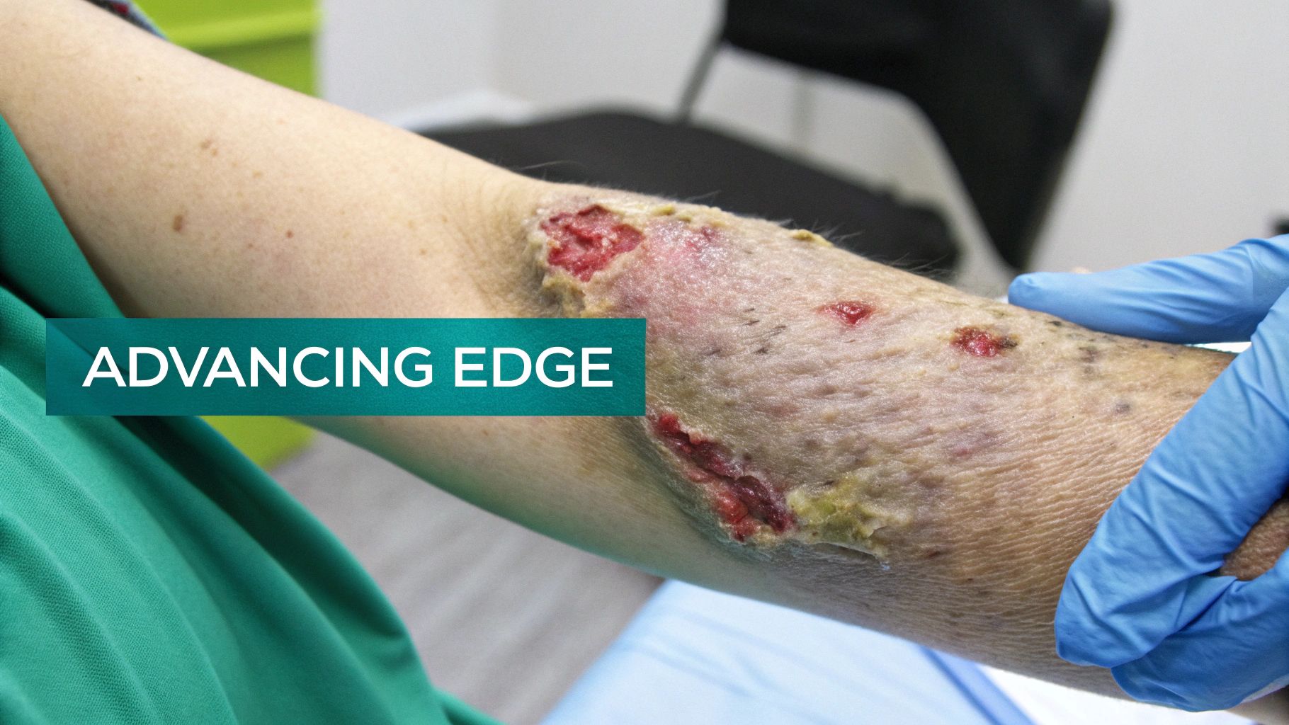
On the other hand, advancing edges—sometimes called epithelializing edges—are visible proof of healing in action. This is where you can actually see new, pale pink or even translucent skin cells (epithelium) actively moving from the wound’s perimeter toward the center, shrinking the wound as they go. This cellular migration is the real engine of wound closure. Observing this delicate, new tissue is one of the most rewarding moments in wound care, as it provides tangible evidence of progress.
Documenting how quickly the edge is advancing gives you an objective way to measure healing. A consistently advancing edge is one of the strongest predictors of a positive outcome.
In fact, research shows that when wound edges advance at a rate of 2-3 mm per week under optimal conditions, it predicts successful closure in 77% of chronic venous leg ulcers. This comes from a meta-analysis covering over 5,000 cases in North America and Europe. You can explore the full study on wound edge advancement findings. This metric provides a quantitative benchmark against which you can measure the effectiveness of your interventions.
Documentation and Annotation Tips
Charting these healing signs accurately is critical for tracking progress and justifying your care plan. Vague notes are insufficient; precision is key.
- Documentation Example: "Wound edges are 100% attached and flush with the granular bed. A 2mm rim of advancing epithelium is noted around the entire perimeter, indicating active healing." This example provides both qualitative and quantitative data, creating a comprehensive picture.
- Photo Annotation: When you take a picture, use arrows or a dotted line to highlight that pale pink, advancing epithelial border. This creates a clear visual distinction between new growth and older tissue, providing solid evidence for AI analysis and future comparisons. Visual documentation is a powerful tool for demonstrating progress over time.
Identifying Detached and Undermined Edges
When you see a wound edge that has pulled away from the tissue beneath it, you're looking at a major roadblock in the healing process. These separations create hidden pockets and shelves—perfect breeding grounds for bacteria—which ramps up the risk of infection and abscesses. Being able to spot, measure, and describe these issues correctly is a non-negotiable part of any solid wound assessment. Failure to identify and manage these features can lead to significant complications and prolonged healing times.
A detached edge is the first sign of trouble. This is where the edge is no longer flush with the wound bed, creating a sort of lip or cliff. Often, this detachment is just the beginning of a bigger problem: undermining. It signals a disruption in the healing cascade and warrants immediate, closer inspection to determine the full extent of the issue.
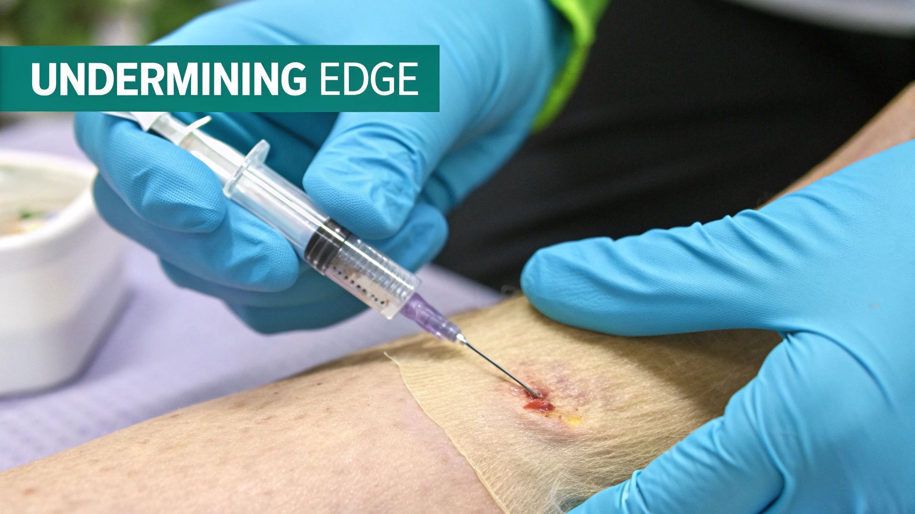
Undermining is what happens when that separation goes deeper, forming a pocket that extends horizontally under the skin at the wound's margin. This hidden cavity can be far more extensive than what you see on the surface, which is why getting an accurate measurement is so critical. It represents a significant area of dead space that must be addressed to allow the wound to heal from the base upwards.
Measuring and Documenting Undermining
Charting undermining isn't something you can just eyeball. You need a systematic approach to be consistent and accurate from one assessment to the next. The gold standard here is the clock method, which gives everyone on the care team a clear, universal map of the wound. This standardized technique ensures that measurements are reproducible and comparable across different clinicians and assessment times.
To measure undermining, a clinician takes a sterile, cotton-tipped applicator and gently probes the wound edge. You'll measure the depth of the pocket and note its location using a clock face, with 12:00 always pointing toward the patient's head. This systematic exploration maps the hidden geography of the wound.
Getting precise measurements and clear documentation for undermining is an absolute must. It directly impacts your treatment plan—like whether you need to pack the wound—and gives you hard data to track if the cavity is getting better or worse.
This process turns a simple observation into real, actionable clinical data. It’s a core skill for any clinician who's serious about high-quality wound care. The accuracy of these measurements directly influences treatment choices and outcomes.
Clinical Implications and Charting Examples
Finding undermining immediately changes your plan of care. That dead space has to be managed to stop fluid from collecting and turning into an infection. This usually means loosely packing the area with the right dressing material to help the wound heal from the bottom up. The goal is to eliminate the dead space and promote granulation from the base of the wound.
Your charting needs to reflect these findings with total clarity. Ambiguity can lead to improper treatment and adverse outcomes.
- Clinical Concern: The presence of undermining dramatically increases the risk of abscess formation and can bring healing to a halt. It also significantly increases the wound's overall volume and complexity.
- Documentation Example: "Wound edges detached from 12:00 to 4:00. Probing reveals a 3.5 cm deep area of undermining extending from the 12:00 position to the 3:00 position. The cavity was loosely packed with antimicrobial ribbon gauze."
- Photo Annotation Tip: When you're taking a picture, place sterile applicators into the undermined areas. This gives a powerful visual of the depth and direction of the pockets, which is invaluable for both documentation and AI-driven analysis. This technique makes the abstract concept of undermining tangible and understandable.
Recognizing Non-Advancing Edges Like Epibole and Fibrosis
When a wound just won't heal, the edges often tell you exactly why. Two of the most common culprits behind a stalled, chronic wound are epibole and fibrosis. Being able to spot these is critical because they're quite literally physical barriers that stop the healing process cold and almost always require intervention. These are not subtle signs; they are clear indicators that the wound has entered a chronic, non-healing state and requires a change in strategy.
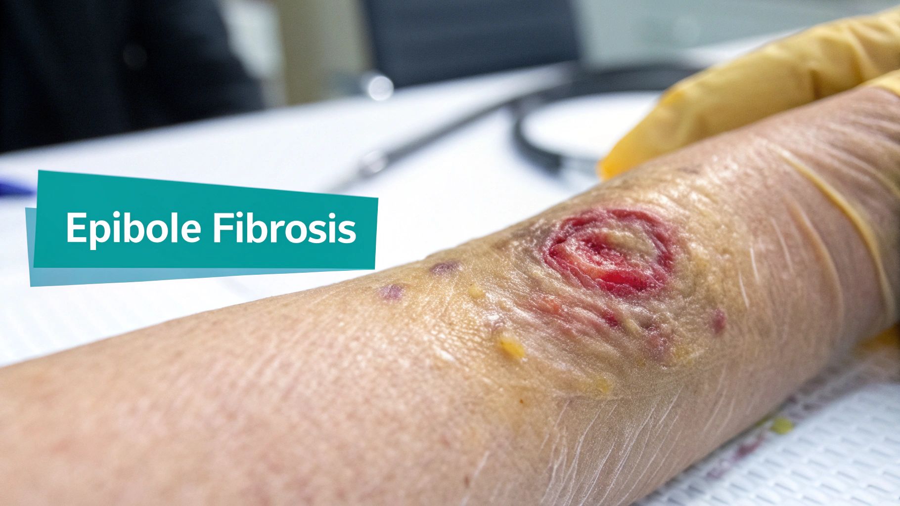
Epibole is what happens when the epidermal cells at the wound margin give up on migrating across the wound bed. Instead, they roll or curl under, creating a distinct raised, rounded, and often calloused rim. The body essentially gets confused and treats this rolled edge as healed, which shuts down any further effort to close the wound, leaving it open indefinitely. This physiological "error" effectively seals the wound's fate as a chronic ulcer unless corrected.
A fibrotic edge is a different beast but just as problematic. You'll know it when you feel it—this type of edge is hard, rigid, and feels non-viable to the touch. It's made of dense, scar-like tissue that physically blocks new epithelial cells from moving forward, building a wall that prevents wound contraction and closure. This barrier is often the result of prolonged inflammation and is a hallmark of chronic wounds.
Clinical Significance and Documentation
The presence of either epibole or fibrotic edges is a clear red flag. It’s a signal that the wound needs more aggressive management, and that usually starts with debridement. You have to remove this non-viable tissue to essentially "re-awaken" the wound, turning it back into an acute wound that can get back on a healthy healing path. Making the right call here is everything; you can explore more about evidence-based wound dressing selection strategies in our related guide.
For multidisciplinary teams, especially in fields like podiatry and vascular surgery, seeing edges described as 'indistinct' or 'fibrotic' signals stalled healing in a staggering 52% of pressure injuries. This kind of data is what drives the use of advanced treatments like NPWT, which was shown to boost healing rates by 40% in a trial of 1,200 patients. You can dig into the impact of these findings on healing rates yourself.
Your documentation has to be razor-sharp to justify these more advanced interventions. Clear, specific notes are what allow the entire care team to understand exactly why a wound is stuck and why a more aggressive approach is medically necessary.
- Epibole Example: "Epibole noted along the entire wound perimeter, with edges rolled and calloused, preventing epithelial migration."
- Fibrosis Example: "A thick, fibrotic rim is present from 9:00 to 12:00, feeling firm and non-pliable. Debridement is indicated to stimulate healing."
- Photo Annotation: When documenting with a photo, use arrows to point directly to the rolled or hardened rim. Label it clearly as "Epibole" or "Fibrotic Edge" so there's no ambiguity. This visual evidence supports your clinical assessment and treatment plan.
Documenting Indistinct and Irregular Edges
When a wound's perimeter gets fuzzy or jagged, it's more than just a measurement challenge—it’s a clinical signal. These edges often point to underlying problems that are actively stalling the healing process. Unlike a clean, well-defined wound, indistinct or irregular edges are early warnings that need precise and descriptive documentation. They suggest that the local wound environment is compromised, which requires further investigation to identify the root cause.
An indistinct or diffuse edge is one where the border seems to melt into the surrounding skin. It lacks a clear line of demarcation. This is a classic sign of maceration from too much moisture or significant inflammation, making it tough to see where the wound truly ends. This ambiguity is a major clinical red flag because it often accompanies periwound skin breakdown, increasing the overall size and complexity of the wound.
Defining Irregular Wound Edges
On the other hand, an irregular edge is one you can clearly see, but it’s not smooth. The shape is uneven, jagged, or asymmetrical instead of a neat circle or oval. Think of a coastline on a map. This often hints at an underlying issue throwing off the healing trajectory, like inconsistent pressure, an lurking infection, or poor blood flow hitting different parts of the wound unevenly. The shape can provide clues about the etiology of the wound or the forces acting upon it.
Noting these edge types is far from a trivial charting task. The well-respected Bates-Jensen Wound Assessment Tool (BWAT), for example, has long treated non-healing edges as a sign of serious trouble. Modern data backs this up, showing that 38% of chronic wounds in Europe have irregular edges. This one characteristic can delay closure by an average of 21 days and dramatically increase infection risk. You can dig deeper into the market impact of chronic wound care to understand the scale of this issue.
Good charting here moves beyond just measuring length and width. When you describe the character of an indistinct or irregular edge, you're building a clinical case for specific interventions, like advanced moisture-wicking dressings or antimicrobials.
Charting Examples for Clarity
Your documentation needs to paint a clear picture for the whole team, connecting what you see to what it means clinically. It's about telling a story that leads to the right action.
- Indistinct Edge Example: "Wound borders are indistinct and diffuse from 10:00 to 2:00 due to significant periwound maceration, obscuring the true wound margin." This documentation links the observation (indistinct edge) to the cause (maceration).
- Irregular Edge Example: "Wound perimeter is irregular with a jagged, sawtooth appearance, particularly along the inferior border, suggesting uneven healing progression." This description provides a vivid mental image of the wound's shape.
- Photo Annotation Tip: For an indistinct edge, use a dotted line in your photo annotation to estimate the true border. For an irregular one, trace the jagged perimeter to draw attention to it. These visual aids help clarify complex wound presentations.
Standardizing Documentation for Billing and AI Integration
The way you describe a wound edge does more than just guide clinical care—it has a direct line to your organization's bottom line and its readiness for the future of healthcare. When you ditch vague notes for standardized, specific terms, you’re building a powerful dataset. This data is what justifies treatments, shores up billing, and paves the way for a smooth integration with artificial intelligence. In an increasingly data-driven healthcare landscape, high-quality documentation is not just good practice; it's a strategic imperative.
This kind of precision is really the backbone of a strong revenue cycle. Payers are looking for undeniable evidence of medical necessity, and this is especially true for procedures like sharp debridement. A note that says "fibrotic rim noted from 1:00 to 4:00, preventing epithelial advancement" gives you concrete justification that a simple "bad wound edge" just can't provide. Getting this level of detail into your charting ensures you're aligned with specific billing requirements and helps cut down on those frustrating claim denials. For a closer look at this connection, our ICD-10 and CPT coding guide for wound care billing is an excellent resource.
Powering AI and Slashing Denials
Beyond billing, standardized documentation is the fuel that runs modern AI platforms. When you and your team consistently use terms like "epibole," "undermining," or "advancing," you're creating structured data. AI systems can then comb through this data to automatically track healing rates, flag wounds that have stalled, and even suggest the right billing codes. This automation reduces administrative burden and enhances clinical decision support.
This is where wound care is headed. In major markets, data shows a clear correlation: North American wound centers using smart sensors see edge advancement rates of 1.5 mm/week in 70% of their cases. This improved data capture and ICD-10 specificity has helped them slash denials by 40%. You can learn more about how precise data impacts financial outcomes by exploring these insights into the wound care market. The transition to value-based care makes this level of data analytics essential for demonstrating quality and efficiency.
Precise language isn't just good practice; it's a strategic asset. It transforms subjective observations into objective, analyzable data that strengthens reimbursement claims and unlocks the full potential of AI-driven workflow automation.
The table below provides a clear snapshot of how specific descriptions connect directly to coding and AI analysis, effectively turning your clinical notes into actionable intelligence. This demonstrates the tangible link between bedside observations and high-level operational efficiency.
Documentation Impact on Coding and AI
This table links the clinical terms you use at the bedside to their downstream impact on reimbursement and AI-powered analytics.
| Wound Edge Description | Example Documentation Phrase | Potential Coding/Billing Implication | Relevance for AI Analysis |
|---|---|---|---|
| Epibole | "Rolled, calloused edges noted along the entire perimeter, preventing epithelial migration." | Justifies the medical necessity for selective debridement to prepare the wound bed. | AI can flag this as a stalled wound and suggest debridement CPT codes for review. |
| Undermining | "4 cm of undermining measured from 12:00 to 3:00, requiring packing." | Supports billing for more complex dressings and additional procedural time. | AI algorithms can calculate wound volume changes and track the resolution of dead space over time. |
| Advancing | "A 2mm rim of advancing epithelium is visible, indicating active healing." | Provides objective evidence of treatment efficacy, supporting continued care authorization. | AI can quantify the rate of advancement, predict closure timelines, and visualize healing progress. |
By adopting these specific descriptors, you're not just documenting—you're creating a robust, data-rich record that serves your patient, your practice, and the advanced tools that will define the future of wound care.
Common Mistakes to Avoid in Wound Edge Documentation
Getting wound edge descriptions right is non-negotiable, but a few common pitfalls can easily trip up even seasoned clinicians. When you avoid these mistakes, your charting becomes clearer, more valuable, and ready to support everything from reimbursement to advanced analytics. Recognizing and correcting these common errors is a simple yet powerful way to improve the quality of care and documentation.
Probably the most frequent error I see is using subjective, fuzzy language. Phrases like "looks better" or "edge appears unhealthy" don't mean anything in a clinical context. They offer zero objective data for the next person to compare against, masking subtle signs of trouble and leading to inconsistent care. This lack of specificity makes it impossible to accurately track changes over time.
Vague vs. Specific Descriptions
Another major misstep is failing to quantify what you see. Just noting the presence of undermining doesn't tell the whole story. To track progress—or regression—you absolutely have to include measurements. Objective measurements are the foundation of evidence-based practice and are essential for demonstrating outcomes.
Think about the difference this makes in a chart:
- What Not to Do: "Wound has some undermining."
- What to Do Instead: "A 3.5 cm deep area of undermining is present from the 1:00 to 4:00 position."
It's also crucial not to confuse distinct clinical terms. Mixing up undermining (a wide, shelf-like pocket) with tunneling (a narrow, linear channel) is a classic example. Each one demands a completely different management strategy, so using precise terminology is critical for effective care. This type of error can lead to inappropriate treatment, such as incorrect dressing selection or packing technique.
Accurate documentation isn't just a clinical best practice; it's a financial necessity. Vague or incomplete descriptions are a primary driver of claim rejections, gumming up the entire revenue cycle.
Globally, poor edge documentation contributes to a staggering 30% of claim denials in outpatient wound care centers. That's a huge piece of the industry's multi-billion dollar annual spend. You can learn more about the staggering costs by reviewing these insights on the wound care market. This statistic underscores the direct financial impact of documentation quality.
By auditing your own charting for these common habits and committing to specificity, you're doing more than just ticking a box. You're directly improving team communication, building ironclad billing claims, and feeding high-quality data into the AI tools that are shaping the future of our field.
Common Questions About Documenting Wound Edges
Once you start getting granular with wound edge descriptions, a few practical questions always pop up. It's one thing to know the definitions, but it's another to apply them consistently in the thick of a busy clinic day. Let's tackle some of the most common queries clinicians have to bridge the gap between theory and practice.
What's the Real Difference Between Undermining and a Tunnel?
This is a classic point of confusion, but the distinction is all about the shape. While both involve tissue loss under the intact skin, they look very different and have different clinical implications. Understanding this difference is crucial for proper wound management.
Undermining is like a cavern or a shelf. It's a wider area of tissue separation that runs parallel to the skin's surface, creating a lip around the wound. A tunnel, on the other hand, is a narrow, linear channel or passageway that dives deeper into the tissue, connecting two distinct points or ending in a blind pouch. When charting, you'll describe undermining using the clock face for location and depth, while a tunnel is all about its direction and total length from a single entry point.
How Often Should I Re-evaluate the Wound Edge?
Simple answer: at every single assessment. The edges tell a huge part of the wound's story, and that story changes constantly. The frequency of assessment is key to detecting changes early and responding appropriately.
How often you assess depends entirely on the setting and the patient's condition.
- Acute Wounds: In the hospital, you might be looking at this daily or even with every dressing change, especially post-op. Frequent monitoring is crucial in the dynamic environment of an acute wound.
- Chronic Wounds: For your clinic or home health patients, weekly assessments are typically the standard. This cadence allows for tracking progress without being overly burdensome.
There's no shortcut here. Consistent, detailed documentation is the only way you can prove healing, spot a wound that's stalled out, and justify your plan of care to payers.
Can a Single Wound Have Multiple Edge Types?
Absolutely. In fact, it's more common than not, especially in large or complex chronic wounds. Don't fall into the trap of describing the entire perimeter with one term. A wound is not a uniform entity; different areas can exhibit different characteristics based on factors like pressure, perfusion, and local tissue health.
It's very typical to see a wound where the superior edge from 10 to 2 o'clock is advancing beautifully, but the inferior edge shows clear signs of epibole or is thick with fibrotic tissue. Documenting this level of detail is what separates a good note from a great one. It paints a complete clinical picture and helps you target interventions to the specific areas that need them most.
At Ekagra Health AI, we get that capturing this level of detail can be time-consuming. Our voice-first platform is designed to turn your expert clinical observations into structured, actionable data—effortlessly. You can capture precise wound edge descriptions every time, which makes charting faster and helps you get paid for the complex work you do. See how our AI-powered solution can cut your documentation time at https://ekagrahealth.ai.
